


Powering scientific intelligence.
Revvity unlocks the full potential of AI with trusted biological intelligence.
Revvity brings together AI-powered informatics, automation, reagents, imaging, analytical technologies, and scientific expertise to power connected Lab-in-the-Loop workflows that transform biological data into actionable scientific insight.
See how AI-enabled science can support your work.
Connecting AI predictive modeling with wet lab experimentation.
Lab-in-the-Loop approaches help close this gap by creating continuous feedback loops between AI-driven prediction and wet lab experimentation.
Revvity supports these next-generation research environments with solutions that help laboratories:
- Capture and manage high-quality R&D data
- Connect in-silico insights with wet lab results
- Scale experimental workflows
- Improve reproducibility across studies
- Accelerate iteration from prediction to validation
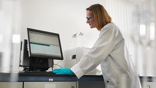

Built for the AI era of discovery.
Revvity enables researchers to turn AI-driven predictions into confident scientific decisions.
Our integrated reagents, platforms, automation, imaging, and informatics solutions connect the full Lab-in-the-Loop workflow, from model creation and experimentation to biological validation and continuous learning.
AI in the news





Enabling the flow between AI insight and scientific validation.
Make
Lay the foundation for AI-enabled discovery by creating biologically relevant models for experimentation.
Revvity provides the tools researchers need to engineer, modify, and prepare biological systems for downstream testing and validation.
Sample prep, automation and plate handling:
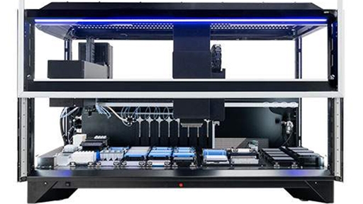
Fontus Workstations
With easy-to-use software, optimized deck access, verified protocols, and reformatting capabilities to simplify your workflows and improve turnaround times
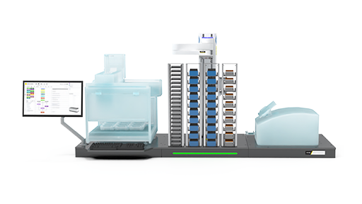
Explorer G3 SmartBench automation solutions
Affordable, scalable laboratory automation with the explorer™ G3 SmartBench workstations, that integrate 150+ instruments with collaborative robotics for seamless workflows.
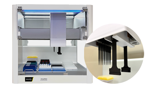
AssayMateワークステーション
あらゆるラボに、自動化性能を:手頃なコストで導入できるベンチトップサイズの自動リキッドハンドラー。幅広いラボアプリケーションを最適化するために設計されています。
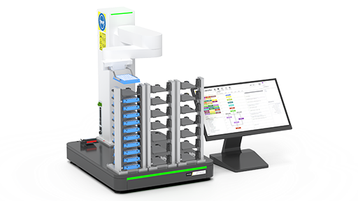
plate::handler Connect
Overcome manual workflow bottlenecks with the plate::handler™ Connect, a collaborative, four-axis SCARA robotic arm for a compact and cost-effective entry point into automated processing.
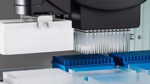
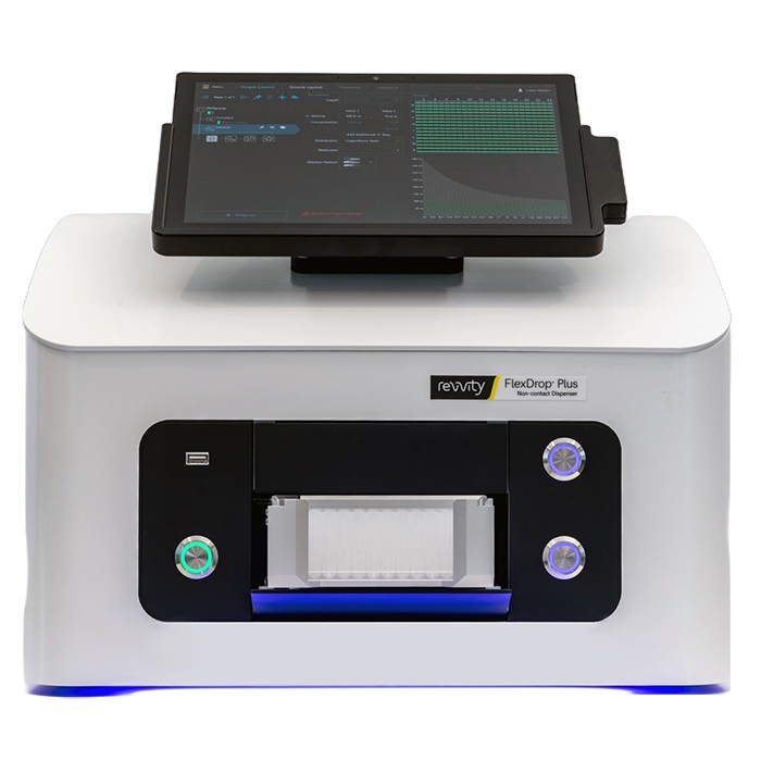
FlexDrop Plus 非接触ディスペンサー
FlexDrop Plusプラットフォームは、ナノリットルレベルの微量液体の自動分注を、これまでにないほど簡単かつ正確に行えるようにしました。統合型タッチスクリーンPCで操作できる使いやすいデバイスで、ボタンを押すだけ最少8 nLの液体を安定して分注できます。あらゆる研究室でお使いいただけます。
FlexDrop Plusプラットフォームは、8つの独立した制御チャネルで陽圧を使用します。各チャンネルは1秒間に最大100滴を分注できます。目標の分注容量を達成するため、8ナノリットル(nL)から50 nLまでの液滴を生成します。システムには液滴検証が搭載されており、分注された液滴数を正確に検証し、実行後に検証レポートを提供します。特許取得済みのソースウェルプレートには、底部に100 µmの穴があり、陽圧の下で液滴を容易に分注します。
タッチスクリーンソフトウェアコントロールはシームレスで簡単に使用可能です。プログラミングや自動化の経験は必要ありません。セットアップから分注開始までわずか数分で完了します。
構成内容:
- FlexDrop Plus機器
- (4)電源コード(EU、英国、米国、オーストラリア)ヒューズの交換が必要になる場合があります。
- エアチューブ – 機器へのクイックコネクトと施設の圧空またはコンプレッサーに接続するためのフィッティングを備えた 6 mm チューブ
- (4)黒色アダプタープレート(1、2、4、8 mm)– ターゲットプレートの高さ調整用
- スタートアップキットに含まれる80 uLウェルのFlexDropソースプレート
- 廃棄用PCRチューブ
Immunoassays:
AlphaLISA™ SureFire® Ultra™
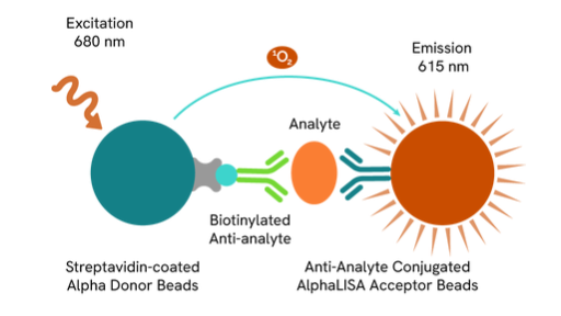
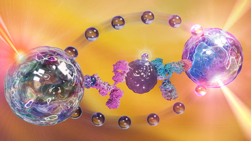
AlphaLISA® SureFire® Ultra™ is a homogeneous, no-wash assay platform designed for the sensitive detection of endogenous and tagged phosphorylated or non-phosphorylated proteins in cell lysates.
HTRF
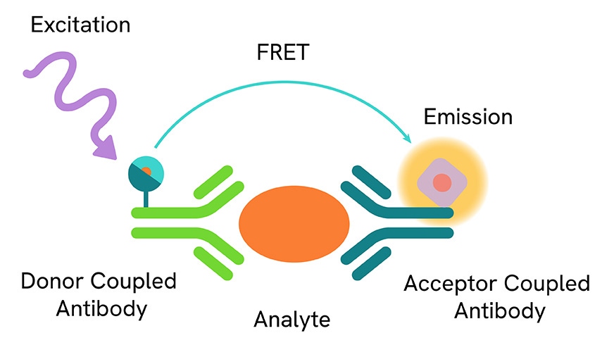
HTRF™ (Homogeneous Time Resolved Fluorescence)は、標準的なFRET技術と蛍光の時間分解測定を組み合わせた無洗浄技術で、短寿命のバックグラウンド蛍光を除去します。
Biological model development:
TotalSeq™ reagents
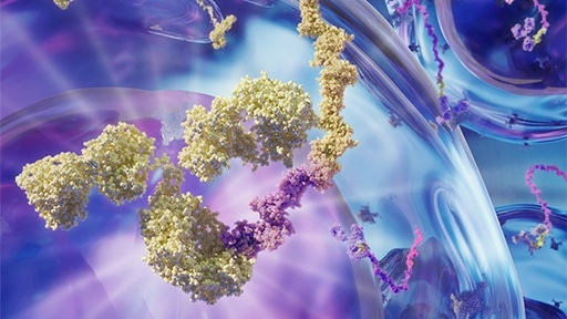
Seamlessly add protein expression to single-cell multiomics workflows with oligonucleotide-conjugated antibodies, hashtags, and cocktails.
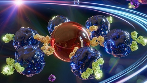
LEGENDplex™
Multiplex protein quantification for 14 analytes in a single sample. Select focused pre-defined panels or customize each target.
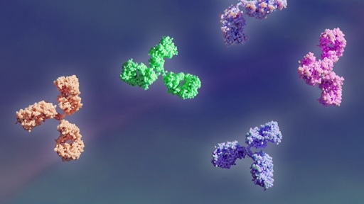
BioLegend Antibodies
Choose from thousands of primary and secondary antibodies. We offer directly conjugated or purified antibodies to match your needs.
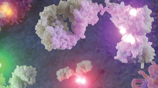
BioLegend Flow Cytometry
Phenotype immune cells by building bigger and better flow cytometry panels with novel fluorophores and highly specific antibodies.
Cell culture reagents:
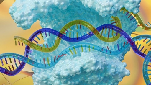
Dharmacon™ CRISPR reagents and libraries
Browse CRISPR based reagents including: sgRNA and Cas9 solutions for CRISPR knock-out, CRISPR knock-in, CRISPR activation, CRISPR interference, and CRISPR derived base editing.
Dharmacon™ RNAi reagents and libraries
Choose from a broad portfolio of synthetic and lentiviral RNAi reagents including: siRNA, shRNA, and microRNA for highly specific and efficient gene silencing.

Pin-point™ base editing reagents
Base editing reagents including custom sgRNAs, CBE or ABE mRNA, validated controls, and non-targeting controls for precise, DSB-free single-nucleotide edits.
Cell monitoring:
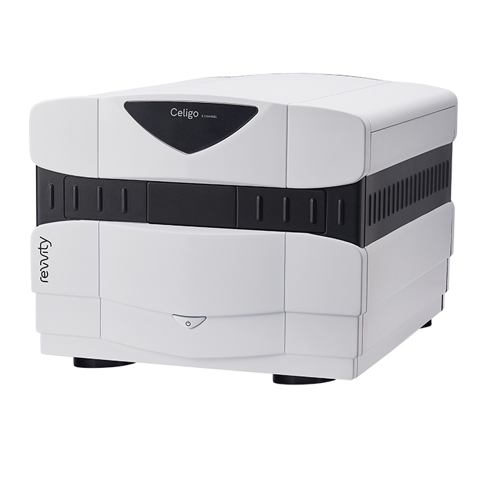
Celigoイメージサイトメーター
Celigoは、プレートベースのベンチトップ型明視野および蛍光イメージングシステムです。ウェル全体での細胞解析と細胞サンプルの特性評価を目的に設計されています。Celigoシステムは、6~1536ウェルプレート、T25、T75フラスコ、10 cmディッシュ、ガラススライドなど、さまざまな容器内の細胞を自然な状態のままイメージングおよび解析します。
個々の細胞レベルでの解析データを簡単に生成できるため、ELISAやタンパク質ベースの生化学的なアッセイとは異なる細胞レベルの情報を提供します。さらに、フローサイトメトリーよりも高速に結果を得られます。Celigoサイトメーターに最適な細胞ベースアッセイ:
- アポトーシス
- 細胞周期
- 蛍光レポーター
- 細胞毒性
- ラベルフリー増殖アッセイ
ウェル全体の高速イメージングと解析
- 正確な細胞集団解析を実現する高感度ウェル全体のイメージング
- 不均一な播種の影響を排除
- 各ウェル内のすべての細胞を正確に検出、イメージング、カウント
- 専用スキャニングミラーによる高速かつ高品質な画像取得
高度な明視野イメージングと最大4チャンネルの蛍光測定
- ラベルフリー細胞解析のための独自アルゴリズム
- 4つの蛍光チャンネルを用いたリアルタイムマルチプレックスアッセイ
生物学者のために設計されたわかりやすいワークフロー
-
複数のオブジェクトベースのアルゴリズムと、さまざまなアッセイベースのアプリケーション
- 直感的に操作できるソフトウェアですぐに使える
- デフォルト設定での測定条件と、サンプルに合わせたカスタマイズが可能
- ゲーティングによる簡単なデータ解析と可視化
- リアルタイムのグラフィックフィードバックにより、複数パラメータを同時測定を直感的にサポート
オートメーションとデータ管理
- オートメーションを容易にするアプリケーションプログラミングインターフェイス(API)
- マイクロプレートの自動搬送によるキネティックエンドポイント解析またはタイムポイント解析
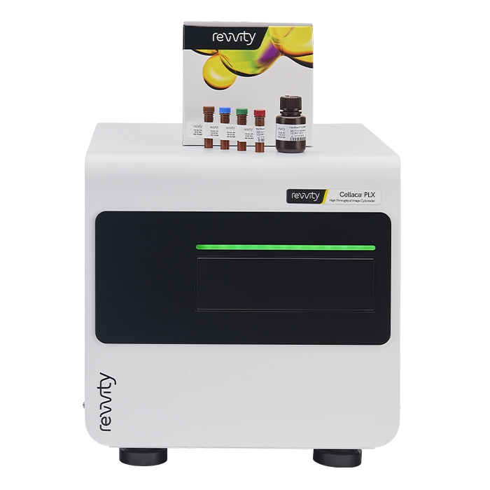
Cellaca PLX ハイスループットイメージサイトメーター
Cellaca PLXイメージサイトメーターは、単純な細胞数、濃度、細胞生存率の測定から、より複雑なマルチプレックスアッセイに至るまで、多様な細胞解析に対応します。わずか15~50 µLのサンプルで実施可能です。システムの感度により、デュアル蛍光および明視野イメージングを使用して24サンプルまでを5分未満で解析可能です。さらに4チャンネルでの単一サンプルのマルチプレックスは1分で行えます。
免疫表現型解析、アポトーシス、蛍光タンパク質発現のためのマルチプレックスパネルは、Cellaca PLX低蛍光スライド、専用試薬、組み込みアッセイ、カスタマイズ可能なデータレポートを使用することで、次の工程までの時間を大幅に短縮します。Cellaca PLXは、ユーザーフレンドリーなソフトウェア、オートメーション互換性を備え、21 CFR Part 11対応です。細胞株の開発、細胞分離、遺伝子導入など、さまざまな研究用途を効率化し、お使いのワークフローにシステムを組み込むための専門的なサポートを行っています。
Cellaca PLXの主な機能と利点:
- ハイスループット性能 - 細胞核と細胞のカウントを蛍光2色および明視野での撮像と解析。最大24のサンプルを5分以内で解析可能
- マルチプレックス機能 - 表面マーカー、アポトーシス、および蛍光タンパク質解析用の専用試薬キットを使用し、ダウンストリーム工程までの時間を短縮(1サンプルあたり4チャンネルで1分)
- 少量のサンプル容量 - わずか15 µLの細胞サンプルで実施可能
- 直感的なワークフロー - カスタマイズされた実験やよく使用するアッセイをすぐに使えるお気に入り機能と、自動データ解析と自動レポート作成に対応
- オートメーション対応 - APIを介した搬送用ロボットの接続が可能
- 21 CFR Part 11対応 - 監査証跡、ユーザーアクセス制御、デジタル署名にアドオンで対応

Cytotoxicity and Cell Proliferation Assays
Cytotoxicity and cell proliferation assays are commonly used in the drug discovery process to assess a compound’s ability to cause or block a biologic activity without having toxic effects on cells. Adenosine TriPhosphate (ATP) – monitoring assays allow for the quantitative evaluation of proliferation and cytotoxicity.
Protein and nucleic acid QC:
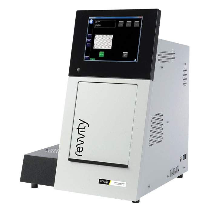
LabChip GXII Touch HTタンパク質特性評価システム
LabChip GXII Touchは、サンプル解析を簡素化します
ユーザーフレンドリーな操作:
- サンプルプレートとチップをロード
- サンプルを選択(1回のランで最大384サンプル)
- アッセイタイプを選択
- 『Run』をタッチして開始
- データは自動的にネットワークやLIMSにエクスポート可能
リアルタイムでランを確認:
- 最短42秒でサンプル析
- サンプルの泳動中にエレクトロフェログラムをリアルタイムで表示
- 取得データを重ねてサンプルプロファイルを比較
- 様々なランタイム解析機能の注釈を選択
データをリアルタイムで確認または後の解析のためにエクスポート:
- 表示形式はエレクトロフェログラム、バーチャルゲル、データテーブルから選択(Figure 1)
- 保存済みプレートを複数呼び出してレビューや比較解析
- 主要属性にフィルターをかけてデータマイニング
- 予想されるピークをハイライト
- FDA 21 CFR Part 11に対応したソフトウェアで関連するユーザーアクセスおよびデータ履歴パラメータを管理
タンパク質および核酸の特性評価のための簡単なセットアップ
LabChip GXII Touchタンパク質特性評価システムの操作コントロールは、ユーザーがわずか3つの簡単なステップで設定とランを行えるよう設計されています。テンプレートは、オペレーター制御機能とともにインポートすることもできます。ランテンプレートには、ウェルの選択、サンプル名、ピークテーブルなどを含めることができ、操作性が向上します。データはネットワークやLIMSディレクトリに自動エクスポートされ、その後の解析に利用できます。すべての装置には、現在のデータや保存済みデータの解析が可能なレビュー用ソフトウェアが標準で付属しています。
品質設計(Quality by Design)をワークフローに統合しやすい設計
LabChip GXII Touchタンパク質特性評価システムは、バイオ医薬品およびゲノム解析のワークフロー全体において、迅速な定量と品質管理を可能にします。たとえば、特性評価プロセスを自動化することで、多くの重要な品質属性を迅速に取得することが可能になります。研究者は、プロセスの初期段階で最適なタンパク質特性をスクリーニングし、品質設計(Quality by Design)イニシアチブをバイオ治療薬の開発ワークフローに統合することが可能です。
LabChip GXII Touchタンパク質特性評価システムにはFDA 21 CFR Part 11に対応したソフトウェアが利用可能
LabChip GXII TouchおよびReviewerソフトウェアにはFDA 21 CFR Part 11規制をサポートするために特別に設計された技術的コントロールおよび機能が内蔵されています。これらの機能には、共有ユーザーアカウントデータベース、アクセス制御、デバイスチェック、ランステップの強制的な順序付け、監査証跡、記録のコピー、記録の保存、システム文書化、および電子署名の制御が含まれます。
お客様のコンプライアンス計画を支援するために、LabChip GxP Security Softwareが利用可能です。このソフトウェアを使用することで、LabChip GXII Touchタンパク質特性解析システムをCFR21 Part 11準拠および検証に対応した環境で運用することができます(図2)。これには、アクセスセキュリティ、データセキュリティと検証、そして完全な監査ログ機能が含まれています。
LabChip CE: その仕組みは?
LabChip CEは小型のマイクロフルイディックチップ上で行われます。解析前に、チップの個別のウェルに試薬をロードします。これらのウェルは、クオーツ製のマイクロフルイディックチップに刻まれた人間の髪の毛の太さ程度の微小チャネルに接続されています。チップをLabChip GXII Touchタンパク質特性評価システムにロードすると、チップのウェルが白金電極と接触し、電圧と電流制御が提供されます。装置はマイクロタイタープレートを直接チップのキャピラリー『シッパー』の下に移動し、約150 nLのサンプルがチップに吸引されます。サンプルの染色および脱染はプラットフォーム上で自動的に実行されます。個々のサンプル解析物は電気泳動的に分離され、チップのキャピラリー内でレーザー誘導蛍光によってバンドが検出されます。各バンドのサイズおよび濃度は、ラダーおよび内部マーカーを使用して決定されます。サンプル間でシッパーをすすぐことでクロスコンタミネーションを最小化します。
Computational design and ideation:
Signals One™ software
AI‑enhanced, unified, cloud‑native R&D solution that helps you reduce complexity and collaborate seamlessly across drug discovery, materials science, and food science.
Test
Transform biological models into meaningful scientific data with Revvity's integrated portfolio of reagents, assays, and imaging solutions.
Rapidly measure cellular responses, characterize biomarkers, and generate reproducible experimental results.
High-content imaging instrument, software and reagents:
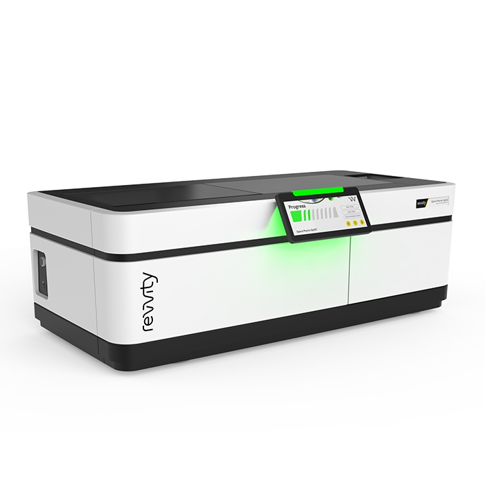
Opera Phenix OptIQハイコンテントスクリーニングシステム
Opera Phenix OptIQハイコンテントスクリーニングシステムは、最難度のハイコンテントアプリケーションに対応します。
- 2カメラまたは4カメラ構成により、同時イメージングで撮像の速度を向上します。
- Synchrony Optics™は、マイクロレンズ強化型スピニングディスクで、厚みのある3Dサンプルに最適化されたピンホール間隔になっています。
- 隣接するスペクトルチャネルを異なる2点から励起するDual-view Excitationで、クロストークを最小限に抑えます。
- 高開口数水浸対物レンズにより、厚みのあるサンプルでもより多くの光を捉え、高解像度の画像取得が可能です。
- 最大100 fpsの高速フレームレートと、オプションのピペッターにより、細胞の高速な応答を捉えることができます。
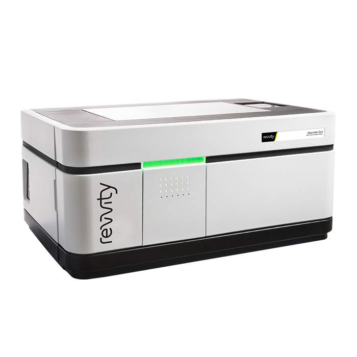
Operetta CLS ハイコンテントアナリシスシステム
Operetta CLS システムは、Operetta プラットフォームで培われた信頼性の高い、直感的で強力なデータ解析機能に、高速性と高感度を組み合わせたシステムです。新しいOperetta CLSは、ハイコンテントアナリシスに求められるあらゆる要件を提供します。さらに、Operetta CLSは、HCSワークフローの一部です。レビティは、HCSシステムや蛍光色素、マイクロプレートをはじめ、自動化やインフォマティクスまで、あらゆるソリューションを提供します。すべてを、信頼できる一社からご提供します。さらに、習得しやすく使いやすい Harmony ハイコンテントイメージングおよび解析ソフトウェアと組み合わせることで、研究者自身による解析が可能となり、日常的な解析から複雑な解析まで、幅広いアプリケーションをすぐに実施できます。
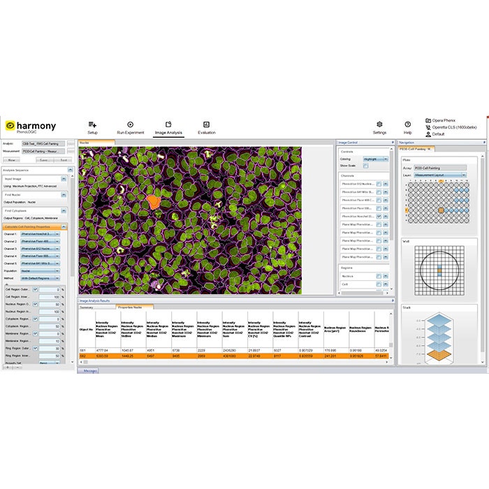
Harmony ハイコンテンツイメージングおよび解析ソフトウェア
生物学研究者向けに設計されたHarmonyのワークフローベースのインターフェースは、顕微鏡やプログラミングの知識が限られているユーザーでも、ハイコンテンツアナリシスのプロセス全体を容易に実行できるよう設計されています。
あらゆるアプリケーションに対応するハイコンテンツアナリシスソフトウェア
- 表現型スクリーニング
- 蛍光強度、形態、テクスチャ、および蛍光分布パラメーターに基づく堅牢な表現型フィンガープリントを取得
- 関連する表現型を認識できるようソフトウェアを容易にトレーニング
- わずかな表現型差も定量化可能
- ライブセルイメージング
- 経時的な表現型変化を追跡
- ラベルフリーのライブセル画像を高精度に定量化
- 個々の細胞の経時的な追跡や創傷治癒アッセイの解析に対応
- 細胞シグナル伝達ダイナミクスの解析(FRETベースアッセイなど)
- 最大100 fpsの撮像速度により、カルシウム流入や心筋細胞拍動速度などの高速応答を実施
- リキッドハンドリングモジュールを備えたOpera Phenix OptIQシステムで「dispense and read」アッセイを設定可能
- 時系列解析機能の向上により、ライブセルアッセイの可能性を拡張
- 3D細胞モデル
- マイクロ組織のターゲットイメージングにより3D画像取得を効率化
- 新しい3DビューアおよびXYZビューアによる細胞モデルの詳細な評価
- 3Dでの形態および体積測定、スフェロイド内の核数カウント
- PlaneMapを使用したMaximum Intensity Projection解析の強化
- 画像取得、3D可視化、解析を1つのソフトウェアで実行
- 稀な細胞表現型解析
- PreciScan™により希少な細胞表現型を迅速に取得
- 低解像度で事前スクリーニングし、対象領域のみを高解像度で取得
- 画像取得時間を短縮
- データ容量を削減
- ルーチンアッセイ
- 標準アッセイ向けに30種類以上の解析ソリューションを提供
- 確立されたプロトコルの実行による再現性の確保
- オプションモジュール
- Phenologic.AIは、事前学習済み深層ニューラルネットワーク(DNN)を活用し、蛍光画像および明視野画像における細胞や細胞核を効率的かつ信頼性高く識別します。
あらゆるユーザーに対応するハイコンテンツ解析ソフトウェア
- 新規ユーザー
- 細胞画像の定量解析を実施
- 顕微鏡やプログラミングの知識が少ないユーザーでも習得可能
- 30種類の既製ソリューションから開始可能
- 論理的なワークフローにより画像取得の各ステップを容易に管理
- 画像解析シーケンスを段階的に構築するためのシンプルなビルディングブロック
- 経験のあるユーザー
- 高度なテクスチャおよび形態指標を用いて複雑な表現型を定量化
- 高度なビルディングブロックを活用し、レシオメトリックFRETイメージングなどの解析に対応
- 複数の視野から「スティッチング」された統合画像を作成
- 複数のスケールでサンプルを評価し、統合画像で特定した領域を元画像の解析に利用可能
- ラボマネージャー向け
- 増加する研究ニーズに対応し、より多くのサンプルを解析
- 新しいユーザーが装置のセットアップおよび画像解析を行える整えられたワークフロー
- 統合された並べ替え可能なデータベースを介して画像、メタデータ、結果を高速に検索
- Harmonyソフトウェアをインストールすることで、どのコンピューターからでもデータの閲覧および解析が可能
- 画像解析の専門知識を備えたスタッフによるアプリケーションサポート

Phenologic.AI
Phenologic.AIモジュールは、細胞イメージング分野における重要な進歩です。事前学習済みのディープニューラルネットワーク(DNN)を活用し、蛍光画像および明視野画像における細胞および細胞核を識別するための効率的で信頼性の高い方法となりました。
Phenologic.AIは、HarmonyおよびImage ArtistソフトウェアにFind Nuclei AIビルディングブロックを追加し、従来は核染色なしでは困難であった明視野画像における細胞核の検出を可能にします。この技術革新により、パラメータの微調整の必要性がなくなり時間を節約できるほか、細胞画像の解析を高速かつ高精度に実施できるようになります。染色を行わずに細胞核を信頼性高く位置特定できるのは、高度なPhenologic.AIアルゴリズムによるものです。
多様な細胞株を用いて学習したPhenologic.AIは、細胞核などの細胞構造を信頼性高く識別するディープニューラルネットワークモデルであり、個々の細胞の細胞質領域を決定するためのフェーズコントラスト画像を提供します。
Phenologic.AIは、画像解析に適したコンピューティングシステムであるGraphics Processing Unit(GPU)に対応しており、AI駆動型ソフトウェアモジュールの処理能力を大幅に向上させることで、生物学研究において重要な高速かつ高精度な解析を可能にします。Phenologic.AIは、Central Processing Unit(CPU)での実行も可能です。
Phenologic.AIは、細胞アナリシスのワークフローを効率化し、細胞の挙動や疾患メカニズムの理解に新たな可能性を提供します。
Phenologic.AIは、Harmonyハイコンテンツイメージングおよび解析ソフトウェア、およびImage Artist画像データ管理および解析システム用のソフトウェアモジュールとして利用可能です。
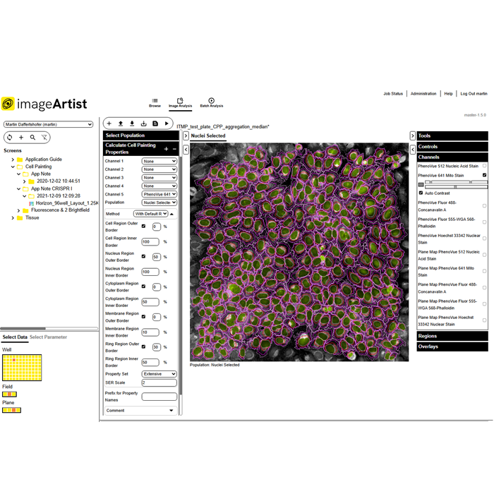
Image Artist
Next generation image analysis and management
Image Artist has powerful capabilities for processing, analyzing, storing, and sharing all your high-content screening and cell imaging data – so you can significantly cut your time to results.
- Fast image data processing and image analysis even for complex assays powered by high performance computing
- Easy-to-use assay building blocks with integrated Artificial Intelligence (AI) that make advanced image analysis
straightforward – now with improved segmentation and analysis capabilities for 3D cell applications - A central location to store all your image data with associated instrument metadata for an enduring picture of
experiments - Compatible with all major high-content screening and cell imaging systems
- Multi-user solution that can support your entire lab without compromising performance
- Scalable data storage to expand with your labs needs over time
- Seamless integration with Revvity Opera Phenix OptIQ™ and Operetta CLS™ high-content screening systems operated
by Harmony™ software, as well as Signals One
for profiling image data, hit selection, and more - NEW! The latest version is compatible with data from a broader range of systems including the Celigo™ image
cytometer - NEW! Wider range of on-premise and cloud options, with AWS S3 support. Now featuring flexible tier-based storage
options, and Intelligent-Tiering for automatic cost-savings. - NEW! Phenologic.AI is an optional module that provides an efficient and reliable method for identifying cells
and cellular nuclei within fluorescent and brightfield images by harnessing the power of pre-trained deep neural
networks (DNNs).
It is also part of our complete workflow solution for high-content screening that includes PhenoVue™ cellular imaging reagents, PhenoPlate™ imaging microplates, HCS instruments, image cytometers, software, and automation.
For all your high-content screening and cellular imaging data
High-content screening and cellular imaging experiments generate huge amounts of image data. With the latest applications such as phenotypic screening, cell painting, and 3D imaging which use more complex,
physiologically-relevant disease models, the volume of data continues to grow.
To maximize all this valuable data, you need a powerful solution for image processing, management and storage, and for sharing data with colleagues.
Image Artist allows you to bring together cell imaging data from a wide range of sources on a single software platform so that you can store, share, analyze, and re-analyze it seamlessly.
Not only does it make managing image data easier for research scientists but also for your IT team. With cloud deployment options (such as AWS S3), your IT team have just one platform and installation to manage and store research image data which can easily be scaled. They can also have peace of mind with enhanced security for cloud deployments. What's more, with AWS S3, you can choose from a range of storage classes based on your cost and data access requirements, and optional Intelligent-Tiering allows automatic transfer of data to more cost-effective tiers, based on access frequency.
Image analysis designed for biologists
Whether you are performing phenotypic screening, cell painting, 3D imaging, live cell imaging, or more routine assays, Image Artist is designed to make it easy for biologists to perform sophisticated image analysis without any coding experience.
The software platform’s image analysis building blocks encapsulate many years of knowledge and expertise in cellular imaging and analysis - so that you can focus on the biology. By simply adding together building blocks such as “Find Nuclei” and “Calculate Cell Painting Properties”, users can quickly create image analysis protocols in just a few simple steps.
With 3D cell models bridging the gap between 2D cell cultures and in vivo animal models, Image
Artist has been further optimized for 3D cell applications, with improved segmentation and analysis capabilities – so you can generate more accurate results than ever.
Built-in Artificial Intelligence (AI) and machine learning technologies allow users to train the software to develop image analysis algorithms. While other systems may require an image analysis expert to create an algorithm, Image Artist uses proprietary machine-learning technology to make it easy for you to do it on your own. Using a learn-by-example approach, in just a few clicks segmented images can be classified quickly and easily.
This is all powered by high performance computing, so you can get your answers faster.
- Get started without extensive training
- Measure complex and subtle phenotypic responses
- Compare multiple samples, plates, or batches to quality check your results
- Kinetic measurement capabilities to track cells, reveal changes in cell properties, and provide information on
cell movement - Use with Signals One to perform screening data analysis and validation, QC analyses,
calculate reliable normalization, multivariate hit stratification, dose response curves, and drug response
profiling
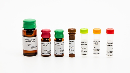
Cellular Imaging Reagents
Revvity's PhenoVue™ suite of cellular imaging reagents – including cell painting kits, organelle and cell compartment stains
Plate-based assay validation:
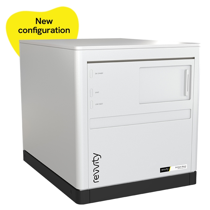
EnVision Nexus マルチモードマイクロプレートリーダー
EnVision Nexusファミリーは、ハイスループットスクリーニングに対応し、複数の検出技術において安定した性能と信頼性の高いデータを提供します。高速測定、ハイスループット性能、各検出技術での高感度測定を実現したEnVision Nexusファミリーは、ワークフローに合わせて柔軟に構成を選択できます。
EnVision Nexus Oneは、シングルディテクター構成で、高度な検出性能をシンプルな構成で提供します。
EnVision Nexusは、デュアルディテクターを搭載しており、2波長の同時測定を可能にします。複雑で要求の高いスクリーニングワークフローを必要とする環境をサポートします。
主な特徴:
- すべての検出技術で感度向上
- マグネット式によるフィルターモジュールの容易な変更
- アプリケーションに合わせて選べる柔軟な検出オプション
主な検出:
- 吸光度
- 蛍光強度
- 発光
- 時間分解蛍光
- 蛍光偏光
高度な検出:
- HTS Alpha
- TRFレーザー
- 超高感度発光
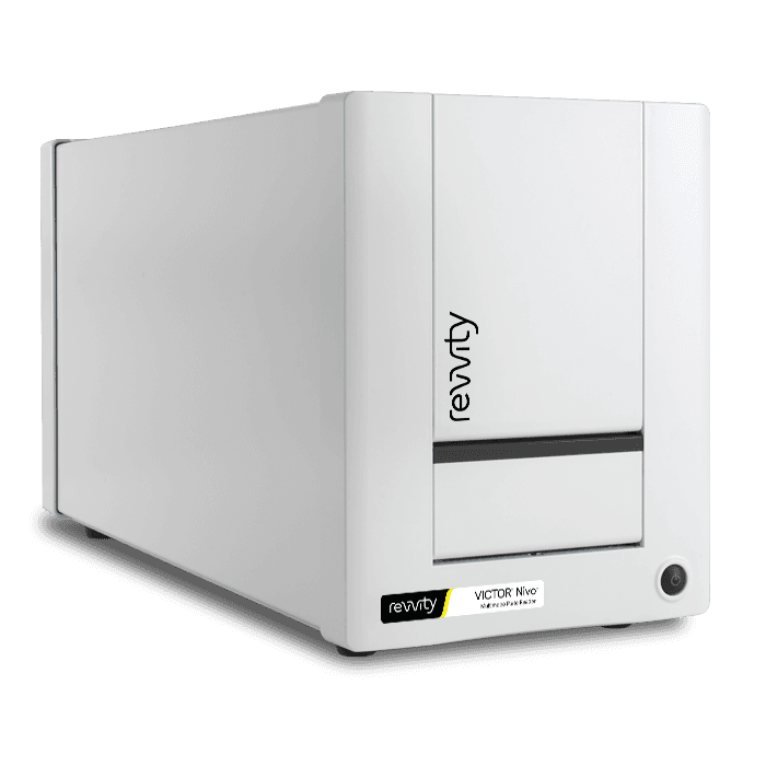
Nivoマルチモードマイクロプレートリーダー
Nivoシステムは、すべての主要な検出技術を備えた高性能フィルターベースのマルチモードマイクロプレートリーダーです。
- 吸光度
- 発光
- 蛍光強度
- 時間分解蛍光(TRF)
- 蛍光偏光
- Alpha
Nivoシステムは、日常的な低スループットアッセイまたは多様なアプリケーション要件に対応したアッセイ開発作業を行うライフサイエンス研究ラボ向けに設計されたコンパクトで軽量なマルチモードプレートリーダーです。スタッカーと組み合わせることで、Nivoのスループットが向上し、ユーザーがプレートの管理ではなく研究に集中できるようにウォークアウェイオートメーションを設定できるため、生産が向上します。
Nivoシステムのソフトウェアは、最新のワークフロー指向のユーザーインターフェースを備えており、学習しやすく使いやすい設計で、事前に作成されたアプリケーションプロトコルを含むため、ユーザーの生産性を迅速に向上します。さらに、アッセイを実行した直後にテスト結果を使用して基本的なデータ解析を行うことができます。複雑な計算のために、研究室やデスクで使用可能なMyAssays® Desktop Pro解析ソフトウェア(オプション)利用できます。Enhanced Securityソフトウェアオプションには、21 CFR Part 11のコンプライアンスの複雑さをガイドする技術ツールが含まれています。
ブラウザベースの制御ソフトウェアはオペレーティングシステムに依存しないため、WindowsやMac OSのPCやタブレットなど、さまざまなタッチスクリーンデバイスで制御できます。
Nivoマイクロプレートリーダーは、WiFiまたはネットワーク接続を介して操作可能で、バイオセーフティキャビネット外や隣接のラボなどから実験をセットアップ、実行、監視できます。
ダイナミック レインボー フィルター エクスチェンジャー
すべてのNivoシリーズは、最大32個のフィルターを収納可能なダイナミックな2層フィルターホイールシステムを備えています。フル装備のシステムは、モノクロメーターよりも優れた感度で、ほぼあらゆるアプリケーションに対応に対応できます。
フィルターは以下の動作を行います:
- 内側と外側のホイール間でフィルターを移動可能
- 同一フィルターを励起光または蛍光パスのいずれかに使用可能
- 位置に固定され紛失しないためのパスワードロック - マルチユーザーラボに最適
検出技術
Nivo マルチモードプレートリーダーを使用した吸光度測定では、フィルターベースまたはスぺクトロメーターベースシステムのいずれかを選択できます。スペクトロメーターベースのプレートリーダーでは、フルスペクトル吸光度測定が超高速で行えます。選択可能な解像度(2.0 nm、5.0 nm、10 nm)で230~1000 nmを1ウェルあたり1秒未満で測定。
最大8つの異なる波長が切り替えなしで1回の測定で検出可能で、スペクトロメーターベースのNivoシステムはさまざまな染料の検出や未知の吸光スペクトルを持つサンプルの測定も可能にします。
機能と利点
- コンパクトで軽量なマルチモードプレートリーダーで、ベンチスペースを節約し、バイオセーフティキャビネットや別の研究室への移動が容易
- 最大32フィルターを収収納可能な内部ダイナミックフィルターホイールシステムで、多くの色素に迅速に対応
- 吸光度測定では、最高感度のフィルターベース検出または波長の柔軟性を持つスペクトロメーターを選択可能
- 時間分解蛍光測定には、HTRF™テクノロジー向けの使用が認証されています。
- すべての標準測定テクノロジー(吸光度、発光、蛍光強度、時間分解蛍光、および蛍光偏光)において、プレート上方からと下方からの測定が可能です。Alpha のみ上方から測定のみに対応しています。1ウェルから1536ウェルまでのプレートフォーマットで高いパフォーマンスを発揮します。
- 最大20枚のマイクロプレートをバッチ処理できるスタッカーオプション
- 規制環境向けのEnhanced Securityソフトウェアは、21 CFR Part 11準拠をサポートする技術制御と機能を提供します。
- 高速反応速度、フラッシュ発光アッセイ、または二重添加アッセイなどのアプリケーション向けのオプションディスペンサー
- 温度制御(最大65°C)およびオプションのガス制御ユニットで、細胞培養条件を保持しながら、長期の測定が可能
- ブラウザベースのソフトウェアにより、PC、ノートパソコン、タブレットなどさまざまなデバイスからプレートリーダーを制御可能
- ネットワークまたはWiFiを介して制御できるため、リモートワークやデータアクセスを容易に
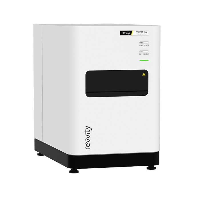
ARVO Kira マルチモードマイクロプレートリーダー
ARVO Kiraは、先進的なモノクロメーターベースの技術により、ワークフローの効率を向上させ、幅広いアプリケーションにに活用できます。直感的に操作できるソフトウェアインターフェイスと内蔵のデータ分析機能を備え、快適なユーザー体験を提供します。さらに、経験豊富なサービスエンジニアおよびアプリケーションサポートの専門スタッフが、お客様をサポートいたします。
ARVO Kira マルチモードプレートリーダーは、3つの検出モードを備えています。温度制御やプレート攪拌機能などが標準搭載されており、細胞ベースおよびカイネティクス測定にも有用です。
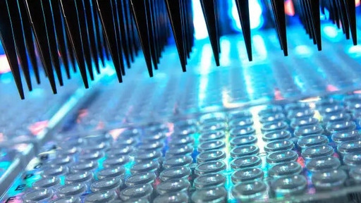
マイクロプレート
適切なマイクロプレートを選択することは、アッセイにおいて非常に重要ですが、見落とされがちです。正しいマイクロプレートは貴重なデータを提供するのに役立ちますが、間違ったマイクロプレートはデータの欠落や不正確さを招き、プロジェクトのタイムラインや最終的なコストの上昇につながります。
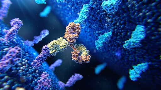
pHSense Reagents
Discover a powerful new plate reader tool specifically designed for cell surface target internalization studies for ADC and GPCR research.
In vivo imaging, software, and reagents:
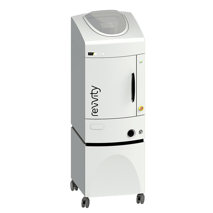
IVIS Spectrum 2 In Vivo Imaging System
Building upon the IVIS Spectrum in vivo optical system with proven 2D bioluminescence and 2D fluorescence imaging and 3D optical tomography capabilities combined in a single system, the IVIS Spectrum 2 is our next generation in optical imaging. This advanced imaging system incorporates a CCD camera with eXcelon® coating that enables detection of more signal at higher efficiency across a broader spectrum of wavelengths. With exclusivity to this innovative camera for in vivo imaging, the IVIS Spectrum 2 preclinical optical imaging system delivers the sensitivity you demand for non-invasive longitudinal imaging to better understand early disease-related biological changes, track disease progression, and help guide the drug development process.

In Vivo Imaging Reagents
Our comprehensive range of IVISbrite™ bioluminescent and IVISense™ fluorescent imaging reagents provides researchers with the necessary tools to better understand early disease-related biological changes, track disease progression, help guide the drug discovery process, and evaluate efficacy and safety of drug candidates.
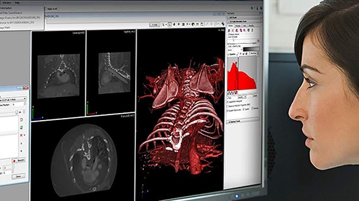
In Vivo Imaging Software
Analyze even the most complex in vivo imaging data with ease using Revvity's portfolio of imaging software. With intuitive interfaces and seamless workflow, our software tools streamline acquisition through analysis simplifying even the most advanced imaging protocol to help facilitate success of your research.
Immunoassays:
AlphaLISA™ SureFire® Ultra™
AlphaLISA® SureFire® Ultra™ is a homogeneous, no-wash assay platform designed for the sensitive detection of endogenous and tagged phosphorylated or non-phosphorylated proteins in cell lysates.
HTRF
HTRF™ (Homogeneous Time Resolved Fluorescence)は、標準的なFRET技術と蛍光の時間分解測定を組み合わせた無洗浄技術で、短寿命のバックグラウンド蛍光を除去します。
Biological insights:
TotalSeq™ reagents
Seamlessly add protein expression to single-cell multiomics workflows with oligonucleotide-conjugated antibodies, hashtags, and cocktails.
LEGENDplex™
Multiplex protein quantification for 14 analytes in a single sample. Select focused pre-defined panels or customize each target.
BioLegend Antibodies
Choose from thousands of primary and secondary antibodies. We offer directly conjugated or purified antibodies to match your needs.
Preclinical services:
Computational design and ideation:
Signals One™ software
AI‑enhanced, unified, cloud‑native R&D solution that helps you reduce complexity and collaborate seamlessly across drug discovery, materials science, and food science.
Validate
Confirm scientific insights and validate AI-generated predictions with trusted experimental evidence.
Revvity's integrated imaging, detection, assay, and biological analysis solutions help researchers confirm targets, characterize biological responses, and generate reproducible results for confident decisions.
Sample prep, automation, and plate handling:
Fontus Workstations
With easy-to-use software, optimized deck access, verified protocols, and reformatting capabilities to simplify your workflows and improve turnaround times
Explorer G3 SmartBench automation solutions
Affordable, scalable laboratory automation with the explorer™ G3 SmartBench workstations, that integrate 150+ instruments with collaborative robotics for seamless workflows.
AssayMateワークステーション
あらゆるラボに、自動化性能を:手頃なコストで導入できるベンチトップサイズの自動リキッドハンドラー。幅広いラボアプリケーションを最適化するために設計されています。
plate::handler Connect
Overcome manual workflow bottlenecks with the plate::handler™ Connect, a collaborative, four-axis SCARA robotic arm for a compact and cost-effective entry point into automated processing.
FlexDrop Plus 非接触ディスペンサー
FlexDrop Plusプラットフォームは、ナノリットルレベルの微量液体の自動分注を、これまでにないほど簡単かつ正確に行えるようにしました。統合型タッチスクリーンPCで操作できる使いやすいデバイスで、ボタンを押すだけ最少8 nLの液体を安定して分注できます。あらゆる研究室でお使いいただけます。
FlexDrop Plusプラットフォームは、8つの独立した制御チャネルで陽圧を使用します。各チャンネルは1秒間に最大100滴を分注できます。目標の分注容量を達成するため、8ナノリットル(nL)から50 nLまでの液滴を生成します。システムには液滴検証が搭載されており、分注された液滴数を正確に検証し、実行後に検証レポートを提供します。特許取得済みのソースウェルプレートには、底部に100 µmの穴があり、陽圧の下で液滴を容易に分注します。
タッチスクリーンソフトウェアコントロールはシームレスで簡単に使用可能です。プログラミングや自動化の経験は必要ありません。セットアップから分注開始までわずか数分で完了します。
構成内容:
- FlexDrop Plus機器
- (4)電源コード(EU、英国、米国、オーストラリア)ヒューズの交換が必要になる場合があります。
- エアチューブ – 機器へのクイックコネクトと施設の圧空またはコンプレッサーに接続するためのフィッティングを備えた 6 mm チューブ
- (4)黒色アダプタープレート(1、2、4、8 mm)– ターゲットプレートの高さ調整用
- スタートアップキットに含まれる80 uLウェルのFlexDropソースプレート
- 廃棄用PCRチューブ
Plate-based assay validation:
EnVision Nexus マルチモードマイクロプレートリーダー
EnVision Nexusファミリーは、ハイスループットスクリーニングに対応し、複数の検出技術において安定した性能と信頼性の高いデータを提供します。高速測定、ハイスループット性能、各検出技術での高感度測定を実現したEnVision Nexusファミリーは、ワークフローに合わせて柔軟に構成を選択できます。
EnVision Nexus Oneは、シングルディテクター構成で、高度な検出性能をシンプルな構成で提供します。
EnVision Nexusは、デュアルディテクターを搭載しており、2波長の同時測定を可能にします。複雑で要求の高いスクリーニングワークフローを必要とする環境をサポートします。
主な特徴:
- すべての検出技術で感度向上
- マグネット式によるフィルターモジュールの容易な変更
- アプリケーションに合わせて選べる柔軟な検出オプション
主な検出:
- 吸光度
- 蛍光強度
- 発光
- 時間分解蛍光
- 蛍光偏光
高度な検出:
- HTS Alpha
- TRFレーザー
- 超高感度発光
Nivoマルチモードマイクロプレートリーダー
Nivoシステムは、すべての主要な検出技術を備えた高性能フィルターベースのマルチモードマイクロプレートリーダーです。
- 吸光度
- 発光
- 蛍光強度
- 時間分解蛍光(TRF)
- 蛍光偏光
- Alpha
Nivoシステムは、日常的な低スループットアッセイまたは多様なアプリケーション要件に対応したアッセイ開発作業を行うライフサイエンス研究ラボ向けに設計されたコンパクトで軽量なマルチモードプレートリーダーです。スタッカーと組み合わせることで、Nivoのスループットが向上し、ユーザーがプレートの管理ではなく研究に集中できるようにウォークアウェイオートメーションを設定できるため、生産が向上します。
Nivoシステムのソフトウェアは、最新のワークフロー指向のユーザーインターフェースを備えており、学習しやすく使いやすい設計で、事前に作成されたアプリケーションプロトコルを含むため、ユーザーの生産性を迅速に向上します。さらに、アッセイを実行した直後にテスト結果を使用して基本的なデータ解析を行うことができます。複雑な計算のために、研究室やデスクで使用可能なMyAssays® Desktop Pro解析ソフトウェア(オプション)利用できます。Enhanced Securityソフトウェアオプションには、21 CFR Part 11のコンプライアンスの複雑さをガイドする技術ツールが含まれています。
ブラウザベースの制御ソフトウェアはオペレーティングシステムに依存しないため、WindowsやMac OSのPCやタブレットなど、さまざまなタッチスクリーンデバイスで制御できます。
Nivoマイクロプレートリーダーは、WiFiまたはネットワーク接続を介して操作可能で、バイオセーフティキャビネット外や隣接のラボなどから実験をセットアップ、実行、監視できます。
ダイナミック レインボー フィルター エクスチェンジャー
すべてのNivoシリーズは、最大32個のフィルターを収納可能なダイナミックな2層フィルターホイールシステムを備えています。フル装備のシステムは、モノクロメーターよりも優れた感度で、ほぼあらゆるアプリケーションに対応に対応できます。
フィルターは以下の動作を行います:
- 内側と外側のホイール間でフィルターを移動可能
- 同一フィルターを励起光または蛍光パスのいずれかに使用可能
- 位置に固定され紛失しないためのパスワードロック - マルチユーザーラボに最適
検出技術
Nivo マルチモードプレートリーダーを使用した吸光度測定では、フィルターベースまたはスぺクトロメーターベースシステムのいずれかを選択できます。スペクトロメーターベースのプレートリーダーでは、フルスペクトル吸光度測定が超高速で行えます。選択可能な解像度(2.0 nm、5.0 nm、10 nm)で230~1000 nmを1ウェルあたり1秒未満で測定。
最大8つの異なる波長が切り替えなしで1回の測定で検出可能で、スペクトロメーターベースのNivoシステムはさまざまな染料の検出や未知の吸光スペクトルを持つサンプルの測定も可能にします。
機能と利点
- コンパクトで軽量なマルチモードプレートリーダーで、ベンチスペースを節約し、バイオセーフティキャビネットや別の研究室への移動が容易
- 最大32フィルターを収収納可能な内部ダイナミックフィルターホイールシステムで、多くの色素に迅速に対応
- 吸光度測定では、最高感度のフィルターベース検出または波長の柔軟性を持つスペクトロメーターを選択可能
- 時間分解蛍光測定には、HTRF™テクノロジー向けの使用が認証されています。
- すべての標準測定テクノロジー(吸光度、発光、蛍光強度、時間分解蛍光、および蛍光偏光)において、プレート上方からと下方からの測定が可能です。Alpha のみ上方から測定のみに対応しています。1ウェルから1536ウェルまでのプレートフォーマットで高いパフォーマンスを発揮します。
- 最大20枚のマイクロプレートをバッチ処理できるスタッカーオプション
- 規制環境向けのEnhanced Securityソフトウェアは、21 CFR Part 11準拠をサポートする技術制御と機能を提供します。
- 高速反応速度、フラッシュ発光アッセイ、または二重添加アッセイなどのアプリケーション向けのオプションディスペンサー
- 温度制御(最大65°C)およびオプションのガス制御ユニットで、細胞培養条件を保持しながら、長期の測定が可能
- ブラウザベースのソフトウェアにより、PC、ノートパソコン、タブレットなどさまざまなデバイスからプレートリーダーを制御可能
- ネットワークまたはWiFiを介して制御できるため、リモートワークやデータアクセスを容易に
ARVO Kira マルチモードマイクロプレートリーダー
ARVO Kiraは、先進的なモノクロメーターベースの技術により、ワークフローの効率を向上させ、幅広いアプリケーションにに活用できます。直感的に操作できるソフトウェアインターフェイスと内蔵のデータ分析機能を備え、快適なユーザー体験を提供します。さらに、経験豊富なサービスエンジニアおよびアプリケーションサポートの専門スタッフが、お客様をサポートいたします。
ARVO Kira マルチモードプレートリーダーは、3つの検出モードを備えています。温度制御やプレート攪拌機能などが標準搭載されており、細胞ベースおよびカイネティクス測定にも有用です。
マイクロプレート
適切なマイクロプレートを選択することは、アッセイにおいて非常に重要ですが、見落とされがちです。正しいマイクロプレートは貴重なデータを提供するのに役立ちますが、間違ったマイクロプレートはデータの欠落や不正確さを招き、プロジェクトのタイムラインや最終的なコストの上昇につながります。
In Vivo Imaging, software and reagents:
IVIS Spectrum 2 In Vivo Imaging System
Building upon the IVIS Spectrum in vivo optical system with proven 2D bioluminescence and 2D fluorescence imaging and 3D optical tomography capabilities combined in a single system, the IVIS Spectrum 2 is our next generation in optical imaging. This advanced imaging system incorporates a CCD camera with eXcelon® coating that enables detection of more signal at higher efficiency across a broader spectrum of wavelengths. With exclusivity to this innovative camera for in vivo imaging, the IVIS Spectrum 2 preclinical optical imaging system delivers the sensitivity you demand for non-invasive longitudinal imaging to better understand early disease-related biological changes, track disease progression, and help guide the drug development process.
In Vivo Imaging Reagents
Our comprehensive range of IVISbrite™ bioluminescent and IVISense™ fluorescent imaging reagents provides researchers with the necessary tools to better understand early disease-related biological changes, track disease progression, help guide the drug discovery process, and evaluate efficacy and safety of drug candidates.
In Vivo Imaging Software
Analyze even the most complex in vivo imaging data with ease using Revvity's portfolio of imaging software. With intuitive interfaces and seamless workflow, our software tools streamline acquisition through analysis simplifying even the most advanced imaging protocol to help facilitate success of your research.
Immunoassays:
AlphaLISA™ SureFire® Ultra™
AlphaLISA® SureFire® Ultra™ is a homogeneous, no-wash assay platform designed for the sensitive detection of endogenous and tagged phosphorylated or non-phosphorylated proteins in cell lysates.
HTRF
HTRF™ (Homogeneous Time Resolved Fluorescence)は、標準的なFRET技術と蛍光の時間分解測定を組み合わせた無洗浄技術で、短寿命のバックグラウンド蛍光を除去します。
Biological insights:
TotalSeq™ reagents
Seamlessly add protein expression to single-cell multiomics workflows with oligonucleotide-conjugated antibodies, hashtags, and cocktails.
LEGENDplex™
Multiplex protein quantification for 14 analytes in a single sample. Select focused pre-defined panels or customize each target.
BioLegend Antibodies
Choose from thousands of primary and secondary antibodies. We offer directly conjugated or purified antibodies to match your needs.
Preclinical services:
Computational design and ideation:
Signals One™ software
AI‑enhanced, unified, cloud‑native R&D solution that helps you reduce complexity and collaborate seamlessly across drug discovery, materials science, and food science.
Generate
Transform scientific knowledge into the next breakthrough.
Revvity's computational design and ideation solutions help generate hypotheses, design experiments to loop AI-driven insights with the next cycle of discovery.
Computational design and ideations:
Signals One™ software
AI‑enhanced, unified, cloud‑native R&D solution that helps you reduce complexity and collaborate seamlessly across drug discovery, materials science, and food science.
Signals ChemDraw™ software
The #1 Trusted Chemical Drawing Solution. A complete suite for creating, managing, sharing, and presenting your chemistry research and discoveries.
Predict
Transform scientific data into predictive intelligence.
Revvity's AI-enabled analytics and modeling solutions help identify patterns, prioritize opportunities, and design smarter experiments to loop computational insights with experimental discovery.
Data modeling and predictive analysis:
Signals Xynthetica™ software
AI-augmented molecular design service combining generative design, property prediction, and virtual screening.
vScreen
Virtual screening helps identify the most promising opportunities before moving into experimental workflows.
Revvity's informatics solutions loop computational screening with wet lab validation, for faster iteration and efficient discovery.
Virtual screening workflows:
Signals One™ software
AI‑enhanced, unified, cloud‑native R&D solution that helps you reduce complexity and collaborate seamlessly across drug discovery, materials science, and food science.
Explore our solutions


Featured resources
Pin-point™ base editing reagents are available for research use only and are not for diagnostic use or direct administration into humans or animals. The Pin-point™ base editing platform technology is available for clinical or diagnostic study and commercialization under a commercial license from Revvity.
For research use only. Not for use in diagnostic procedures.

How can we help you?
Contact us to see how AI-enabled science can support your work.
