


JP
Revvity Sites Globally
Select your location.
*e-commerce not available for this region.

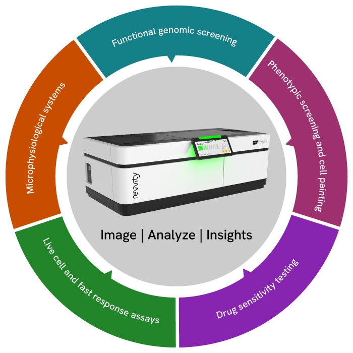
Opera Phenix OptIQハイコンテントスクリーニングシステム
Opera Phenix OptIQ™ハイコンテントアナリシスシステムは、複雑でハイスループットなアプリケーションに対応する最先端の共焦点テクノロジーを提供します。20年以上にわたる技術革新により、3Dオルガノイドやorgan-on-chip等を含む多様な生物学的モデルでの表現型スクリーニングや高度なイメージングアプリケーションを実現します。高評価な画像取得・解析ソフトウェアのHarmony™およびPhenologic.AI™により、Opera Phenix OptIQは、取得から解析、グラフ化まで直感的な操作で実行できます。
What's new:
- >95%量子効率カメラ
- 最新のレーザー式オートフォーカス
- 低酸素環境下でのイメージング
- 高開口数10x対物レンズ
- Phenologic.AI分類ツール
- Find Organoidsビルディングブロック
本製品は研究用です。診断用にはご使用いただけません。
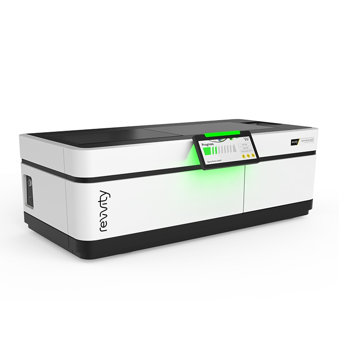

Opera Phenix OptIQハイコンテントスクリーニングシステム
Opera Phenix OptIQ High-Content Screening System
Part #:
HH25000000
Imaging Modality:
AI-based phase contrast, 明視野, 共焦点, デジタル位相差, 蛍光
Loading...
Video overview
Key features
複数台カメラ
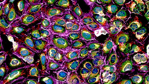
最大4台の高量子効率カメラ(量子効率95%以上)を使用した同時多色取得により、特に3Dモデル撮像時の大量のスタック取得で撮像速度が向上します。
マイクロレンズ強化型スピニングディスク共焦点
ピンホールディスクとマイクロレンズディスクを同期回転させることで光効率を高め、3Dサンプルに適したピンホール間隔によって迷光を低減します。
自動水浸対物レンズ
より多くの光を取り込み、シグナルの取り込み量とZ分解能を改善することで、画像品質とデータ精度を高めます。
独自のSynchrony Optics
多色同時取得時のスペクトルクロストークを低減。速度と感度を向上します。
機械学習(Phenologic)
専門的な画像解析知識がなくても、Phenologic の機械学習機能を用いて解析アルゴリズムを容易に作成できます。
インテリジェント画像取得
必要な対象だけを高倍率で取得します。特に3Dモデルのイメージングでは、撮像を効率化し、取得時間やデータ量を大幅に削減します。
シンプルで強力な解析
Harmonyソフトウェアは、画像解析を効率化します。すぐに使える豊富なテンプレートだけではなく、簡単な操作で解析をカスタマイズすることができます。
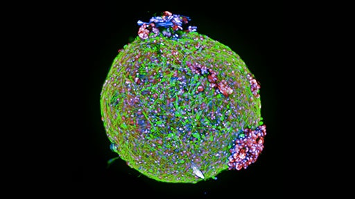
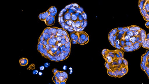
3D解析
3DビューやXYZビューを用いて細胞モデルを3D再構築し、体積解析などの3D特有の指標を簡単に定量できます。
Find Organoids ビルディングブロック
明視野スタック画像から2D/3Dでオルガノイドを検出します。
Applications
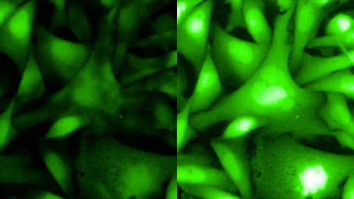
低酸素条件下でのイメージング
オンボード環境制御チャンバーにより、低酸素条件下で細胞を観察できます。生体により近い生理的条件下でのイメージングが可能です。
Opera Phenix OptIQ ファミリー:代表的な構成
Opera Phenix OptIQ Single
Phenixシリーズと同等の感度と解像度を備えます。後からカメラを追加してアップグレードすることができます。
Opera Phenix OptIQ Simultaneous
高速化を実現したデュアルカメラ構成により、マルチカラーの同時共焦点イメージングが可能です。
Opera Phenix OptIQ FRET
5本のレーザーと4台のカメラ構成により、CFP/YFP FRETアプリケーションに対応し、タンパク質間相互作用の解析をサポートします。
Opera Phenix OptIQ Screener
4台のカメラと高出力レーザー4基を備え、高いスループット性能で大規模ライブラリーのスクリーニングをサポートします。
構成の詳細
| Single | Simultaneous | FRET | Screener | ||
|---|---|---|---|---|---|
| レーザー | 375/425 nm | Unavailable | Optional | ✓ | Unavailable |
| 405 nm | ✓ | ✓ | Optional | ✓ | |
| 488 nm | ✓ | ✓ | ✓ | ✓ | |
| 561 nm | ✓ | ✓ | ✓ | ✓ | |
| 640 nm | ✓ | ✓ | ✓ | ✓ | |
| システムオプション | Number of cameras (confocal) | 1 | 2 | 4 | 4 |
| 環境コントロール | Optional | Optional | ✓ | Optional | |
| 水浸対物レンズ | ✓ | ✓ | ✓ | ✓ | |
| 透過光 | ✓ | ✓ | ✓ | ✓ | |
| オンボードリキッドハンドリング | Optional | Optional | Optional | Optional | |
| ロボット/自動化対応 | ✓ | ✓ | ✓ | ✓ | |
| イメージングモード | Minimal crosstalk imaging | ✓ | ✓ | ✓ | ✓ |
| レシオメトリックFRET | Suitable | Suitable | Suitable (CFP/YFP) | ||
| 3Dイメージング | ✓ | ✓ | ✓ | ✓ | |
| マルチカラー蛍光イメージング | ✓ | ✓ | ✓ | ✓ | |
| マルチカラー同時共焦点イメージング | Unavailable | ✓ | ✓ | ✓ | |
| 明視野およびデジタル位相差 | ✓ | ✓ | ✓ | ✓ | |
| 蛍光広視野イメージング | ✓ | ✓ | ✓ | ✓ | |
| 高速フレームレートイメージング | ✓ | ✓ | ✓ | ✓ |
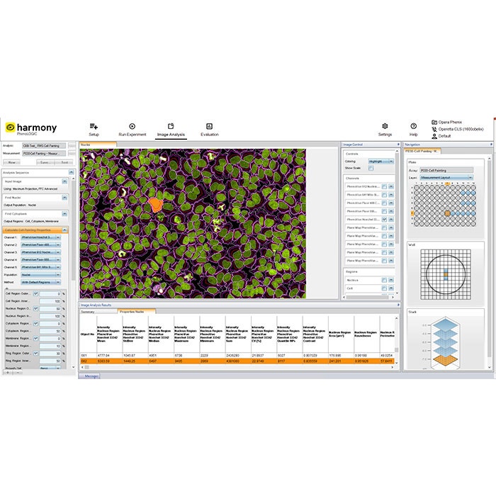
Harmony ソフトウェアで実現するシンプルな画像取得と解析
直感的なワークフロー
Harmony ソフトウェアは、画像取得から解析・グラフ化まで実現し、わかりやすく直感的なユーザーインターフェースを備えています。
クイックセットアップ用テンプレート
テンプレートを使用することで、画像取得のチャネルや各種パラメーターを効率よく設定できます。
すぐに使える解析ソリューション
よく使われるアプリケーションには、あらかじめ構築された画像解析ソリューションを選ぶことで、ワークフローを簡素化できます。
カスタマイズ可能なビルディングブロック
画像解析用のビルディングブロックを用いて、簡単な解析から複雑なハイコンテント解析アプリケーションまで作成・調整・カスタマイズできます。
最新の解析機能
テクスチャ解析や STAR 形態解析など、細胞形態の特徴を詳細に記述し、表現型の判別を強化する最新の解析機能を備えています。
データ管理
解析結果やメタデータ(アッセイレイアウト、装置設定、ユーザー定義のキーワードや注釈など)を自動的に保存します。
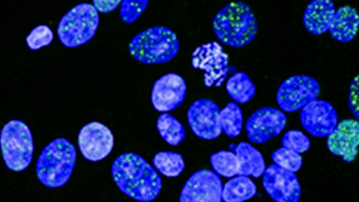
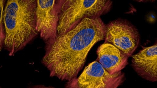
AI ベースの画像解析
事前学習済みの深層学習モデルを使用し、Phenologic.AI が明視野および蛍光画像における細胞・核のセグメンテーションや分類を実行します。これにより、生細胞および固定細胞アッセイの撮像と解析が容易になります。
認証済みソリューションで研究を加速
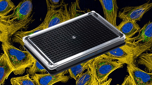
ハイコンテントアナリシス向けマイクロプレート
ハイコンテントアナリシスに最適化されたPhenoPlate™ 384ウェルマイクロプレートを使用できます。3DモデルのイメージングにはCellCarrier Spheroid ULAプレートもご用意しています。

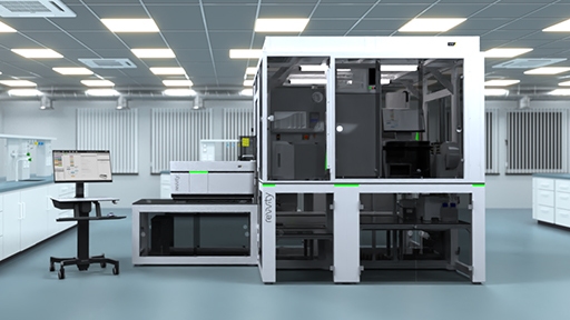
自動化およびワークステーション
スループットや生産性の向上、ばらつきや試薬コストの低減が実現できます。explorer G3ワークステーションは、セルペインティング、3D細胞培養、表現型スクリーニングの自動化ワークフローを実現します。
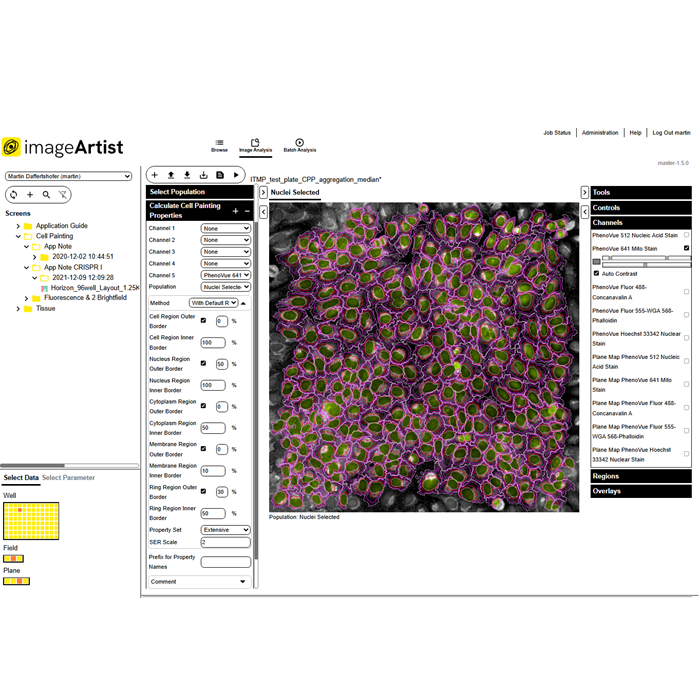
効率的なデータ管理
解析結果をImage Artist™プラットフォームに自動的にエクスポートできます。高性能コンピューティングと業界標準的なオブジェクトストレージを用いた、スケーラブルなマルチユーザー対応ソリューション環境が構築できます。

Product information
Overview
Opera Phenix OptIQハイコンテントスクリーニングシステムは、最難度のハイコンテントアプリケーションに対応します。
- 2カメラまたは4カメラ構成により、同時イメージングで撮像の速度を向上します。
- Synchrony Optics™は、マイクロレンズ強化型スピニングディスクで、厚みのある3Dサンプルに最適化されたピンホール間隔になっています。
- 隣接するスペクトルチャネルを異なる2点から励起するDual-view Excitationで、クロストークを最小限に抑えます。
- 高開口数水浸対物レンズにより、厚みのあるサンプルでもより多くの光を捉え、高解像度の画像取得が可能です。
- 最大100 fpsの高速フレームレートと、オプションのピペッターにより、細胞の高速な応答を捉えることができます。
Specifications
| Automation Compatible |
Yes
|
|---|---|
| Brand |
Opera Phenix OptIQ
|
| Imaging Modality |
AI-based phase contrast
明視野
共焦点
デジタル位相差
蛍光
|
| Unit Size |
1 unit
|
Citations
Resources
Are you looking for resources, click on the resource type to explore further.
Brochure
3D cell culture workflow solutions
More than ever, researchers are turning to 3D cell cultures, microtissues and organoids to bridge the gap between 2D cell cultures...
Technical Note
3D volumetric analysis of luminal spaces inside cysts or organoids
High-content assays using 3D objects such as cysts or organoids can be challenging from the perspectives of both image acquisition...
Technical Note
3D volumetric and zonal analysis of solid spheroids
Multicellular 3D “oids” (tumoroids, spheroids, organoids) have the potential to better predict the effects of drug candidates...
Whitepaper
A brand-new modality on the horizon: how targeted protein degradation can address the unmet need in drug discovery and development
Targeted protein degradation (TPD) is an emerging drug discovery modality that offers the potential to probe biological pathways...
Technical Note
A scalable and reproducible workflow for high-content analysis of cytotoxic effects in RASTRUM 3D cell cultures
Explore our Technical Note unveiling a robust workflow for advanced cytotoxicity analysis in 3D cell cultures. This method...
Case Study
A scalable method to monitor protein levels and localizations in cells
In this case study, we present a novel method developed by researchers at the CeMM Research Center for studying protein levels and...
Loading...


How can we help you?
We are here to answer your questions.

