


JP
Revvity Sites Globally
Select your location.
*e-commerce not available for this region.

Celigoイメージサイトメーター
Celigo™画像サイトメーターは、接着細胞および浮遊細胞を使用した2Dおよび3D培養向けのマイクロプレートベースのマルチチャネル明視野および蛍光イメージングサイトメーターです。21 CFR Part 11モジュールが利用可能です。
本製品は研究用です。診断用にはご使用いただけません。
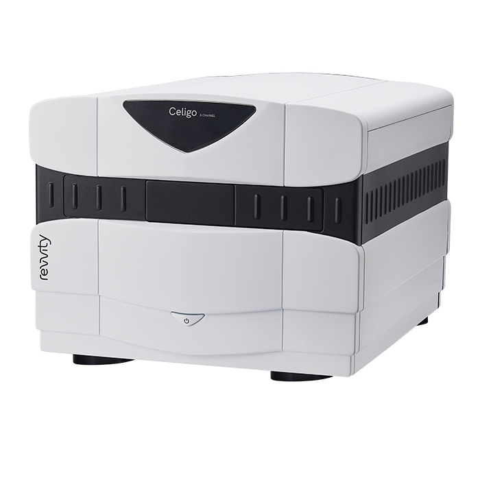
Celigoイメージサイトメーター
The Celigo features brightfield plus 4 fluorescent channels for comprehensive cell analysis and multi-parameter assays.
Part #:
200-BFFL-5C
Imaging Modality:
蛍光・明視野
Loading...
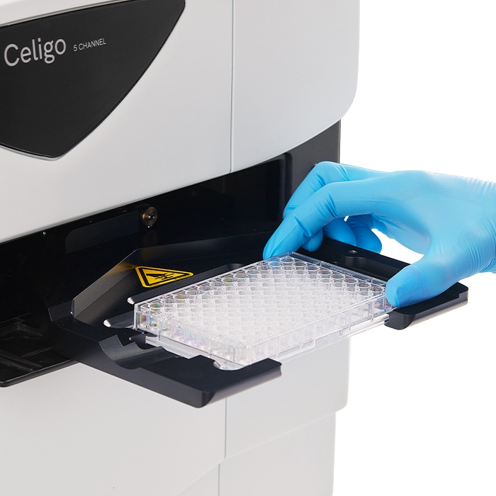
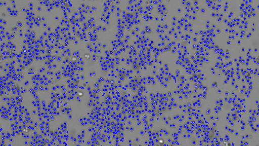
Celigo image cytometer - rapid whole-well analysis
Label-free cell counting
Performs direct cell quantification without trypsinization for adherent cells or cell removal, maintaining experimental conditions.
Versatile plate compatibility
Supports microplates from 6-well to 1536-well formats as well as T-flask and petri dish compatibility for maximum experimental flexibility.
Whole-well imaging capability
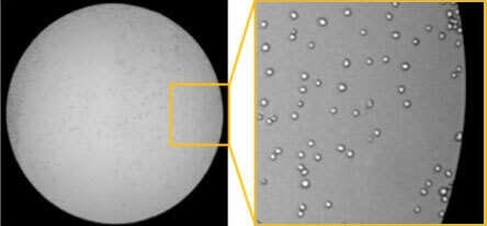
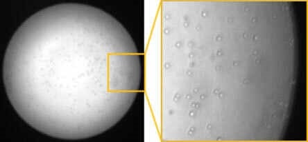
Captures complete well images with proprietary optics providing uniform illumination and excellent edge-to-edge contrast for cells in plate.
High-speed automated analysis
Images and analyzes entire 384-well plates in less than 2 minutes with minimal plate movement, ensuring sample integrity.
Multi-channel imaging system
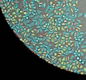
Features brightfield plus 4 fluorescent channels (Blue, Green, Red, Far-Red) for comprehensive cell analysis and multi-parameter assays.
Automation-ready design
Easily integrates with robotic systems, plate stackers, and liquid handlers for high-throughput screening workflows.
The Celigo image cytometer highlights
Research
Enrich your applications
Discover a wide range of applications and protocols supporting the Celigo image cytometer.
Brochure
The Celigo at a glance
Discover the features and benefits of the Celigo image cytometer in detail.
Reagents
For your workflow
Revvity offers fluorescent reagents & kits for cell counting, cell viability and cell-based assays.
Demos
See for yourself
Schedule an expert-guided demonstration showcasing the Celigo image cytometer.
The Celigo image cytometer - offers a wide range of application capabilities
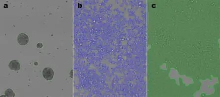
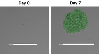
3D tumor model analysis
Specialized capabilities for spheroid growth tracking, viability assessment, and drug screening in physiologically relevant 3D cultures.
Immuno-oncology applications
Performs cell-mediated cytotoxicity assays (ADCC, NK cell killing, CAR-T) with individual cell-level sensitivity.
Cell line development tools
Provides single cell detection, single cell to single colony verification, and transfection/transduction efficiency monitoring.
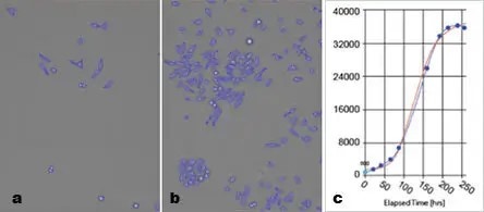
Kinetic analysis capabilities
Performs time-lapse studies for growth tracking, migration assays, and real-time monitoring of cellular processes.
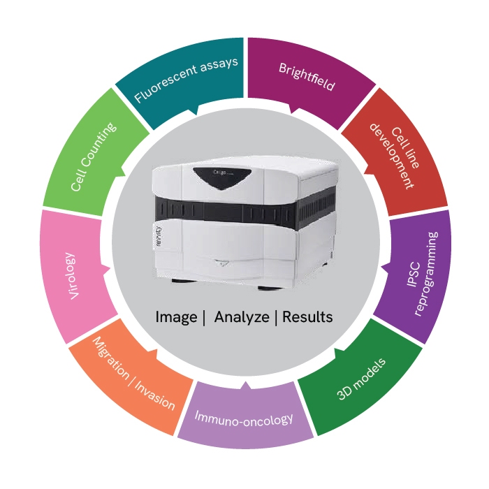
Celigo image cytometer application spotlights
Fluorescent image applications
Cell cycle, cell health, internalization & phagocytosis, co-culture, surface proteins & antibodies, transfection/ transduction, apoptosis, migration.

Cell counting applications
Adherent cell counting, suspension cell counting, fluorescent cell counting, T-cells, splenocyte.
Brightfield image applications
Adherent cell growth tracking, suspension cell counting, embryoid bodies, colonies, spheroids, wound healing, morphology.

Immuno-oncology applications
Direct cell counting, visualization and documentation of all cells, using gating interface, ADCC, direct NK cell killing, CAR-T, CDC.
Migration | Invasion applications
Chemotaxis, wound healing, transwell invasion, 3D migration, 3D invasion.
Virology applications
Viral titer, viral infection, antibody neutralization, transduction efficiency, cytopathic effect, CAR T cell-mediated cytotoxicity.
3D models applications
Growth inhibition, apoptosis, tumor spheroid viability, invasion into matrigel, migration onto ECM, tumor sphere formation & clonogenic survival, EBs & PDOs, 3D confrontation assay.
iPSC reprogramming applications
Fibroblast doubling, iPSC colony counting, embryoid body formation, immunostaining for differentiation.
Cell line development application types
Single colony identification, single tumor sphere identification, single cell per well verification, transfection/transduction.
Your questions answered
-
What makes the Celigo a unique imaging tool?
The Celigo has unique hardware which includes a large f-theta scan lens and galvanometric scanning mirrors, which allows it to rapidly capture whole well images with even, flat illumination. The Celigo is designed for rapid speed-to-data, providing not only very fast high-quality imaging, but built-in, concurrent quantitative analysis as well. Easy whole well imaging and analysis make the Celigo ideal for screening applications in 2D and 3D, looking for rare populations or viral infections, documenting a single cell for colony outgrowth assays, label-free cell analysis in brightfield, or fluorescent cell-based assays directly in a plate without the need to trypsinize.
-
How fast can Celigo scan a plate?
While the exact time it takes the Celigo to scan a plate will depend on how many channels you capture, exposure times, and other settings, a good rule of thumb is about 5 minutes per channel for whole well images for the entire plate. Because scanning and analyzing happen concurrently, you will see quantitative results for the first well as soon as that well is captured and analyzed, before the rest of the plate is even finished scanning!
-
Can I analyze my Celigo images with AI software?
One of the helpful features of the Celigo is that images can be easily analyzed within the Celigo software, or are completely open format and can be exported to be analyzed in any software of your choice! You can automatically export .jpg or .tif files of every individual image or the stitched whole well image, and can apply rules to which wells get exported (for example, only export wells that have more than 10 colonies, etc). With images in a very accessible, full-quality format, you are never limited in how you can use the image data you capture.
-
What is the benefit of using Celigo for drug screening?
Celigo directly counts and analyzes cells in a plate. For a drug screening assay, you can track proliferation of the cells label-free over the course of treatment, then could do endpoint functional assays such as viability staining, cell cycle analysis, or apoptosis directly in the plate without trypsinizing. Celigo can also be used for drug screening in 3D models, with readouts for size, migration, invasion, viability, apoptosis, and more. Celigo's rapid imaging and analysis enable the use of multiple replicates for a high-quality screening assay.
-
Is the Celigo compatible with automation?
Yes, the Celigo is compatible with automation platforms and has an optional automation license add-on, making it easy to scale up your throughput to over 100 plates per day. The Celigo has been integrated into Revvity automated workstations that include an automated incubator, robotic arm, and scheduling software for streamlined assays. Celigo is also compatible with third-party automation solutions and is frequently integrated into custom platforms.
Product information
Overview
Celigoは、プレートベースのベンチトップ型明視野および蛍光イメージングシステムです。ウェル全体での細胞解析と細胞サンプルの特性評価を目的に設計されています。Celigoシステムは、6~1536ウェルプレート、T25、T75フラスコ、10 cmディッシュ、ガラススライドなど、さまざまな容器内の細胞を自然な状態のままイメージングおよび解析します。
個々の細胞レベルでの解析データを簡単に生成できるため、ELISAやタンパク質ベースの生化学的なアッセイとは異なる細胞レベルの情報を提供します。さらに、フローサイトメトリーよりも高速に結果を得られます。Celigoサイトメーターに最適な細胞ベースアッセイ:
- アポトーシス
- 細胞周期
- 蛍光レポーター
- 細胞毒性
- ラベルフリー増殖アッセイ
ウェル全体の高速イメージングと解析
- 正確な細胞集団解析を実現する高感度ウェル全体のイメージング
- 不均一な播種の影響を排除
- 各ウェル内のすべての細胞を正確に検出、イメージング、カウント
- 専用スキャニングミラーによる高速かつ高品質な画像取得
高度な明視野イメージングと最大4チャンネルの蛍光測定
- ラベルフリー細胞解析のための独自アルゴリズム
- 4つの蛍光チャンネルを用いたリアルタイムマルチプレックスアッセイ
生物学者のために設計されたわかりやすいワークフロー
-
複数のオブジェクトベースのアルゴリズムと、さまざまなアッセイベースのアプリケーション
- 直感的に操作できるソフトウェアですぐに使える
- デフォルト設定での測定条件と、サンプルに合わせたカスタマイズが可能
- ゲーティングによる簡単なデータ解析と可視化
- リアルタイムのグラフィックフィードバックにより、複数パラメータを同時測定を直感的にサポート
オートメーションとデータ管理
- オートメーションを容易にするアプリケーションプログラミングインターフェイス(API)
- マイクロプレートの自動搬送によるキネティックエンドポイント解析またはタイムポイント解析
Additional product information
Brightfield imaging for all well sizes
Excellent optics for enhanced image quality. Improves brightfield optical image quality at the edge of wells and reduces edge optical distortion by using an F-Theta lens for superior well edge-to-edge image contrast.
Measure adherent cells without trypsinization
Analyze your cell sample without trypsinization to help avoid losing cells and look at cells right where they grow over multiple scan times.
Series of customized applications for each assay - ready-to-use
Label-free brightfield cell analysis: Take advantage of label-free brightfield applications to avoid staining cells with toxic dyes or transfecting with fluorescent reporters.
Numerous cell characterization assays: Live cell analysis of images for cell counting, confluence, colonies, and 3D-spheroids.
Monoclonality verification and screening: Detect and evaluate single cell derived colonies over the course of multiple timepoints.
Ability to export brightfield and fluorescent images for publications
Capitalize on the Celigo brightfield and fluorescent image quality to take images of your cells for your records and strengthen your publications.
Expand your lab's image analysis & data management capabilities
Storing, managing and sharing large volumes of image data can be a challenge. That's why we've made the Image Artist™ image analysis and management platform from Revvity compatible with Celigo image data for use alongside the Celigo system's own powerful acquisition, visualization and analysis software.
With the addition of the Image Artist platform to your lab, you'll be able to quickly process, store, and share all your image data from the Celigo image cytometer and other major cell imaging systems in a single central location. Its powerful image processing capabilities and ready-made analysis building blocks will provide you with more options for exploring your data and gaining new insights. Benefit from machine learning and artificial intelligence (AI) capabilities to train the software to develop analysis algorithms - and get answers faster. You can even access data and perform analysis remotely via the browser login.
*Image Artist platform is sold separately - it is not sold as part of the Celigo system.
Instrument details
| Software |
|
|---|---|
| Illumination/optics |
|
| Plate compatibility |
|
| High-speed imaging |
|
| Power requirements |
|
| Regularity compliance |
|
Fluorescent channels
| Channel | Excitation | Dichroic | Emission | Typical dyes |
|---|---|---|---|---|
| Blue | 377/50 | 409 | 470/22 | Hoechst, DAPI |
| Green | 483/32 | 506 | 536/40 | FITC, Calcein, GFP,AlexaFluor® 488 |
| Red | 531/40 | 593 | 629/53 | R-PE, PI, Texas Red, AlexaFluor 568 |
| Far-Red | 628/40 | 660 | 688/31 | DRAQ5®, AlexaFluor 647 |
Specifications
| Dimensions | 20.0 in (W) x 17.0 in (D) x 25.0 in (H) |
|---|---|
| Weight |
53.0 kg
|
| 21CFR Compatible |
Yes
|
|---|---|
| Automation Compatible |
Yes
|
| Brand |
Revvity
|
| Imaging Modality |
蛍光・明視野
|
| Model |
Celigoイメージサイトメーター
|
| Technology |
Celigo Pro
|
| Unit Size |
1ユニット
|
Citations
Resources
Are you looking for resources, click on the resource type to explore further.
eBook
10 key factors to improve your cell counting results
Cell counting plays a crucial role in the development and manufacturing of cell and gene therapies as well as regenerative...
Brochure
3D cell culture workflow solutions
More than ever, researchers are turning to 3D cell cultures, microtissues and organoids to bridge the gap between 2D cell cultures...
Application Note
3D tumor spheroid analysis method for HTS drug discovery using Celigo imaging cytometer
Inhibition of cancer cell proliferation in drug discovery research has not translated well from traditional two dimensional (2D)...
Scientific Poster
A high-throughput image cytometry-based screening method for the detection of IL2-induced peripheral blood mononuclear cell-mediated cytotoxicity
Celigo instrument poster describing A high-throughput image cytometry-based screening method for the detection of IL2-induced...
Technical Note
A modern approach to traditional virology research using the Celigo image cytometer.
Application Note
A rapid and label-free in situ assay method for cell proliferation and drug toxicity using the Celigo image cytometer.
Loading...


How can we help you?
We are here to answer your questions.

