


JP
Revvity Sites Globally
Select your location.
*e-commerce not available for this region.

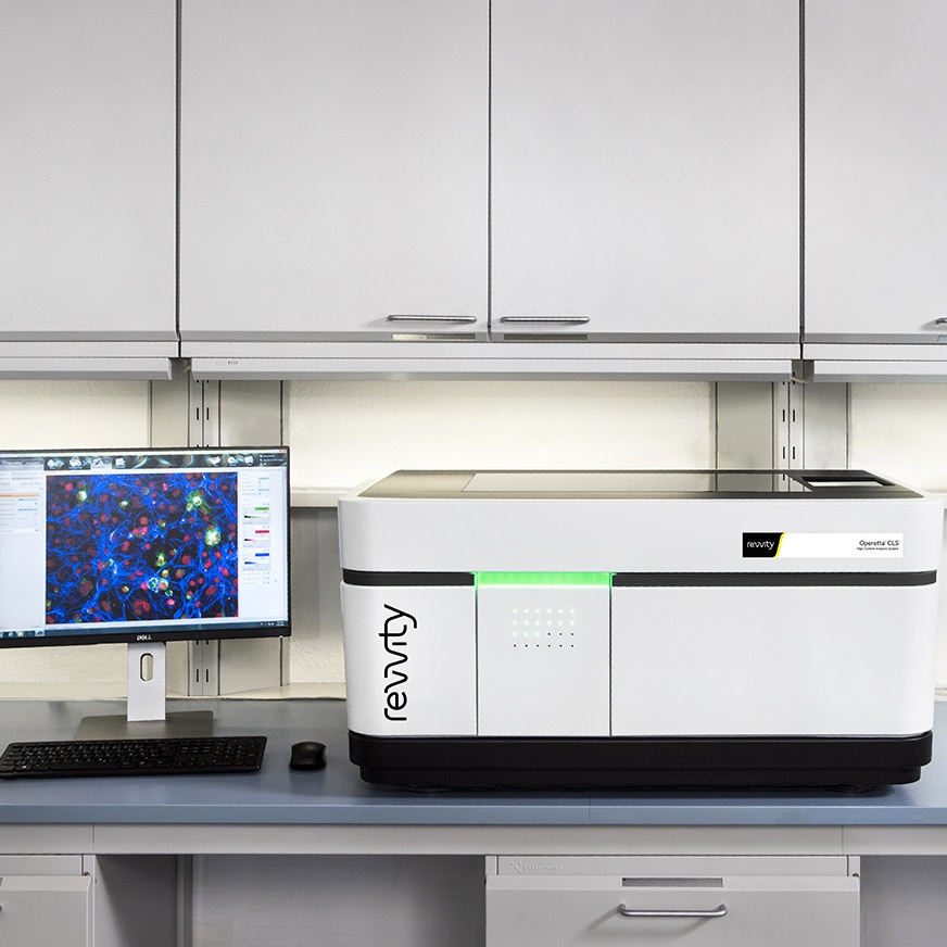
Operetta CLS ハイコンテントアナリシスシステム
Operetta CLS™ ハイコンテントアナリシスシステムの導入は、日常的なアッセイや革新的なアプリケーションにおいて、新しい生物学的知見を得ることができます。ユニークな技術を備えたOperetta CLSは、表現型解析のために必要な速度、感度、解像度を備えており、さらに、シンプルながら強力なHarmonyソフトウェアによって、Operetta CLSは微細な表現型の差異も見つけ出すことができます。
本製品は研究用です。診断用にはご使用いただけません。
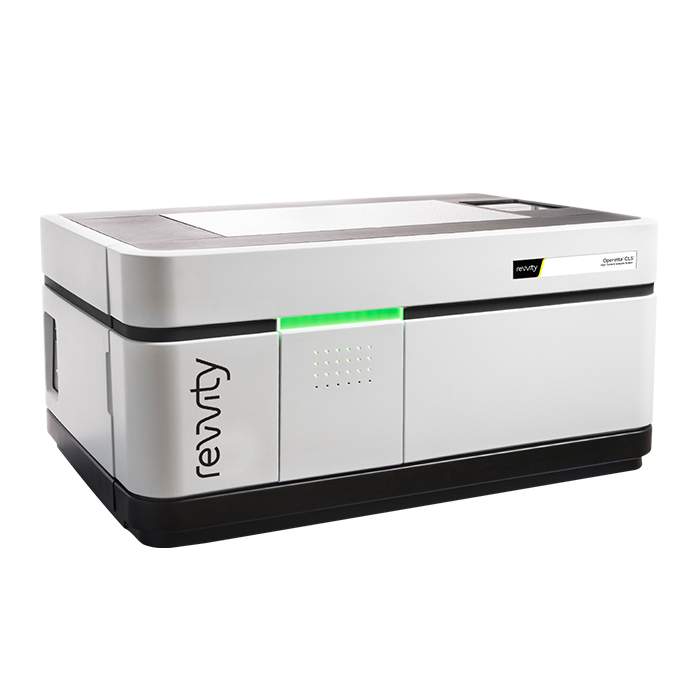


Operetta CLS ハイコンテントアナリシスシステム
Operetta CLS High-Content Analysis System
Part #:
HH16000020
Imaging Modality:
明視野, 共焦点, デジタル位相差, 蛍光
Loading...
完全なHCSワークフロー
Operetta CLSは、速度と感度に直感的な解析を組み合わせたシステムです。包括的なHCSワークフローおよびHarmonyソフトウェアと統合されており、生物学研究者が複雑な解析を独立して実行できる環境を提供します。導入後すぐに使用可能です。
ハイコンテントアナリシス向けに設計されたPhenoPlate™マイクロプレートにより、より優れた結果が得られます。
セルペインティングキットを含むPhenoVue™細胞イメージング試薬と組み合わせて使用できます。
Operetta CLSシステムを自動化することで、スループットと生産性を向上させることができます。また、自動化された細胞ワークフローや創薬ワークフローの活用も可能です。
結果は、Image Artist™画像解析・管理プラットフォームへ自動でエクスポート可能です。Operetta CLSおよびその他のHCSシステムから取得したすべての細胞画像データにアクセスし、再解析、保存、共有が可能です。
Signals One™と組み合わせることで、強力な多変量統計手法および教師なし機械学習技術を活用し、特徴的な細胞フィンガープリントを最もよく定義するパラメーターを特定できます。
Video overview
主な特長
インテリジェントな画像取得
特に3Dマイクロティッシュやレアイベント解析において、関心のある対象をより正確に特定することで、画像取得および解析時間を大幅に短縮します。
機械学習
Revvity独自のPhenoLOGIC機械学習技術により、画像解析の専門知識がなくても容易に解析シークエンスを作成できます。
複雑な3Dモデルにおける細胞表現型の定量
3DビューおよびXYZビューアで細胞モデルを可視化し、体積などの3D関連表現型を定量化できます。
高機能かつシンプルな解析機能
Harmonyハイコンテントアナリシスおよび解析ソフトウェアは、習得しやすく使いやすいソフトウェアです。画像ベースのアプリケーションの生産性が向上します。
高効率励起設計
ダイレクトカップリングにより、ライトガイドや光ファイバーに特有の光損失を抑制します。
高速フレームレートイメージング
最大105fpsのフレームレートによるイメージングで、細胞の迅速な変化を捉えます。
自動水浸対物レンズ
20年以上の実績に基づく技術により、露光時間の短縮、分解能の向上、光毒性の低減を実現します。
共焦点スピニングディスク技術
特にライブセル実験に適した穏やかな励起条件を可能にし、効率的なバックグラウンド除去を実現します。
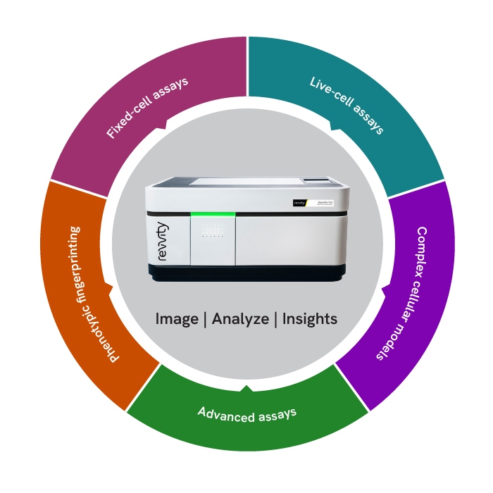
Operetta CLSのアプリケーション
Fixed-cell assays(固定細胞アッセイ)
最大8基の高出力励起光源とユーザーがアクセス可能なエミッションフィルターにより、蛍光染色およびラベルの柔軟性を最大化します。さらに、広視野およびスピニングディスク蛍光イメージングに対応しています。
Live-cell assays(ライブセルアッセイ)
スピニングディスク共焦点光学系と同期LED励起により、安定した励起と、光毒性および退色を最小限に抑えます。明視野またはデジタル位相差イメージングモードも選択できます。
Complex cellular models(複雑な細胞モデル)
水浸対物レンズとラージフォーマットsCMOSカメラの組み合わせにより、高感度かつ高分解能の撮像を実現します。付属のHarmonyソフトウェアは、複雑な細胞モデルにおけるイメージングおよび解析を実行できるプラットフォームです。
Advanced assays(高度なアッセイ)
FRETは、タンパク質間相互作用や構造変化を解析するための有効な手法です。Operetta CLSシステムの高感度イメージングと、レシオメトリックイメージング向けの専用解析ツールにより、再現性の高い結果が得られます。
Phenotypic fingerprinting(表現型フィンガープリンティング)
Operetta CLSは、高解像度イメージングと高度なソフトウェアを組み合わせることで、表現型の微細な差異を捉え、信頼性の高い表現型フィンガープリントの作成を支援します。
Operetta CLSを活用した研究事例
Operetta CLSの構成

Operetta CLS Quattro
感度と分解能が求められる一般的なアプリケーションに適しており、ニーズの拡大に応じて拡張できる構成です。

Operetta CLS FLEX
多様で高度なアプリケーションに対応するため、励起およびイメージングモードの柔軟性を提供します。さらに、より高い性能へアップグレードすることも可能です。

Operetta CLS LIVE
穏やかな照射条件でありながら高感度なライブセルイメージングを実現する構成です。
Configuration details
| Quattro | FLEX | LIVE | ||
|---|---|---|---|---|
| System Options | カメラ | sCMOS | sCMOS | sCMOS |
| LED数 | 4 | 8 | 8 | |
| 共焦点ディスク | º | • | • | |
| 環境制御 | º | º | • | |
| 水浸 | º | º | • | |
| 透過光 | • | • | • | |
| ロボティクス/オートメーション対応 | • | • | • | |
| Imaging Modes | 蛍光広視野イメージング | • | • | • |
| 共焦点蛍光イメージング | º | • | • | |
| 明視野およびデジタル位相差 | • | • | • | |
| レシオメトリックFRET | • | • | • | |
| 3Dイメージング | º | • | • | |
| ライブセルイメージング | º | º | • |
• 標準 º オプション
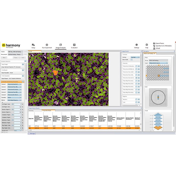
Harmonyソフトウェアによる画像取得および解析の簡素化
直感的なワークフロー
Harmonyソフトウェアは、画像取得から解析、評価までをガイドする直感的なユーザーインターフェースを提供します。
迅速なセットアップ用テンプレート
テンプレートを使用することで、画像取得チャネルや各種パラメーターを効率的に設定できます。
すぐに使えるソリューション
一般的な画像解析タスク向けに事前構築された解析シークエンスを選択することで、ワークフローを簡素化できます。
カスタマイズ可能なビルディングブロック
画像解析用ビルディングブロックを使用して、独自のハイコンテント解析アプリケーションを作成、設定、カスタマイズできます。
高度な解析機能
Harmonyには、テクスチャ解析やSTAR形態解析などの高度な解析機能が含まれており、細胞形態の詳細な記述と表現型の高い識別性を提供します。
データ管理
ソフトウェアは、アッセイレイアウト、装置設定、ユーザー定義のキーワードや注釈を含む解析結果およびメタデータを自動的に保存します。
Operetta CLSハイコンテント解析システムの主な特長
研究の深化
Enrich your research
Operetta CLSシステムは、3D細胞培養を解析することで、より生理学的に意義のあるデータを取得し、より適切な判断を支援します。
Image gallery
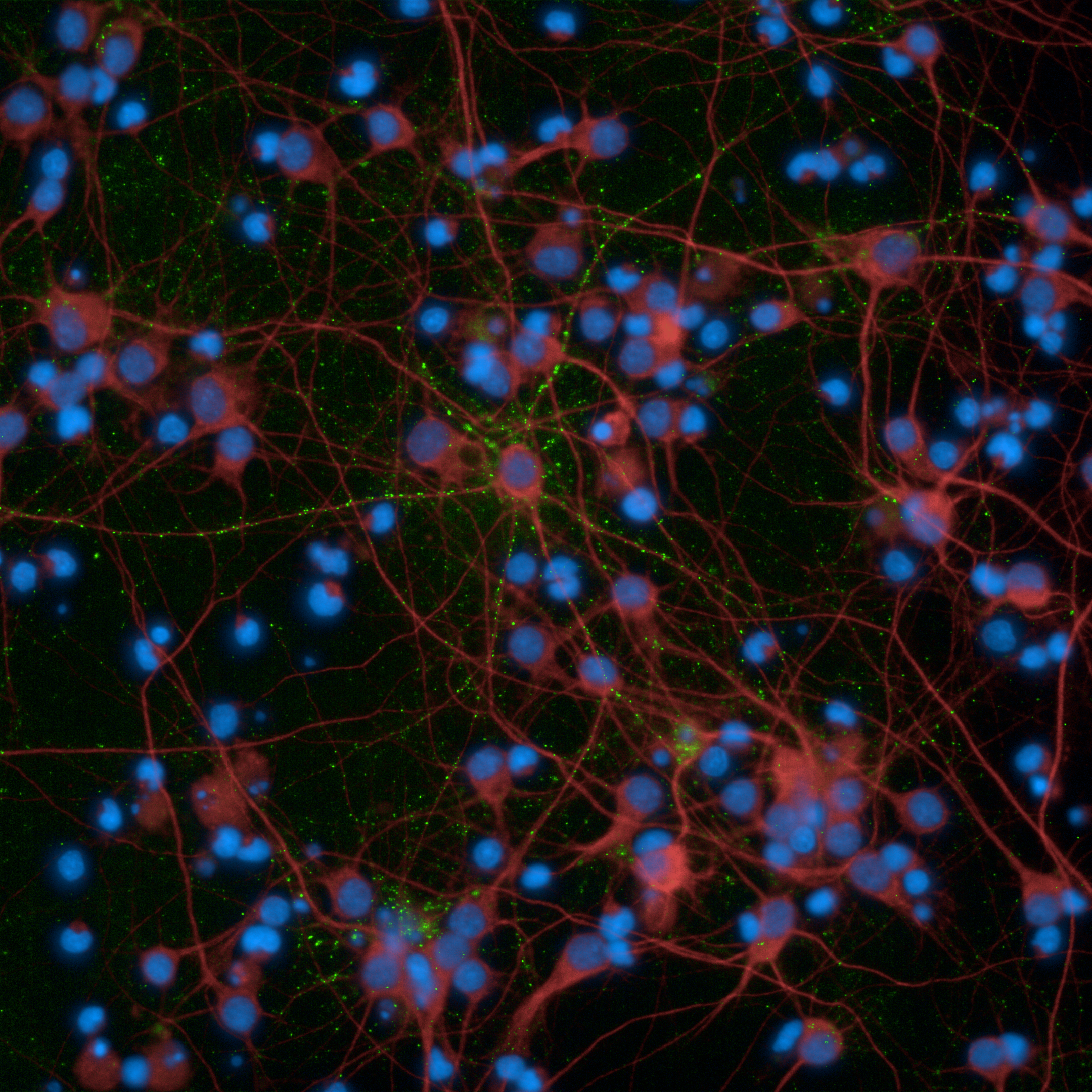
Primary rat neurons imaged on the Operetta CLS high-content analysis system
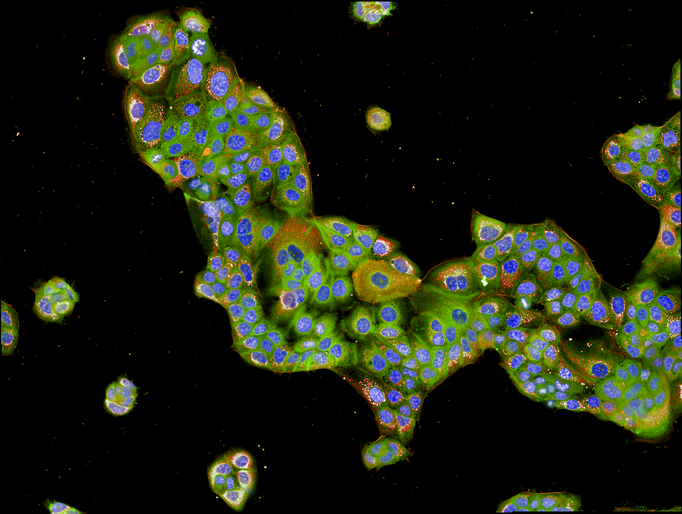
Human ovarian granulosa tumor cell line imaged on the Operetta CLS high-content analysis system
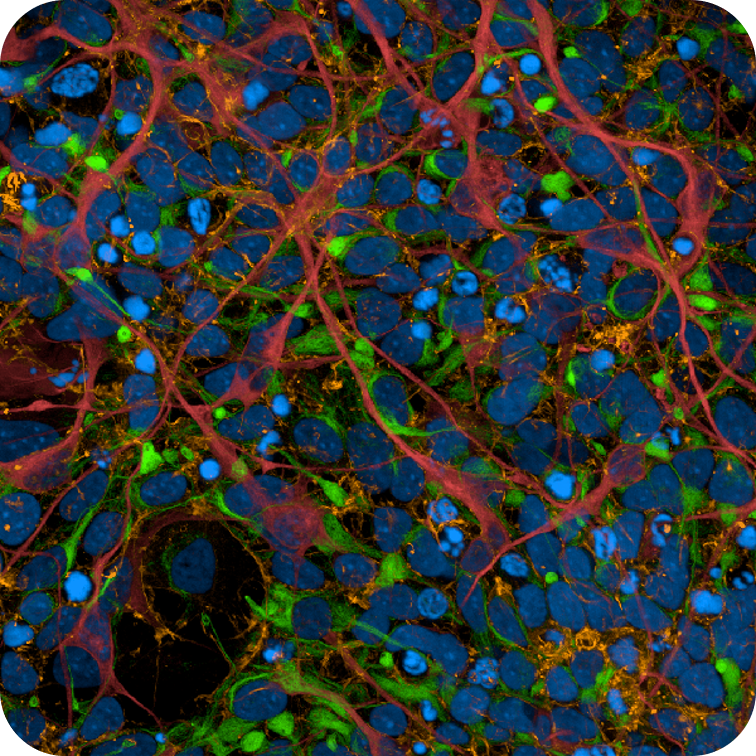
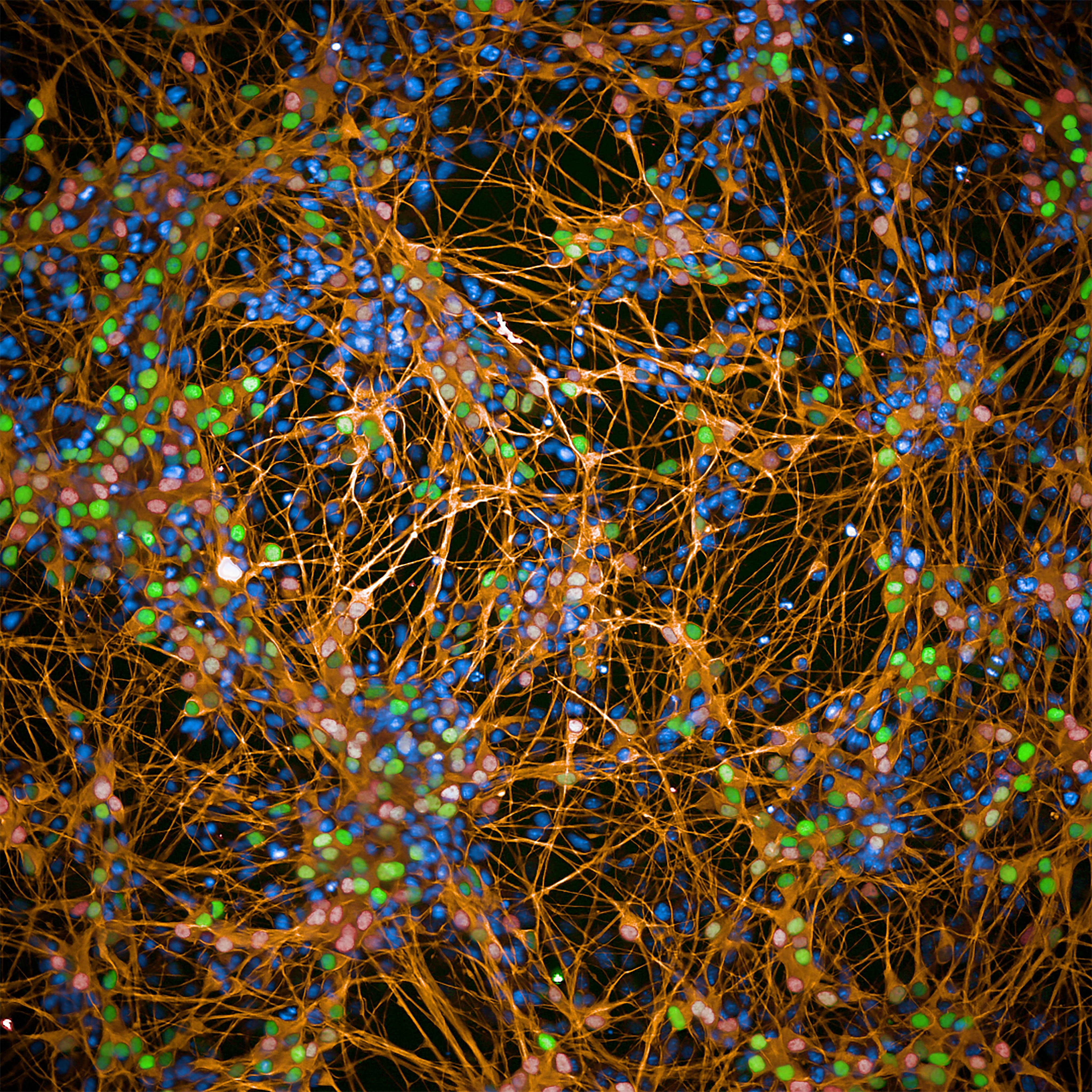
iPSC-derived human cortical neurons imaged on the Operetta CLS high-content analysis system
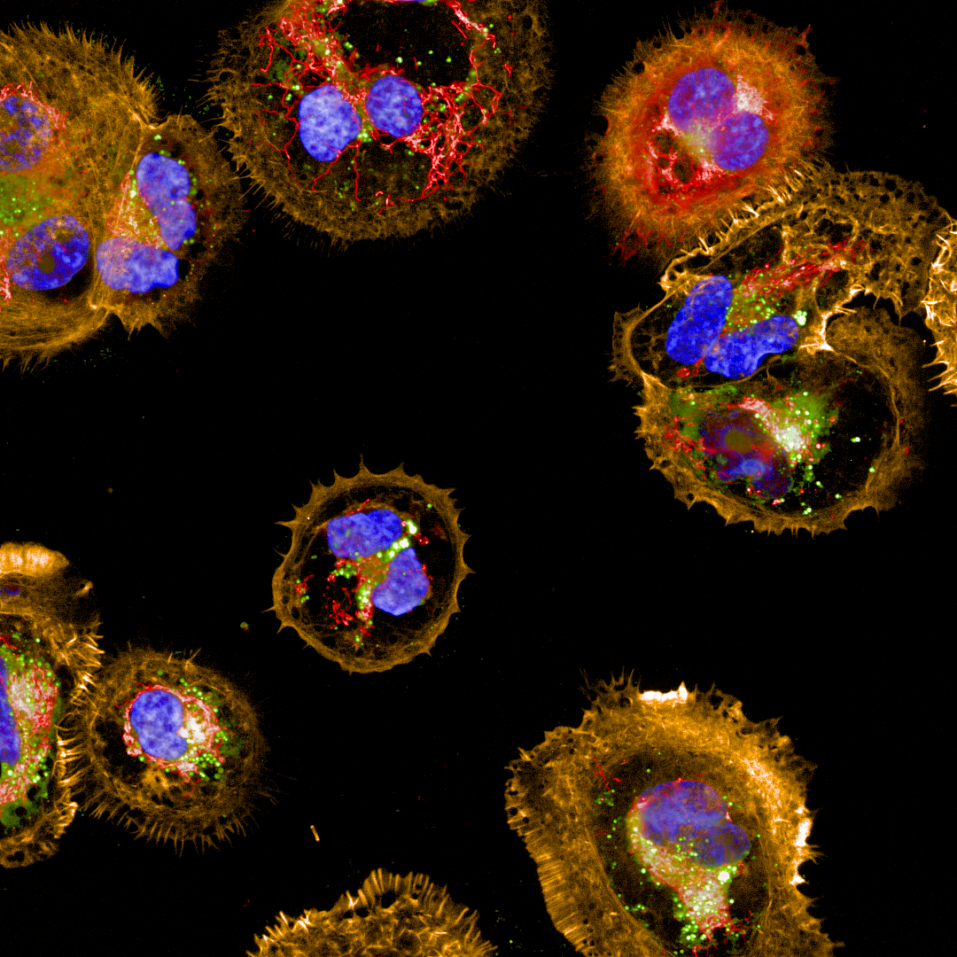
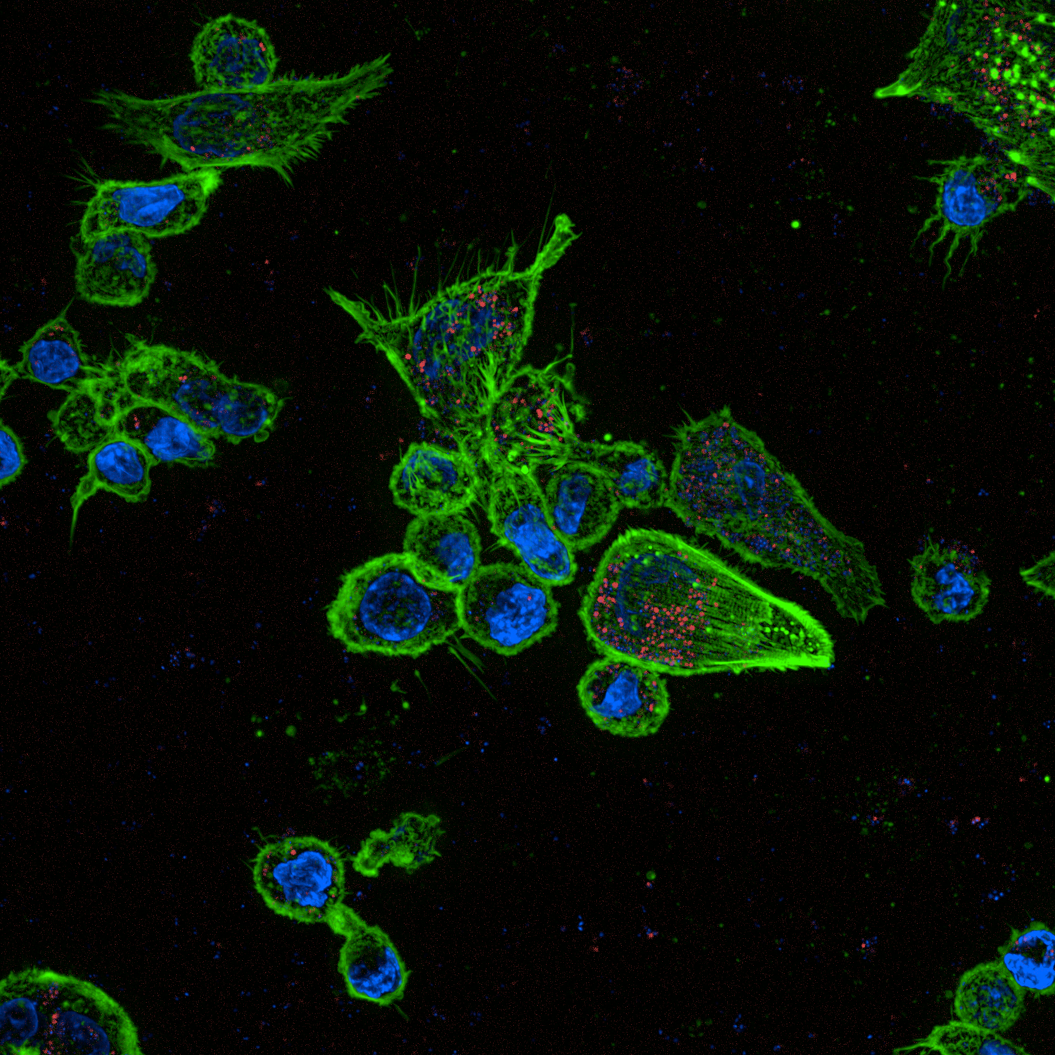
Macrophages imaged on the Operetta CLS high-content analysis system
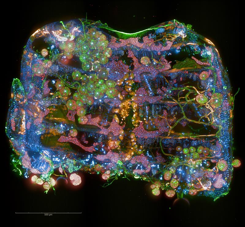
Drosophila abdomen imaged on the Operetta CLS high-content analysis system
iPSC-derived motor neurons imaged on the Operetta CLS high-content analysis system
HPAF-II cells imaged on the Operetta CLS high-content analysis system.
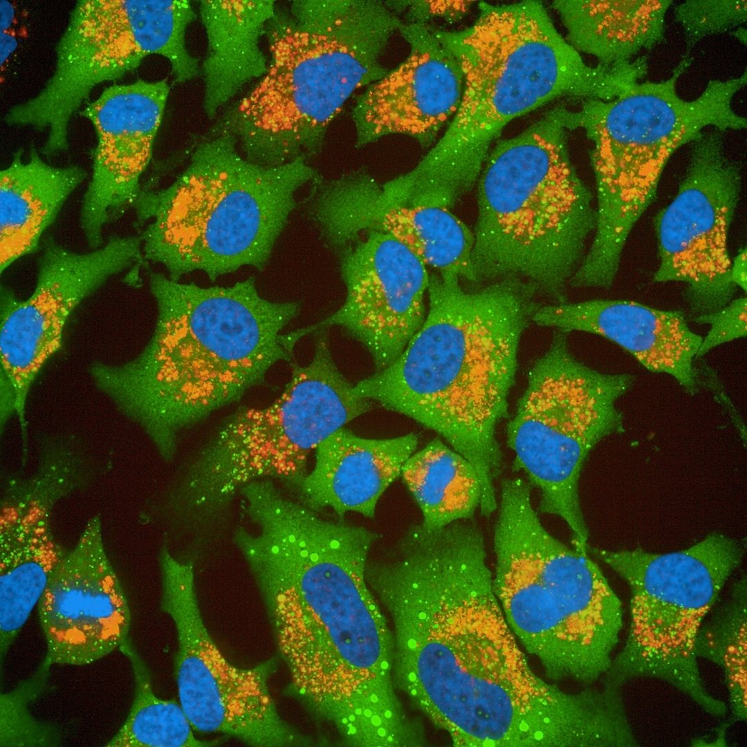
HeLa cells imaged on the Operetta CLS high-content analysis system.
×
Product information
Overview
Operetta CLS システムは、Operetta プラットフォームで培われた信頼性の高い、直感的で強力なデータ解析機能に、高速性と高感度を組み合わせたシステムです。新しいOperetta CLSは、ハイコンテントアナリシスに求められるあらゆる要件を提供します。さらに、Operetta CLSは、HCSワークフローの一部です。レビティは、HCSシステムや蛍光色素、マイクロプレートをはじめ、自動化やインフォマティクスまで、あらゆるソリューションを提供します。すべてを、信頼できる一社からご提供します。さらに、習得しやすく使いやすい Harmony ハイコンテントイメージングおよび解析ソフトウェアと組み合わせることで、研究者自身による解析が可能となり、日常的な解析から複雑な解析まで、幅広いアプリケーションをすぐに実施できます。
Specifications
| Dimensions | 66.0 cm (W) x 98.0 cm (D) x 45.0 cm (H) |
|---|
| Automation Compatible |
Yes
|
|---|---|
| Brand |
Operetta CLS
|
| Imaging Modality |
明視野
共焦点
デジタル位相差
蛍光
|
| Unit Size |
1 unit
|
Video gallery
Operetta CLS ハイコンテントアナリシスシステム
Citations
Resources
Are you looking for resources, click on the resource type to explore further.
Application Note
3D Analysis of Cell Invasion using Operetta
Here, we present a method for analyzing cell invasion into a 3D extracellular matrix using the Operetta® high-content analysis...
Brochure
3D cell culture workflow solutions
More than ever, researchers are turning to 3D cell cultures, microtissues and organoids to bridge the gap between 2D cell cultures...
Technical Note
3D volumetric analysis of luminal spaces inside cysts or organoids
High-content assays using 3D objects such as cysts or organoids can be challenging from the perspectives of both image acquisition...
Technical Note
3D volumetric and zonal analysis of solid spheroids
Multicellular 3D “oids” (tumoroids, spheroids, organoids) have the potential to better predict the effects of drug candidates...
Application Note
A multiparametric live-cell cytotoxicity analysis using the Operetta High-content Analysis System
The detection of compound cytotoxicity is an essential part of drug discovery. In this work we describe a rapid and flexible image...
Case Study
A workflow to characterize and benchmark human induced pluripotent stem cells
The UK-based Human Induced Pluripotent Stem Cell Initiative (HipSci) aims to offer the scientific community access to a vast panel...
Loading...


How can we help you?
We are here to answer your questions.

