


JP
Revvity Sites Globally
Select your location.
*e-commerce not available for this region.

Cellometer Auto T4明視野セルカウンター
This product has been discontinued
Cellometer™ Auto T4自動細胞カウンターは、明視野イメージングを用いて、細胞株の濃度測定およびトリパンブルー法による生存率評価を実施できます。凝集した細胞にも対応し、必要に応じてGMP/GLP対応ソフトウェアを利用できます。
本製品は研究用です。診断用にはご使用いただけません。
Cellometer Auto T4明視野セルカウンター
Cellometer Auto T4 Brightfield Cell Counter
Discontinued
Part #:
CMT-AT4P
Imaging Modality:
明視野
Loading...
Product information
Overview
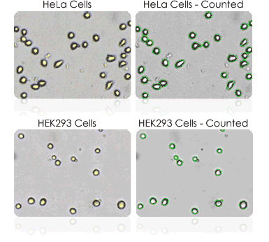
Cellometer Auto T4は、明視野イメージングとパターン認識ソフトウェアを利用して、個々の細胞を高速かつ正確に識別・カウントします。細胞数、濃度、直径、および生存率(%)が自動的に計算され、報告されます。
Cellometer Auto T4セルカウンターの特長:
- 個々の細胞レベルの情報を画面上で確認可能
- 精度の高いデータ取得のために、複数視野の撮像に対応
- 豊富な細胞タイプライブラリ
- 凝集した細胞や形状が不規則な細胞のカウントにも対応
- IQ/OQ バリデーションおよび GMP/GLP ソフトウェアのオプション
Additional product information
One-step concentration & viability
The Cellometer Auto T4 simultaneously calculates cell concentration and % viability for cultured cells stained with trypan blue.
Trypan blue viability is a dye exclusion method that utilizes membrane integrity to identify dead cells. The dye is unable to penetrate healthy cells, so they remain unstained. Dead cells have a compromised cell membrane that is permeable to the trypan blue dye. Dead cells are stained blue and display as dark cells in the Cellometer software with brightfield imaging.
Within 10 seconds, the Cellometer Auto T4 instrument reports:
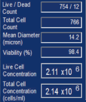
- Live, dead, and total cell count
- Live and total cell concentration
- Mean Cell Diameter
- % Viability
Counted brightfield image
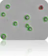
Live cells = green circle
Dead (stained) cells = red circle
Multiple cell images for data verification
With the Cellometer Auto T4 Cell Counter, cell morphology can be immediately viewed on-screen.
Two images with four fields of view each are captured per cell count. This is equivalent to the area of four quadrants of a hemocytometer. Counted cells are indicated on-screen for further verification that cells in the sample are being imaged and analyzed properly.
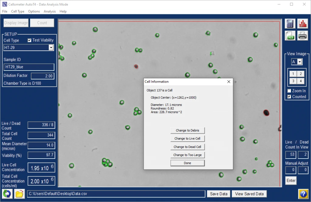
Users can confirm that:
- Cells are counted correctly, based on size and shape
- Debris is excluded
- Cells within clumps are being counted individually
- Cell images can be archived and exported for use in publications and presentations
- Saved images can be re-counted using default or user-optimized analysis settings
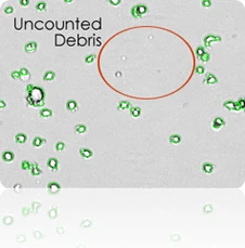
Exclusion of debris
The Cellometer distinguishes cells from cellular debris based on size, brightness, and morphology. This allows for increased accuracy with cell counts.
- Cell size parameters can be modified to optimize exclusion of debris from results and enhance the accuracy of counting for a wide range of cell sizes.
- The counted image can be viewed to verify exclusion of debris from results.
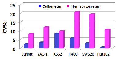
Cellometer precision
The Cellometer %CV (Coefficient of Variation), including sample preparation, was well under 5% for most cell lines tested. The CVs for manual counts ranged from 7% to 20%. Automated cell counters remove inter-operator variability and subjectivity from cell counts to generate more precise data.
Improve data accuracy & consistency
- Eliminate wash steps
- Reduce judgment errors
- Reduce recording & calculation errors
- Reduce counting time... Run more experiments
| Cellometer Auto T4 | Hemacytometer | ||||
|---|---|---|---|---|---|
| Concentration | n | CV | Concentration | n | CV |
| 1.17 x 10⁶ | 4 | 2.27% | 1.11 x 10⁶ | 3 | 8.04% |
| 1.36 x 10⁶ | 4 | 3.17% | 1.41 x 10⁶ | 3 | 11.89% |
| 6.36 x 10⁵ | 4 | 8.29% | 5.58 x 10⁵ | 3 | 9.66% |
| 1.32 x 10⁶ | 4 | 5.57% | 1.16 x 10⁶ | 3 | 20.65% |
| 3.81 x 10⁵ | 4 | 2.79% | 3.70 x 10⁵ | 3 | 19.49% |
| 9.18 x 10⁵ | 4 | 0.39% | 7.68 x 10⁵ | 3 | 10.60% |
Cellometer Auto T4 brightfield cell counter specifications
| Includes |
|
|---|---|
| Available accessories |
|
| Imaging performance |
|
| Instrument specifications |
|
| PC / Laptop requirements: (If purchasing Cellometer without PC laptop) |
|
Specifications
| Dimensions | 3.5 in (W) x 4.2 in (D) x 12.6 in (H) |
|---|---|
| Weight |
4.7 kg
|
| Brand |
Revvity
|
|---|---|
| Imaging Modality |
明視野
|
| Model |
Cellometer Auto T4明視野セルカウンター
|
| Technology |
Cellometer Software
|
| Unit Size |
1 each
|
Citations
Resources
Are you looking for resources, click on the resource type to explore further.
eBook
10 key factors to improve your cell counting results
Cell counting plays a crucial role in the development and manufacturing of cell and gene therapies as well as regenerative...
Application Note
An alternative image-based technique for phytoplankton cell counts in shellfish aquaculture.
Brochure
Automated cell counting and image cytometry solutions brochure
With expertise and a pioneering spirit, Revvity boldly leads the way in cell imaging and analysis. As creators of advanced...
Application Note
Cell counting repeatability and consistency using the Cellometer Auto T4 brightfield cell counter.
Loading...


How can we help you?
We are here to answer your questions.

