Optical Imaging
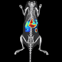
Sensitivity to image down to a single cell with the IVIS™ platform
Whether your research involves better understanding molecular processes of disease, tracking disease progression, or evaluating therapeutic effectiveness of drug candidates, your cutting-edge research commands high-sensitivity and reliable optical imaging data.
Bioluminescence and fluorescence imaging offer their own unique characteristics for small animal imaging. Bioluminescence imaging (BLI) uses luciferase genes and offers minimal background signals from the animal tissues, provides high specificity and precise quantification, and can be used to detect and monitor biological events such a tumor growth deep within the tissue. Fluorescence imaging (FLI) is ideal for monitoring and quantifying cell behavior of biological targets.
We are here to help you achieve your research goals with our leading IVIS molecular imaging platforms and diverse range of IVISbrite™ & IVISsense™ optical reagents. From single mode 2D optical and 3D tomography to multimode integrated systems, we have the tools you need to help you get the answers you are searching for.
In vivo optical imaging is a fast, cost-efficient, easy-to-use, and powerful technique to help you non-invasively study molecular and biological processes of disease, or help drive discovery and development of novel drug candidates using bioluminescent or fluorescent reporters.
For research use only. Not for use in diagnostic procedures.

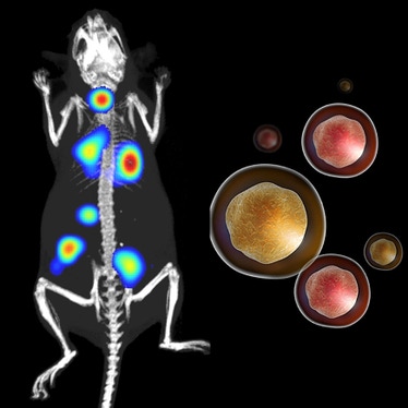
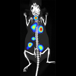
Bioluminescence
Exquisitely sensitive, cost-effective, easy-to-use. Superior signal to noise ratio, high sensitivity, short acquisition times and ease-of-use make bioluminescence imaging an excellent non-invasive tool to better understand the mechanisms of disease biology. Accelerate your in vivo imaging research studies or propel your drug discovery development process using IVIS, the most widely published bioluminescence imaging platform in the industry.
Exquisitely sensitive, cost-effective, easy-to-use. Superior signal to noise ratio, high sensitivity, short acquisition times and ease-of-use make bioluminescence imaging an excellent non-invasive tool to better understand the mechanisms of disease biology. Accelerate your in vivo imaging research studies or propel your drug discovery development process using IVIS, the most widely published bioluminescence imaging platform in the industry.
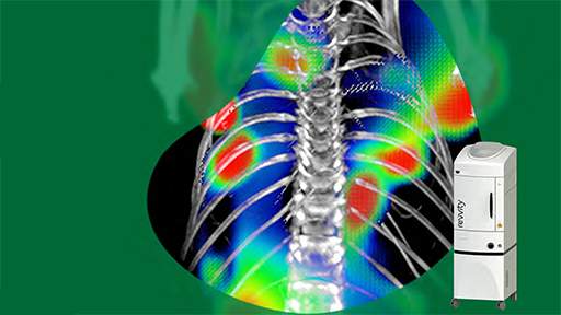
Fluorescence
Obtain more information from your animal study. Fluorescence imaging is a powerful tool that allows you to visualize and quantify biological targets, pathways, and processes in your animals models. In vivo fluorescence imaging utilizes fluorescent reporters such as proteins, dyes, or nanoparticles, which emit photons when excited at a specific wavelength to produce light. Whether your biology focus is to monitor cellular or genetic activity, track gene expression, track disease progression, or evaluate the effect of new drug candidate, Revvity offers a wide range of fluorescence imaging systems to meet your research requirements.
Obtain more information from your animal study. Fluorescence imaging is a powerful tool that allows you to visualize and quantify biological targets, pathways, and processes in your animals models. In vivo fluorescence imaging utilizes fluorescent reporters such as proteins, dyes, or nanoparticles, which emit photons when excited at a specific wavelength to produce light. Whether your biology focus is to monitor cellular or genetic activity, track gene expression, track disease progression, or evaluate the effect of new drug candidate, Revvity offers a wide range of fluorescence imaging systems to meet your research requirements.

Optical reagents
Obtain more information from your target and get more insightful research results with our wide-range of imaging reagents. Built around your applications our optical reagents are optimized for use on our full range of IVIS in vivo imaging systems, including:
- IVISense fluorescent agents, labeling kits, dyes and panels
- IVISbrite bioluminescent substrates
- IVISbrite luciferase labeled oncology cell lines and bacteria


Which IVIS system is best for your research?
|
|
|
|
|
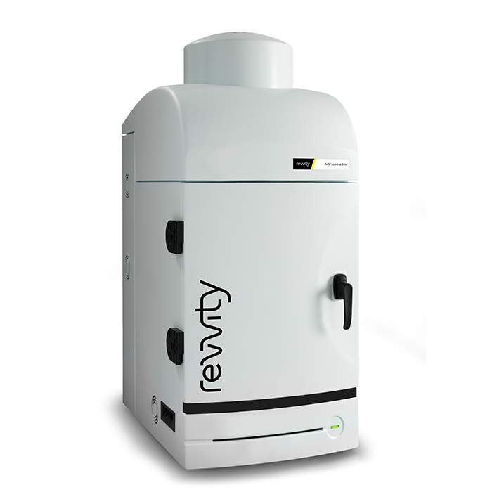
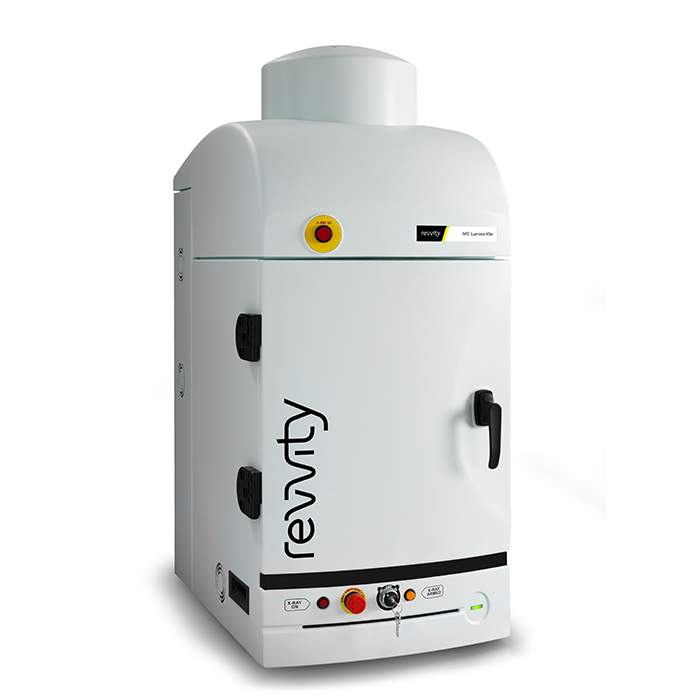
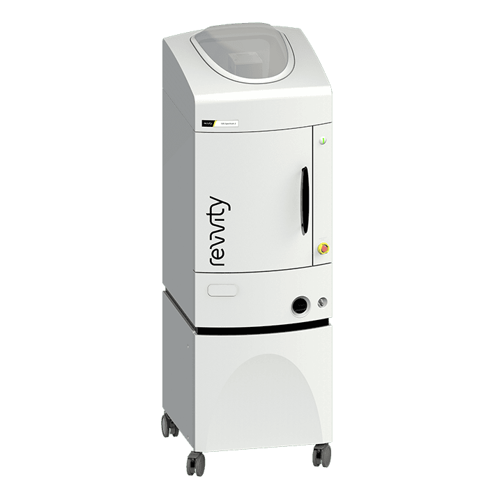
| IVIS Lumina S5 | IVIS Lumina X5 | IVIS Spectrum 2 | IVIS SpectrumCT 2 | |
| Capacity | Up to 10 mice* | Up to 10 mice* | Up to 10 mice* | Up to 10 mice* |
| Benchtop format | ✔ | ✔ | ||
| 2D Bioluminescence / Fluorescence | ✔/✔ | ✔/✔ | ✔/✔ | ✔/✔ |
| 3D Bioluminescence / Fluorescence | ✔/✔ | ✔/✔ | ||
| Enhanced Fluorescence capabilities | ✔ | ✔ | ✔ | ✔ |
| Integrated standard x-ray | ||||
| Integrated high resolution x-ray | ✔ | |||
| Integrated CT | ✔ | |||
| Spectral Unmixing | ✔ | ✔ | ✔ | ✔ |
| 3D Multimodal Co-Registration | ✔ | ✔ | ||
| Optical FOV (cm) (Nominal) | 10–22.5 | 10–22.5 | 4–22.5 | 4–22.5 |
| Wavelength range (nm) | 410–865 | 410–865 | 415–850 | 415–850 |
| NIST Calibration | ✔ | ✔ | ✔ | ✔ |
| Relative Cost | $$$$$ | $$$$$ | $$$$$ | $$$$$ |
Please contact your sales representative for more detailed information on our IVIS optical imaging systems.
*Using optional manifold kit
**Using expansion lens
Featured Resources