メニュー
Revvity logo
Contact us
JP
Revvity Sites Globally

Select your location.

*e-commerce not available for this region.

australia.webp Australia
austria.webp Austria
belgium.webp Belgium
brazil.webp Brazil *
canada.webp Canada
china.webp China *
denmark.webp Denmark
finland.webp Finland
france.webp France
germany.webp Germany
hong-kong.webp Hong Kong (China) *
india.webp India *
ireland.webp Ireland
italy.webp Italy
japan.webp Japan *
luxembourg.webp Luxembourg
mexico.webp Mexico *
netherlands.webp Netherlands
norway.webp Norway
philippines.webp Philippines *
republic of korea.webp Republic of Korea *
singapore.webp Singapore *
spain.webp Spain
sweden.webp Sweden
switzerland.webp Switzerland
thailand.webp Thailand *
uk.webp United Kingdom
usa.webp United States
Breadcrumb
...
  • ホーム
  • Products
  • プラットフォームと自動化
  • イメージング
    • 核酸分離
    • 自動液体分注機
    • 統合ラボオートメーション
    • マイクロ流体分析
    • 検出システムソリューション
    • イメージング
    • Cell Counting and Image Cytometry
    • サンプルのホモジナイゼーション
    • IVDプラットフォーム&オートメーション
    • 消耗品&アクセサリー
    • Instrument Service & Maintenance
  • In vivo イメージング
    • Cellular Imaging & Analysis
    • In vivo イメージング
  • MicroCT Imaging
    • in vivoイメージングアクセサリー
    • In Vivo Imaging Instruments
    • In Vivo Imaging Reagents
    • In Vivo Imaging Software
    • MicroCT Imaging
    • Multimodality Imaging
    • Nimbus AI Accelerated Analysis Pilot Program
    • Optical Imaging
    • Ultrasound Imaging

MicroCT Imaging

Biology doesn't happen in 2-dimensions

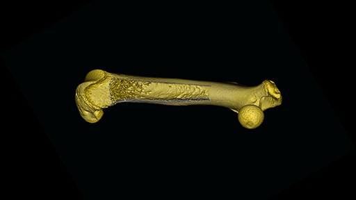
Being able to study 3-dimensional (3D) structure and morphology of tissues with high resolution imaging is becoming increasingly important in biomedical research. Micro-computed tomography is a unique imaging modality that enables you to image 3D structure rapidly and non-invasively at high resolution providing valuable anatomical information for your basic research or drug discovery and development programs.

Micro-CT offers excellent cross-sectional images of skeletal structures and has long been an established tool for evaluating bone structure. It is often used to study bone diseases such as osteoporosis as well as evaluating the efficacy of therapeutics such as bisphosphates. In addition to bone research, microCT is commonly used to study cancer biology. With the development of new contrast agents, microCT scanning of soft tissue is an emerging area for this modality particularly in the area of cardiovascular and pulmonary research.

Whether you are studying tumor biology, bone, cardiovascular or pulmonary disease research, or evaluating drug efficacy, our microCT imaging solutions can help you get the answers you need.

Micro-computed tomography uses x-rays to create a 3D image composed of 2D planar images that are reconstructed and processed into 3D models.

Often referred to as 3D x-ray, microtomography, or microCT imaging, it’s similar to CT scans performed in hospitals but on a smaller scale and offers greatly enhanced resolution.


For research use only. Not for use in diagnostic procedures.
 

in vivo trade in banner
MicroCT imaging 872x872

Featured Resources

quantum-gx3-512 (1)

Video

The Quantum GX3 microCT imaging system

The Quantum™ GX3 offers high spatial resolution down to 5 microns.

Read now

Featured products

of 0
line

ご質問がございましたら、
お気軽にお問い合わせください。

お問合せ
Revvity Logo

©2025 Revvity - All rights reserved

Revvity is a trademark of Revvity, Inc. All other trademarks are the property of their respective owners.