


JP
Revvity Sites Globally
Select your location.
*e-commerce not available for this region.

pHSense Eu-Labeled Anti-FLAG Antibody, 10 x 96 Assay Points
pHSense Eu-Labeled Anti-FLAG Antibody, 10 x 96 Assay Points

Generic pHSense
The pHSense Eu Anti-FLAG carries a pH-sensitive probe and is designed for the detection of FLAG-tagged receptor and membrane protein internalization. It is especially suited for monitoring GPCR internalization.
| Feature | Specification |
|---|---|
| Application | 内部化 |
| Sample Volume | 50 µL |
The pHSense Eu Anti-FLAG carries a pH-sensitive probe and is designed for the detection of FLAG-tagged receptor and membrane protein internalization. It is especially suited for monitoring GPCR internalization.
Product variants
Unit Size: 96 assay points
Part #:
81FL1EU1AA
Unit Size: 2 x 96 wells
Part #:
81FL1EU1AB
Unit Size: 10 x 96 wells
Part #:
81FL1EU1AC
For research use only. Not for use in diagnostic procedures. All products to be used in accordance with applicable laws and regulations including without limitation, consumption and disposal requirements under European REACH regulations (EC 1907/2006).
pHSense Eu-Labeled Anti-FLAG Antibody, 10 x 96 Assay Points
Generic pHSense
Loading...
Product information
Overview
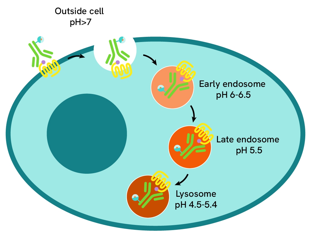
pHSense™ Eu Anti-FLAG is non cell permeant, and is labeled with a pH sensitive europium complex. This Monoclonal Anti-FLAG antibody is designed to monitor receptor and membrane protein internalization when tagged with the FLAG® sequence (DYKDDDDK) on the extracellular part. The antibody recognizes the FLAG® sequence fused at the N-terminus, intra or C-terminus and remains minimally fluorescent at neutral extracellular pH (≥7). Once internalized, the pHSense™ Eu Anti-FLAG encounters increasingly acidic compartments such as early and late endosomes and lysosomes, where the europium signal becomes progressively stronger. This new pH sensitive europium anti-FLAG is compatible with a time-resolved fluorescence (TRF) detection, effectively eliminating most fluorescence background and significantly enhancing the signal-to-background ratio. Its unique photophysical properties enable simple and robust no-wash detection of receptor-mediated endocytosis in plate-based assays with live-cells.
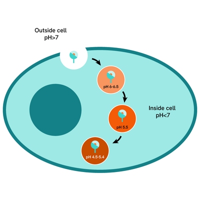
How it works
pHSense Eu-Anti-FLAG assay principle
pHSense Eu Anti-FLAG is non cell permeant, and is labeled with a pH sensitive europium complex. This Monoclonal Anti-FLAG antibody is designed to monitor receptor and membrane protein internalization when tagged with the FLAG® sequence. The antibody recognizes the FLAG sequence and remains minimally fluorescent at neutral extracellular pH (≥7). Once internalized, the pHSense Eu Anti-FLAG encounters increasingly acidic compartments such as early and late endosomes and lysosomes, where the europium fluorescent signal becomes progressively stronger.
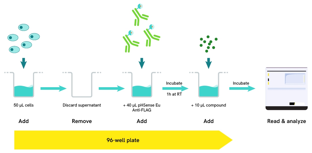
pHSense Eu Anti-FLAG Assay Protocol
The assay begins by culturing cells in a 96-well plate. The pHSense Eu Anti-FLAG is then added to the cells and incubated for 1 hour at room temperature. Following this incubation, cells are stimulated with a pharmacological compound. Fluorescence is then measured either kinetically or at endpoint using an HTRF-compatible plate reader.
Assay validation
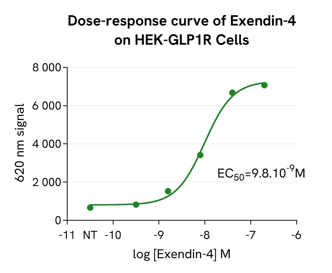
pHSense-Based Detection of GLP1R Internalization Following Exendin-4 Stimulation
Tag-Lite® GLP1R cells (stable cell line, Part #: C1SU1GLP1, Revvity) were seeded in a 96-well white, culture-treated plate at a density of 80,000 cells per well in complete culture medium and incubated overnight at 37°C with 5% CO₂. After cell supernatant removal, 40 µL of the pHSense Eu Anti-FLAG diluted in cell culture medium (DMEM +10%FBS) were added to the cells, followed by a 1 hour incubation at room temperature.
Exendin-4, a GLP1R agonist, was serially diluted in cell culture medium, and 10 µL of each dilution were added to the wells. After incubation at 37°C, the signal was recorded using an HTRF-compatible plate reader. Results show Exendin-4 induced a dose-dependent increase in signal, with an EC₅₀ value consistent with values described in the scientific literature.
In parallel, cells were pre-incubated with 10 µM of the GLP1R antagonist Exendin 9-39 for 30 minutes at 37°C prior to the addition of Exendin-4. Following a 20 min incubation at 37°C, the signal was recorded. In presence of Exendin 9-39 the signal decreased to a level close to the constitutive internalization, which suggests effective inhibition of agonist induced GLP1R internalization.

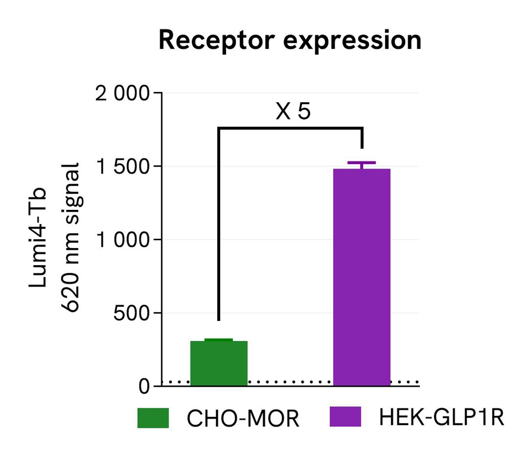
Validation of pHSense Eu Anti-FLAG assay in cell lines expressing different receptor levels
The pHSense Eu Anti-FLAG was tested on 2 different stable cell lines expressing various level of receptors at the membrane: Tag-Lite® GLP1R cells (HEK-GLP1R) and Tag-lite® Mu opioid cells (CHO-MOR).
The relative levels of receptor expression levels were determined using the Tag-lite SNAP-Lumi4-Tb Labeling Reagent (Part#: SSNPTBC, Revvity). Cells were seeded in a 96-well black culture-treated plate at a density of 100,000 cells per well in complete culture medium. After overnight incubation at 37°C with 5% CO₂, the supernatant was removed and 100 µL of 100 nM SNAP-Lumi4-Tb Labeling Reagent were added to the cells, followed by a 1h incubation at 37°C with 5% CO₂. Cells were washed 3 times with Tag-lite SNAP/CLIP Labeling Medium 1X (Part#: LABMED, Revvity) before the addition of 100 µL of the same medium. The 620 nm signal was recorded using an HTRF-compatible plate reader. Results showed Tag-Lite® GLP1R cells exhibit a 5-times higher expression level than Tag-lite® Mu opioid cells.
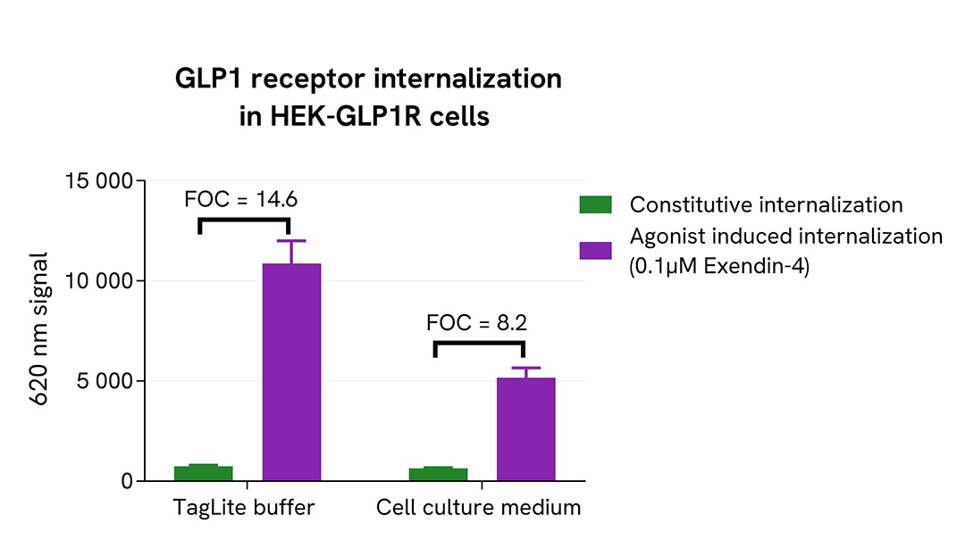
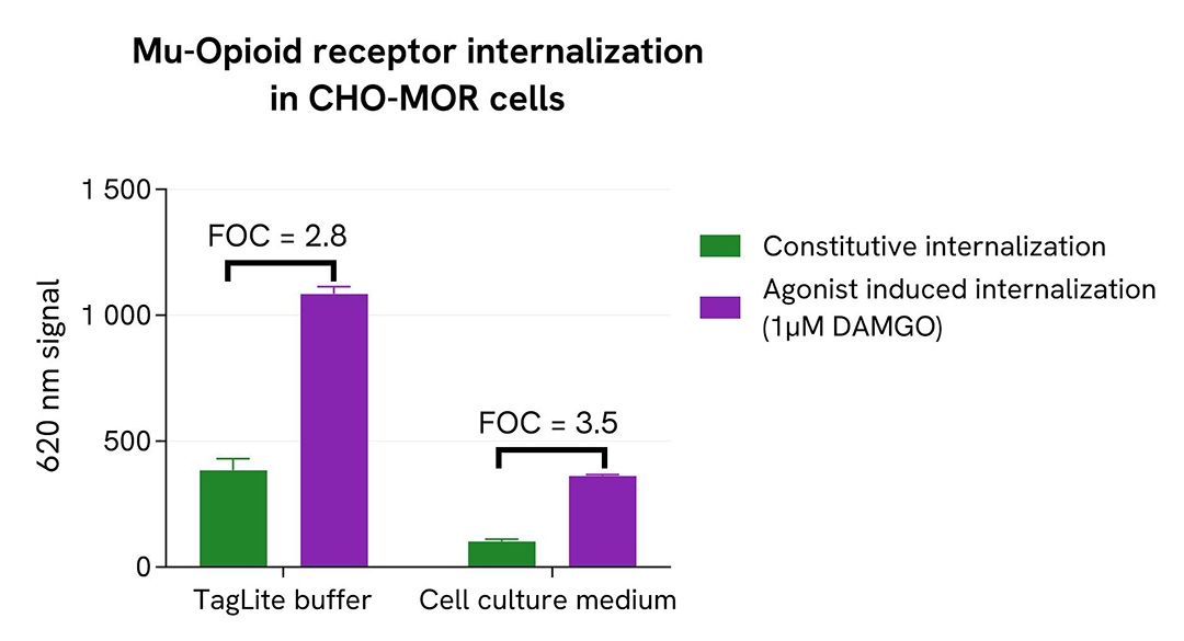
To run the pHSense Eu Anti-FLAG assay, Tag-Lite® GLP1R and Tag-lite® Mu opioid stable cell lines were seeded in a 96-well white culture-treated plate at a density of 80,000 cells per well in complete culture medium, and then incubated overnight at 37°C with 5% CO2.
After cell supernatant removal, 40 µL of the pHSense Eu Anti-FLAG diluted in cell culture medium were added to the cells and incubated for 1 hour at room temperature. Exendin-4, a GLP1R agonist, and DAMGO, a Mu opioid agonist, were diluted in cell culture medium, and 10 µL of agonists or cell culture medium - as constitutive internalization condition - were added to the wells. After incubation at 37°C, the signal was recorded using an HTRF-compatible plate reader.
In parallel, the same assay was performed with the pHSense Eu Anti-FLAG and receptor agonists prepared in Tag-lite SNAP/CLIP Labeling Medium as physiological buffer.
As demonstrated here, the pHSense Eu Anti-FLAG allows the detection of agonist induced and constitutive internalization in cell lines with different receptor expression levels, and is compatible with Tag-lite SNAP/CLIP Labeling Medium as physiological buffer or cell culture medium.
Specifications
| Application |
Internalization
|
|---|---|
| Automation Compatible |
Yes
|
| Brand |
pHSense
|
| Detection Modality |
pH sensitive dye
|
| Product Group |
Fluorescent Reagent
|
| Sample Volume |
50 µL
|
| Shipping Conditions |
Shipped in Dry Ice
|
| Target |
FLAG
|
| Target Class |
Cell surface proteins, antibodies, ADCs
|
| Technology |
TRF
|
| Unit Size |
10 x 96 wells
|
Resources
Are you looking for resources, click on the resource type to explore further.
Flyer
GPCR internalization: simplified detection, clear results with pHSense
pHSense™ probes are plate reader-compatible reagents with time-resolved fluorescence (TRF) readout specifically developed for...
Flyer
GPCR research reagents, from binding to response
Explore Revvity's complete GPCR research reagent portfolio — from ligand binding and G-protein activation to arrestin recruitment...
Brochure
pHSense reagents product list
Discover pHSense™ probes – plate reader-compatible reagents with time-resolved fluorescence (TRF) readout, specifically developed...
Loading...


How can we help you?
We are here to answer your questions.

