


JP
Revvity Sites Globally
Select your location.
*e-commerce not available for this region.

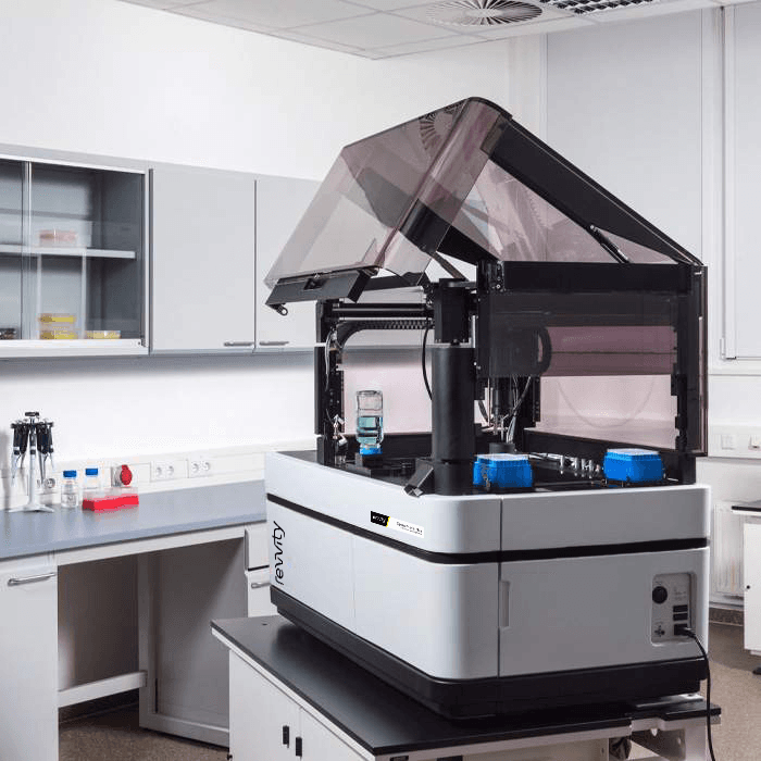
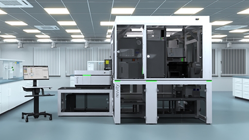
Opera Phenix Plus ハイコンテントスクリーニングシステム
Opera Phenix™ Plus ハイコンテントスクリーニングシステムは、今日の複雑に多様化したアプリケーションにできる最高峰のマルチカメラ共焦点システムです。20年以上の経験に基づき、Opera Phenix Plusは、ハイスループットイメージング、表現型スクリーニング、複雑な疾患モデルを用いたアッセイ(ライブセル、初代細胞、マイクロティッシュなど)や、Ca2+フラックスのような高速応答アッセイなどに対応可能なシステムです。
本製品は研究用です。診断用にはご使用いただけません。
/poster.jpg?format=auto&width=40&height=40)
/poster.jpg?format=auto&width=40&height=40)


Opera Phenix Plus ハイコンテントスクリーニングシステム
Opera Phenix Plus High-Content Screening System
Part #:
HH14001000
Imaging Modality:
明視野, 共焦点, デジタル位相差, 蛍光
Loading...
Opera Phenix Plusハイコンテントアナリシスソリューションは、より深い洞察をもたらします
Revvityの先進的なハイコンテントアナリシスソリューションで、価値ある成果をより早く得られます。
より多くの知見
ハイコンテントアナリシスは、これまでの手法よりもより深い情報を提供し、細胞の応答を詳細に記述することを可能にします。
細部まで定量化
単一細胞から細胞集団全体まで、表現型の変化を様々なスケールで解析します。ハイコンテントアナリシスが細部までの定量を可能にします。
3Dイメージングの強化
水浸対物レンズにより3D画質が向上し、複雑な生物学的構造内の微細な変化を明らかにします。
スループットの向上
複数台のカメラで速度と効率が向上します。ハイコンテンツアナリシスは研究パイプラインを加速します。
Video overview
Key features
複数台カメラ
複数台のカメラの同時撮像で、撮像速度が向上します。3Dモデルのスタック撮像においてより効果的です。
マイクロレンズ付きピンホールディスク
マイクロレンズにより励起効率が向上します。さらに、広いピンホール間隔によって、共焦点撮像時の迷光が減少します。
自動水浸対物レンズ
シグナル強度とZ軸分解能が改善し、画像品質とデータ精度が向上します。
独自のSynchrony™光学系
マイクロレンズ付きピンホールディスクとDual-view共焦点光学系により、励起光と蛍光を分離し、複数台カメラで同時画像取得時のクロストークを最小化。撮像速度と感度を向上させます。
機械学習
Revvity独自のPhenoLOGIC™マシンラーニングにより、画像解析の専門性を問わず、目的に応じた特徴量を簡単に見つけ出すことができます。
インテリジェント画像取得
関心のある撮像対象のみを選択し、高倍率で撮像する過程を全自動化できます。撮像時間とデータサイズを削減します。
高機能かつシンプルな解析
Harmony™ハイコンテント画像解析ソフトウェアにより、生産性が向上します。30を超えるテンプレート解析と、多様な解析を実現するカスタム解析をご用意しています。
3D解析
細胞モデルやオルガノイドを3DビューおよびXYZビューで可視化し、体積やその他の3D特徴量を定量化できます。
Applications
What our clients say about us
Opera Phenix Plus ファミリー – 代表的な構成
Opera Phenix Plus Single
Phenixファミリーと同等の感度と分解能を備え、後からカメラを追加してアップグレードすることも可能です。
Opera Phenix Plus Simultaneous
高速・Dualカメラシステムにより、多色同時共焦点撮像と高速なマルチプレックス解析を実現します。
Opera Phenix Plus FRET
5本のレーザーと4台のカメラ構成により、CFP/YFP FRETアプリケーションに対応。タンパク質間相互作用のマッピングを可能にします。
Opera Phenix Plus Screener
4台のカメラと高出力レーザーにより、最大限のスループットと性能を実現。大規模ライブラリーのスクリーニングをサポートします。
構成仕様
| シングル | スタンダード | FRET | スクリーナー | ||
|---|---|---|---|---|---|
| システムオプション | カメラの台数 | 1 | 2 | 4 | 4 |
| カメラの追加 | 可能 | 可能 | - | - | |
| 自動水浸対物レンズ | ✔ | ✔ | ✔ | ✔ | |
| 明視野撮像 | ✔ | ✔ | ✔ | ✔ | |
| 環境制御 | ✔ | オプション | ✔ | オプション | |
| オンボードリキッドハンドリング | オプション | オプション | オプション | オプション | |
| オートメーション対応 | ✔ | ✔ | ✔ | ✔ | |
| エミッションフィルター | 8 | 最大16 | 最大14 | 最大14 | |
| 画像取得速度 | 2D/3D | +/+ | ++/++ | +++/++++ | +++/++++ |
| 励起レーザー | 375/425 nm | - | - | ✔ | - |
| 405 nm | ✔ | ✔ | - | ✔ | |
| 488 nm | ✔ | ✔ | ✔ | ✔ | |
| 561 nm | ✔ | ✔ | ✔ | ✔ | |
| 640 nm | ✔ | ✔ | ✔ | ✔ | |
| 撮影モード | 同時撮像時のクロストーク最小化(Synchrony光学系) | - | ✔ | ✔ | ✔ |
| マルチカラー蛍光イメージング | ✔ | ✔ | ✔ | ✔ | |
| マルチカラー同時共焦点イメージング | - | ✔ | ✔ | ✔ | |
| 3Dイメージング | ✔ | ✔ | ✔ | ✔ | |
| 明視野およびデジタル位相差 | ✔ | ✔ | ✔ | ✔ | |
| 蛍光広視野イメージング | ✔ | ✔ | ✔ | ✔ | |
| ラジオメトリックFRET* | + | + | ++ | + | |
| 高速フレームレートイメージング | ✔ | ✔ | ✔ | ✔ |
*CFP/YFPまたは類似スペクトルペアに対応
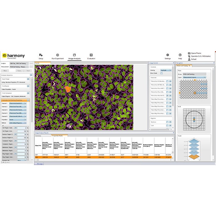
Harmonyソフトウェアで撮像と解析をシンプルに
直感的なワークフロー
Harmony™ソフトウェアは、画像取得から解析・評価までを直感的なユーザーインターフェースでサポートします。
クイックセットアップ用テンプレート
撮像チャンネルやパラメータの設定を効率的に行えるテンプレートを用意しています。
Ready-made解析ソリューション
よく使われる画像解析には構築済みの解析シークエンスを選択することで、ワークフローを簡素化できます。
カスタマイズ可能な解析ビルディングブロック
画像解析用のビルディングブロックを使って、アプリケーションに最適化された解析シークエンスを自由に作成・調整・カスタマイズできます。
高度な解析機能
Harmony™には、テクスチャ解析やSTAR形態解析などの最新の解析アルゴリズムが含まれており、細胞形態の詳細な記述や表現型の高精度な識別が可能です。
データマネジメント
解析結果とメタデータをソフトウェアが自動的に保存します。保存されるデータには、アッセイレイアウト、装置設定、ユーザー定義のキーワードや注釈などが含まれます。
Image gallery


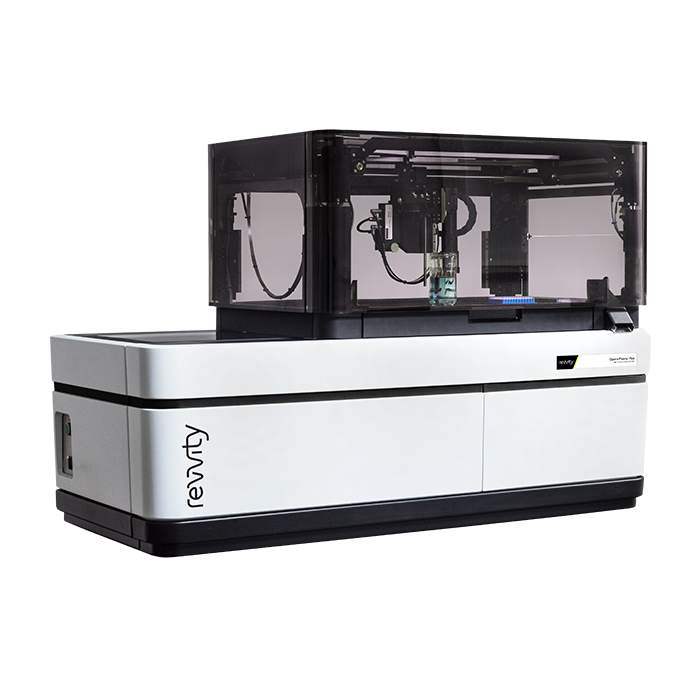
Opera Phenix Plus liquid handling module
Opera Phenix Plus system with liquid handling module
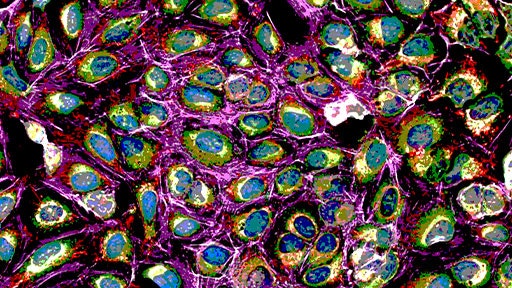
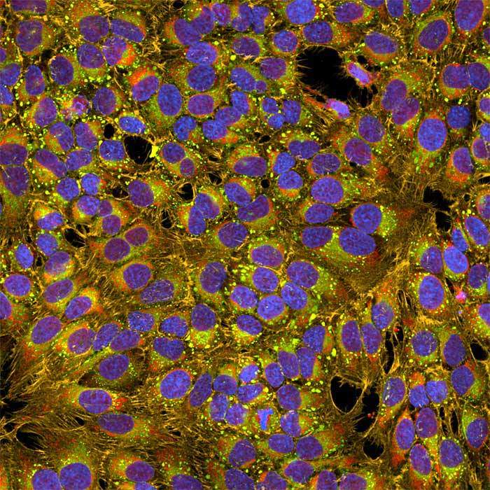
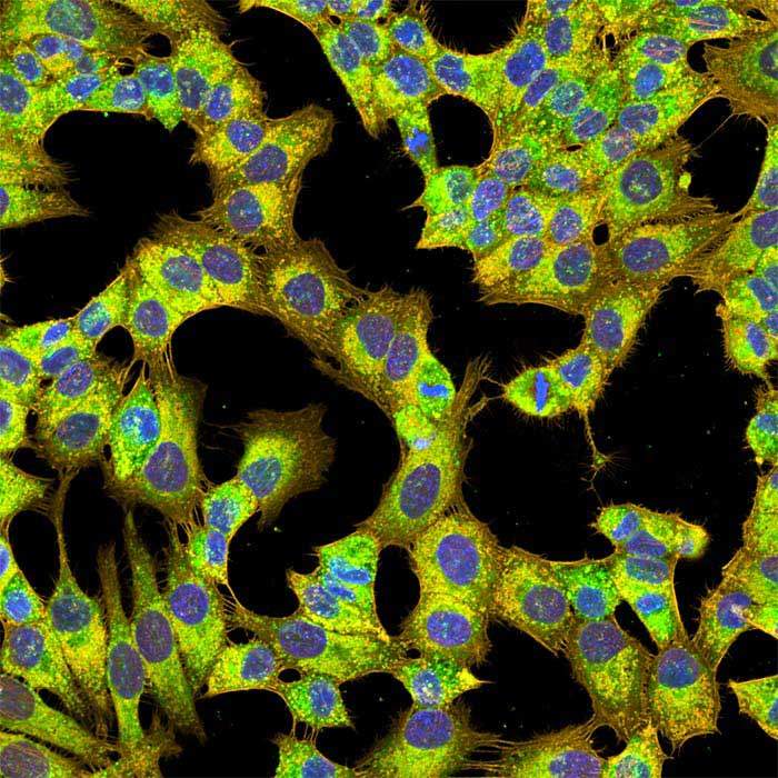
Cell painting assay imaged on the Opera Phenix Plus system
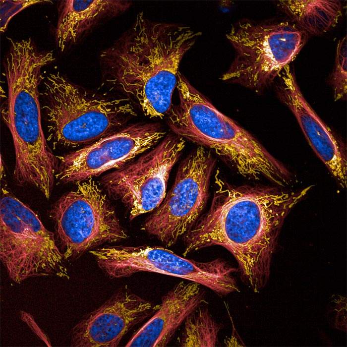
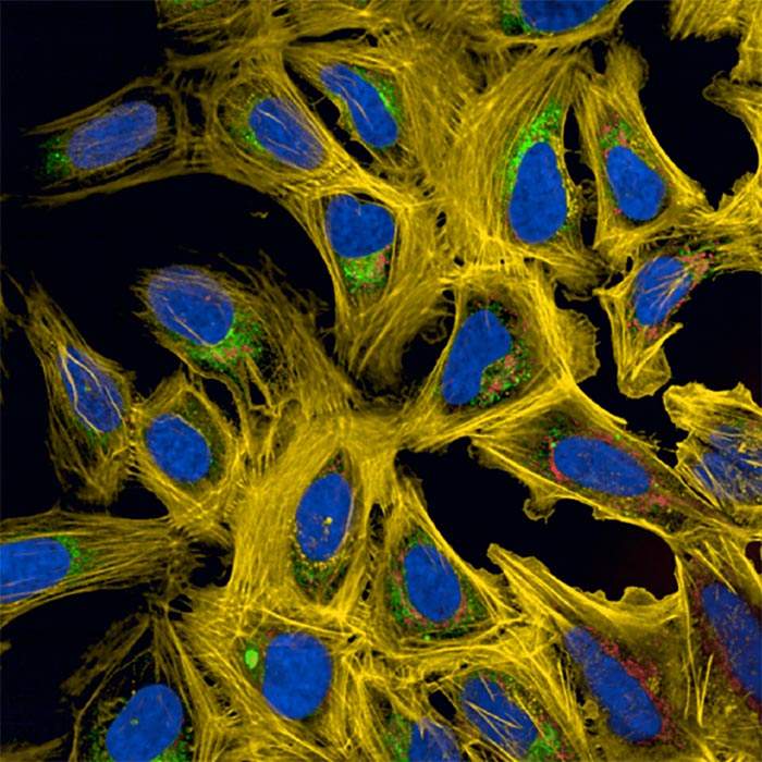
U2OS cell imaging on the Opera Phenix Plus system
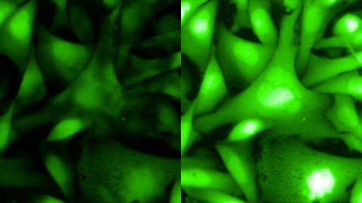
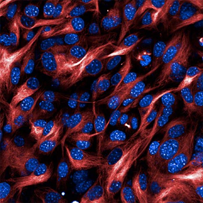
Live cell staining on the Opera Phenix Plus system
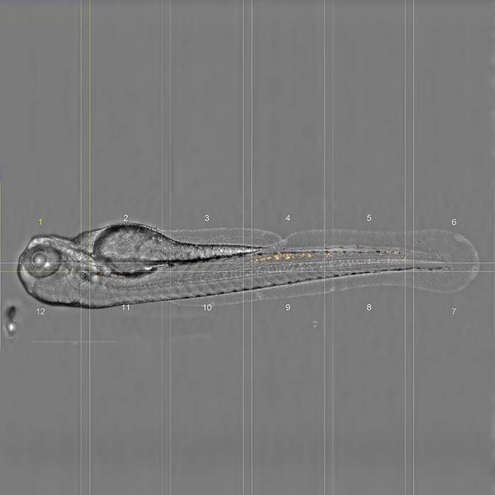
Zebra fish imaging on the Opera Phenix Plus system
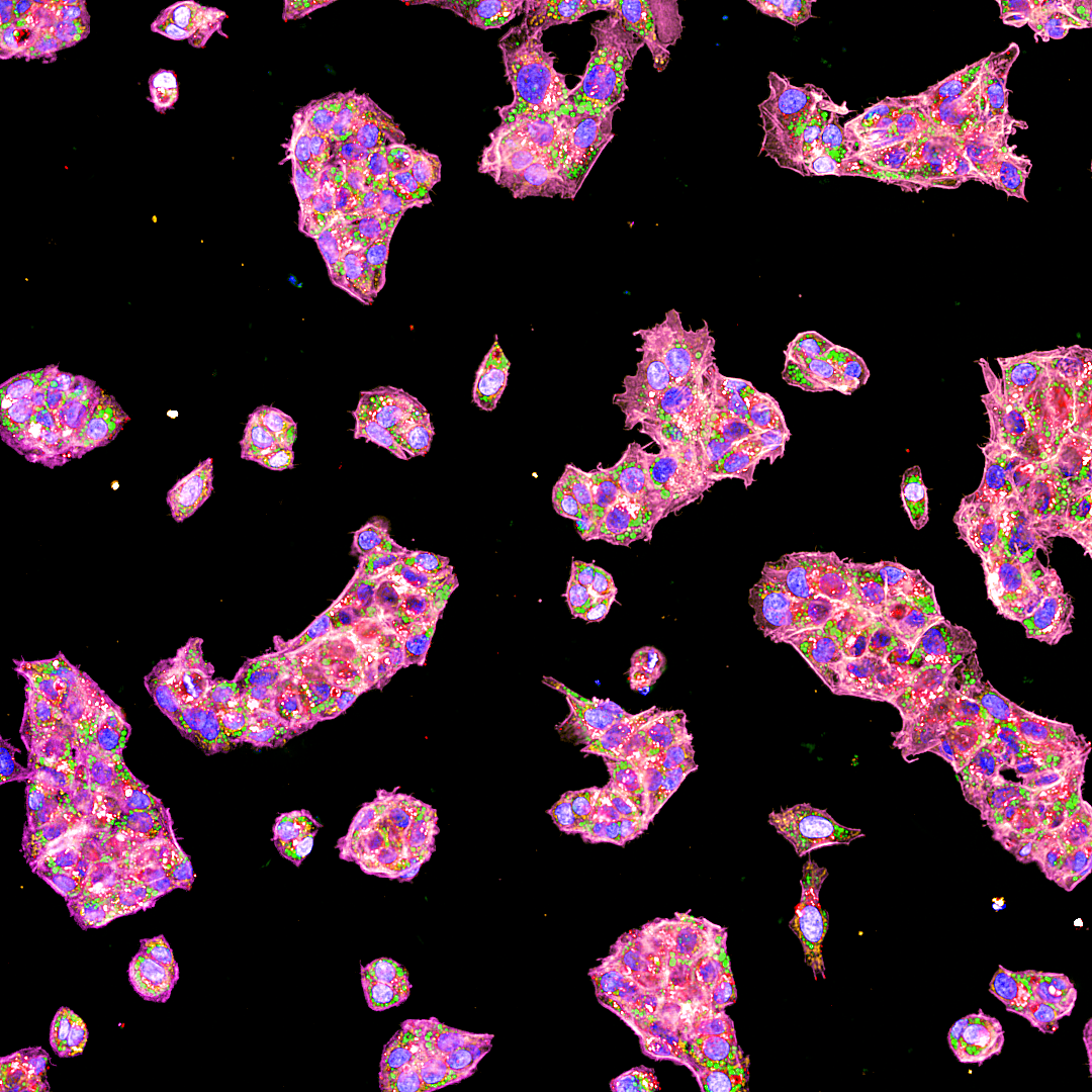
Cell painting assay imaged on the Opera Phenix Plus system
Calcium flux assay imaged on the Opera Phenix Plus system
Cell painting assay imaged on the Opera Phenix Plus system
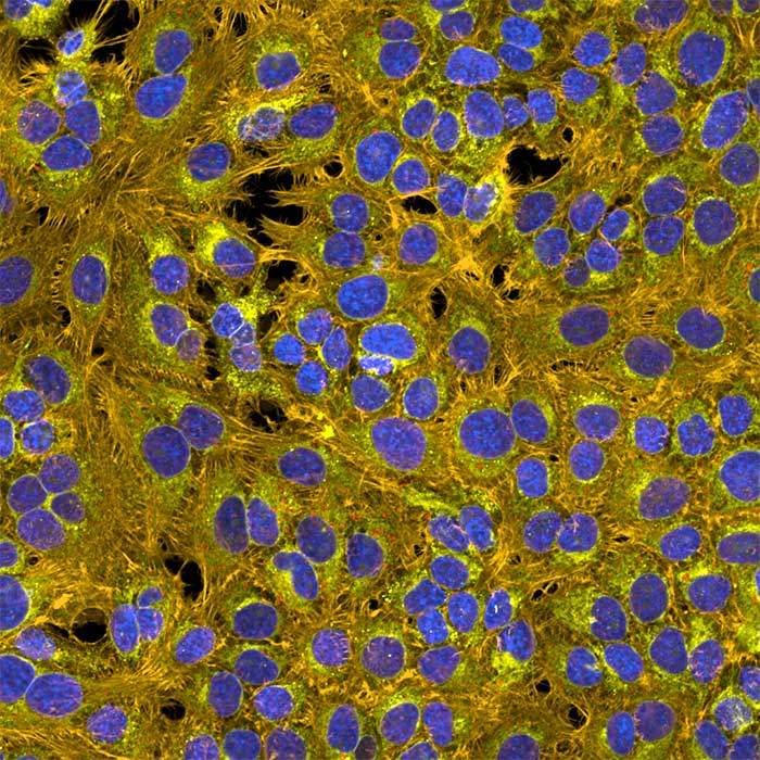
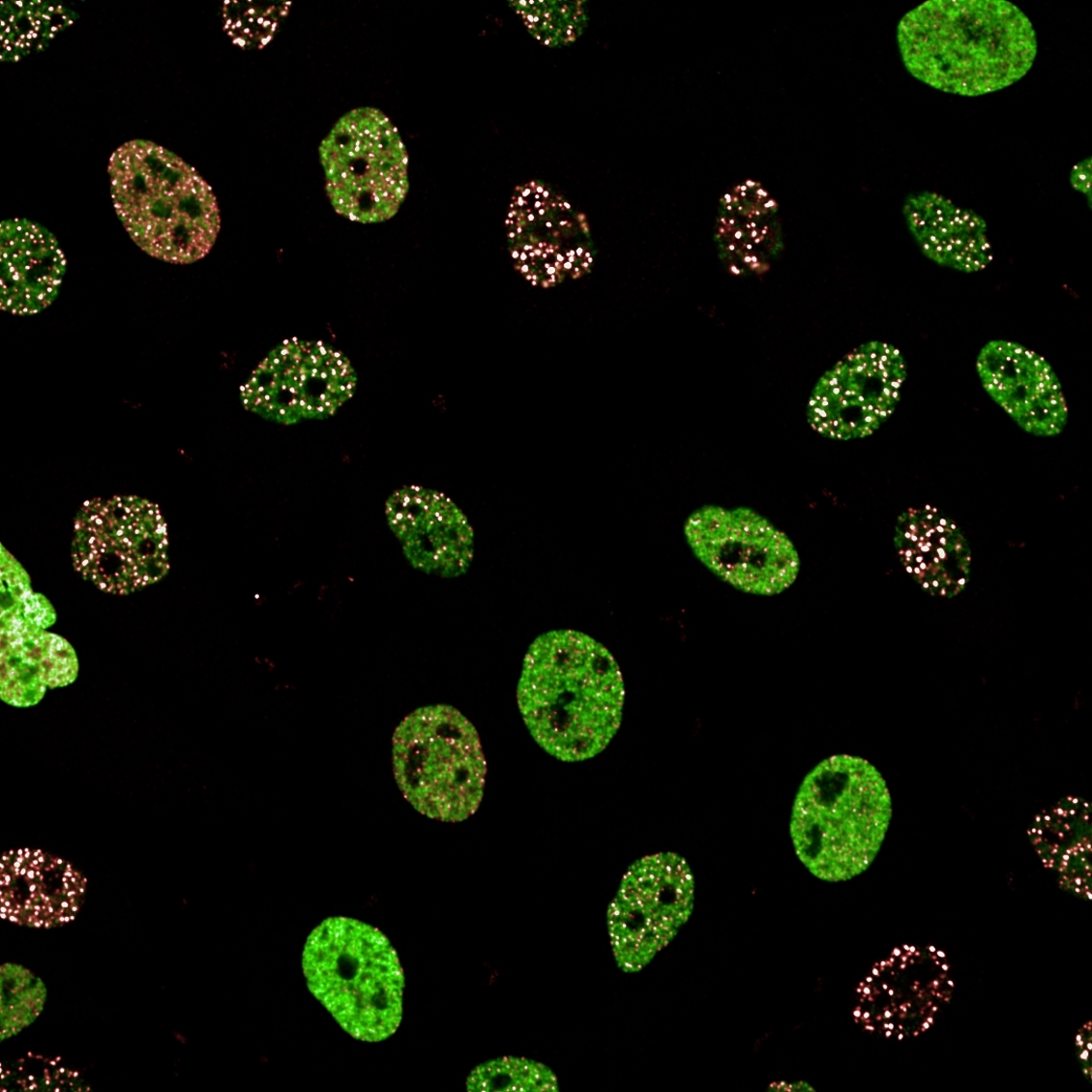
U2OS cell imaging on the Opera Phenix Plus system
5-plex staining on the Opera Phenix Plus system
Cell painting assay imaged on the Opera Phenix Plus system
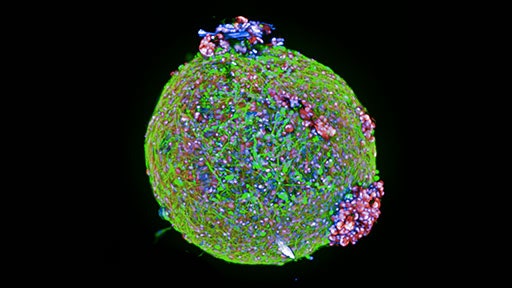
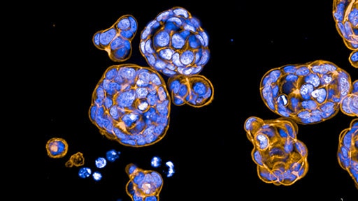
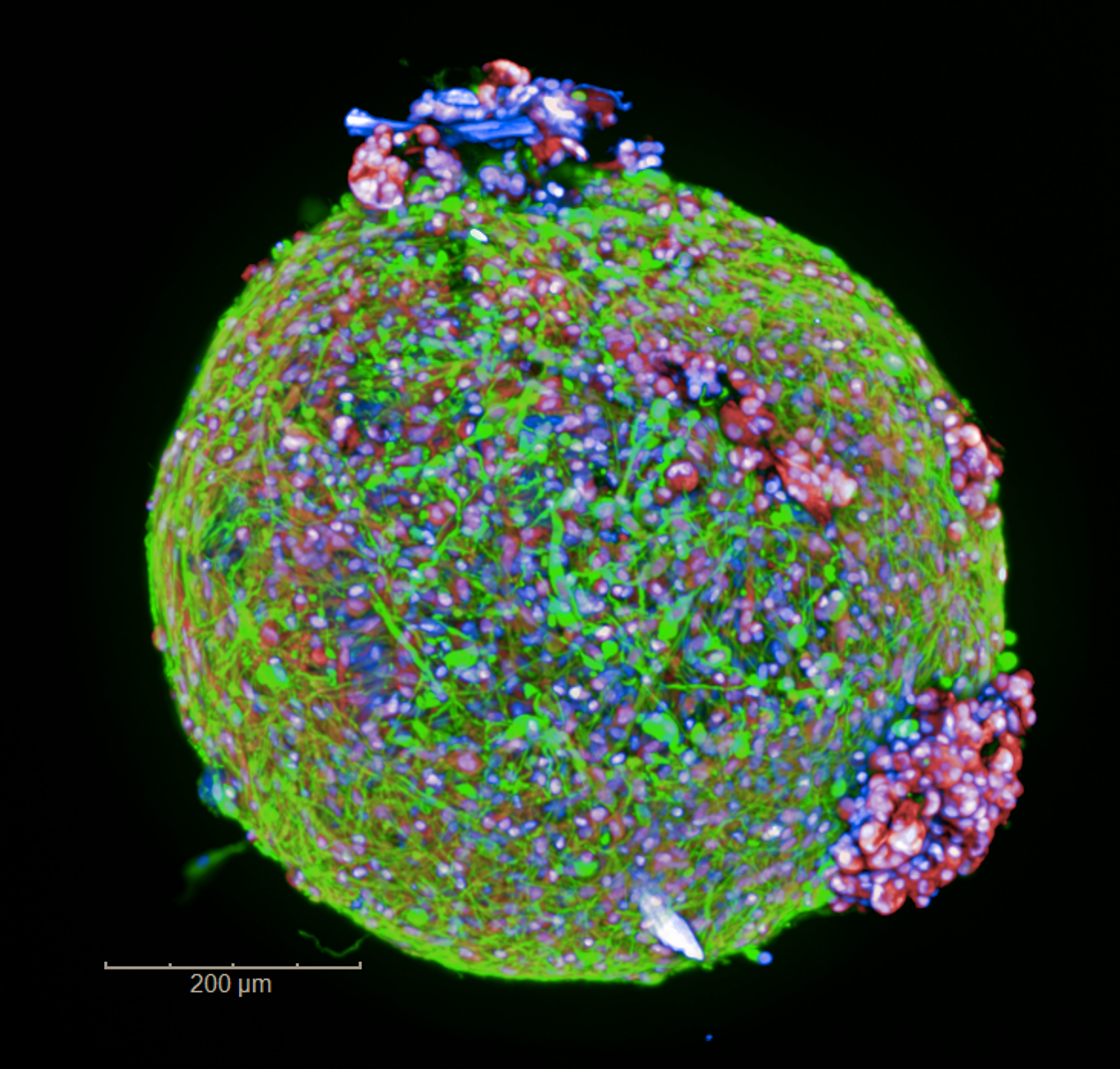
Organoids from pediatric brain tumor imaged on the Opera Phenix Plus high-content screening system
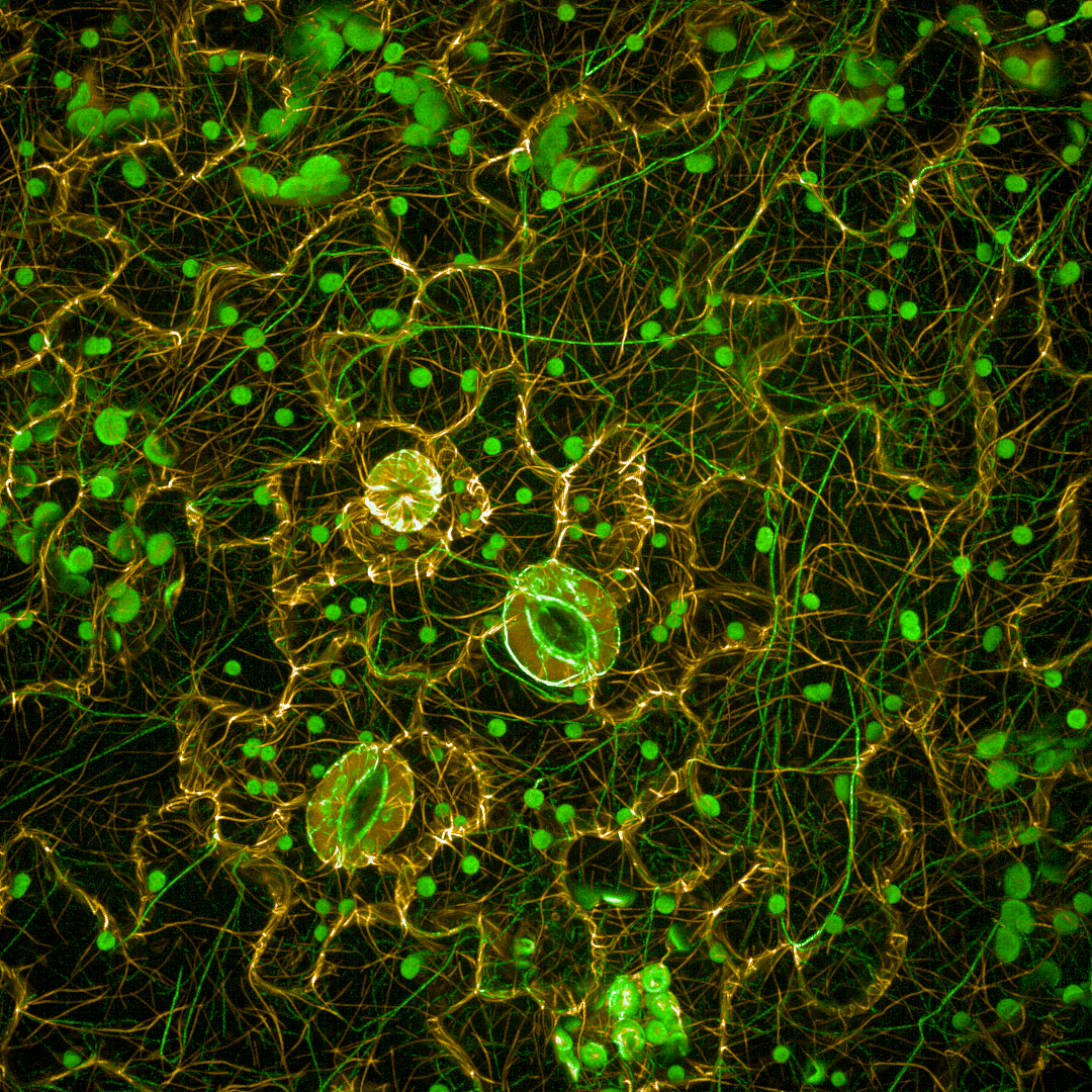
Immortalized human podocytes imaged on the Opera Phenix Plus high-content screening system
Arabidopsis cells imaged on the Opera Phenix high-content screening system
HepG2 spheroids imaged on the Opera Phenix Plus high-content screening system.
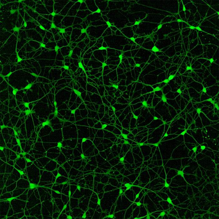
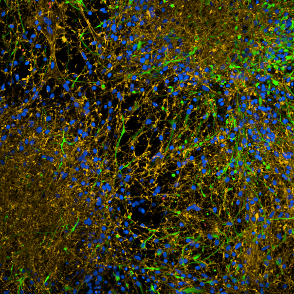
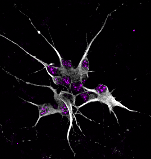
Human iPSC-derived neurons imaged on the Opera Phenix Plus high-content screening system.
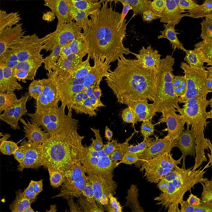
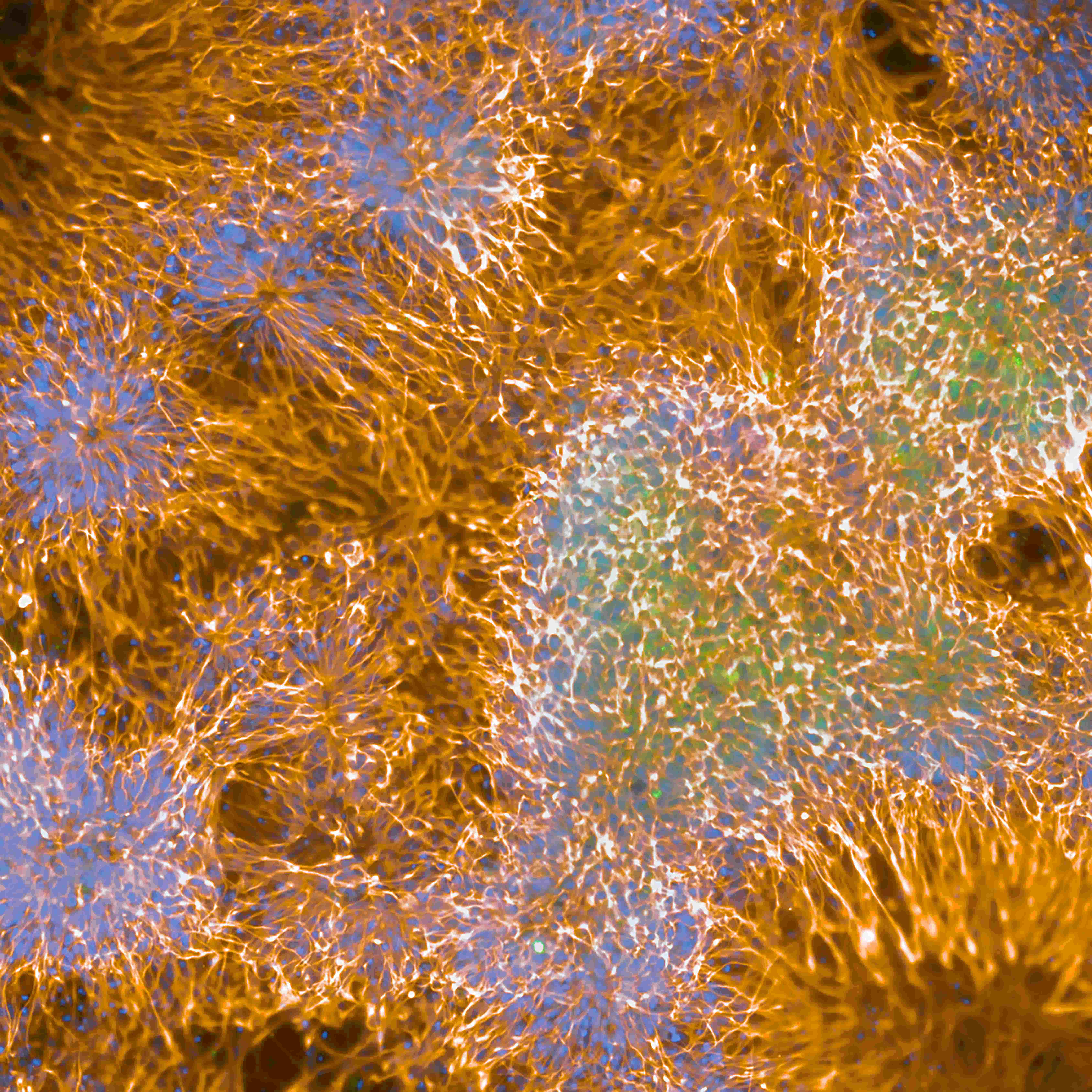
Neuroblastoma SHSY5Y cells imaged on the Opera Phenix Plus high-content screening system.
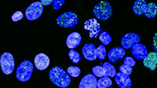
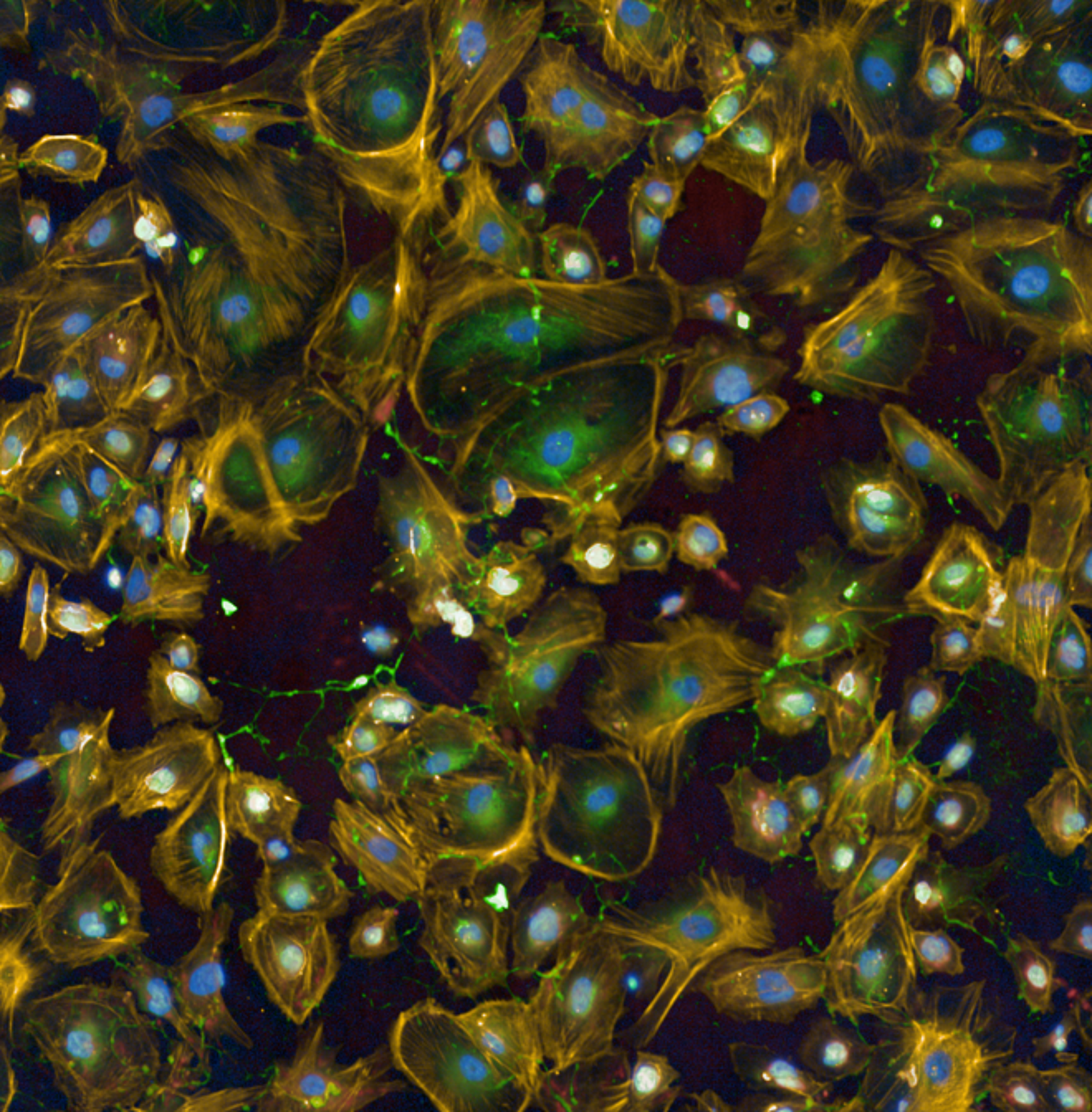
HAP1 cell pool imaged on the Opera Phenix Plus high-content screening system.
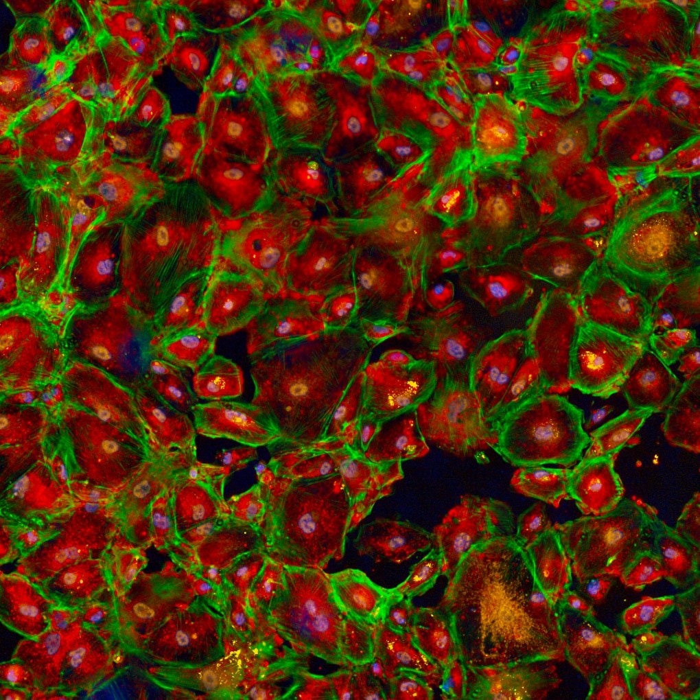
HepG2 cells imaged on the Opera Phenix high-content screening system.
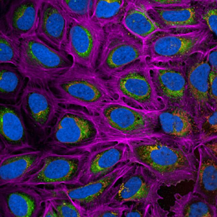
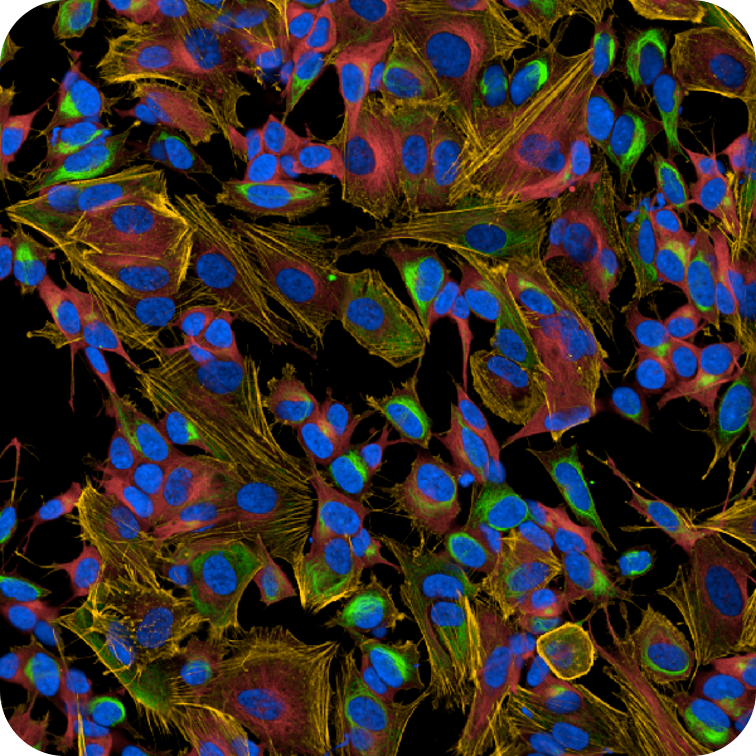
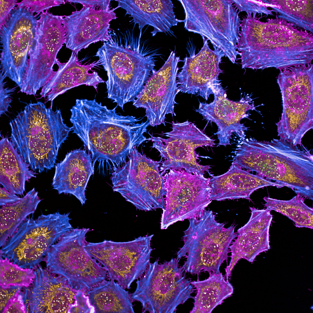
HeLa cells imaged on the Opera Phenix Plus high-content imaging system.
Induced pluripotent stem cells imaged on the Opera Phenix high-content screening system.
Immortalized human podocytes imaged on the Opera Phenix Plus high-content screening system.
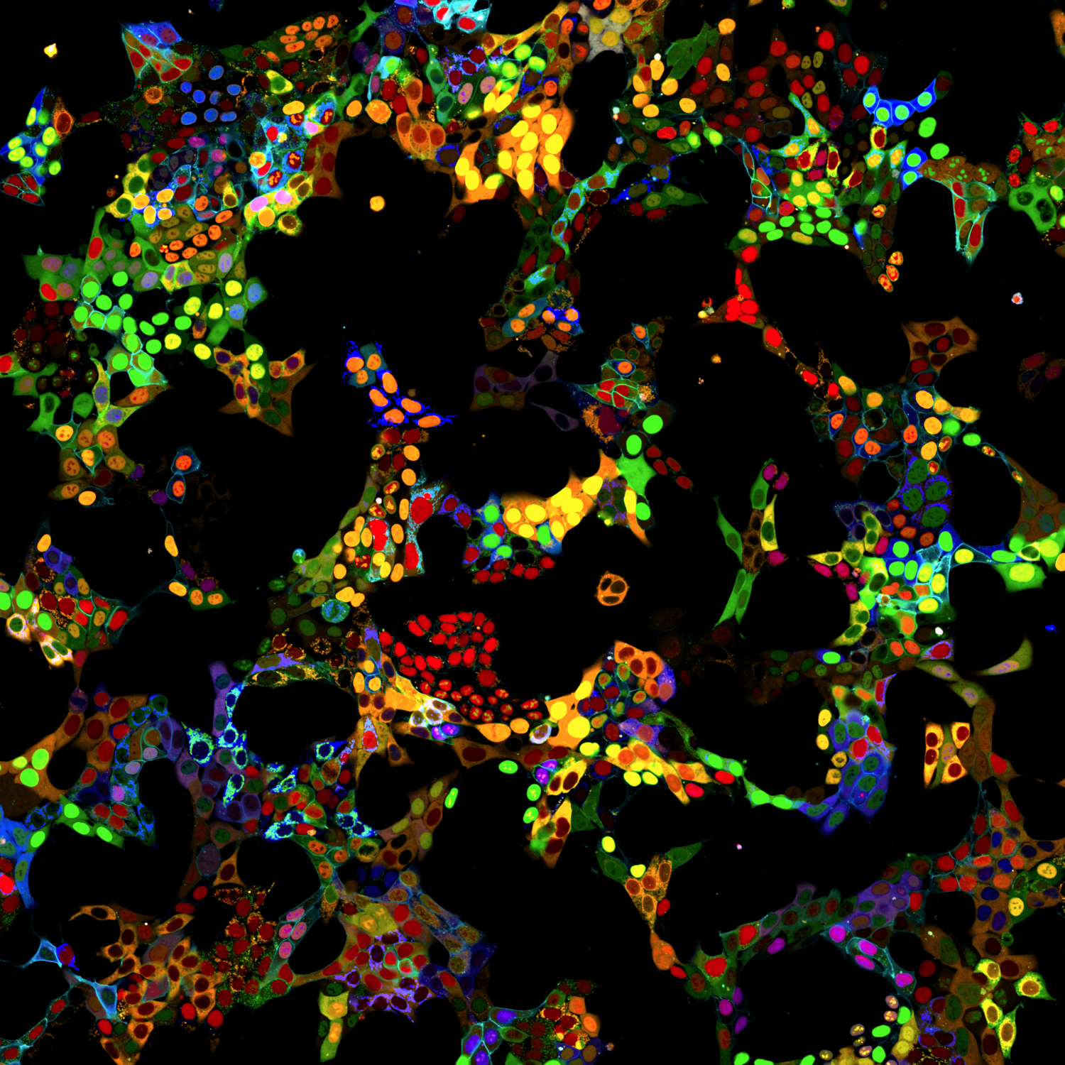
5-plex phenotypic assay imaged on the Opera Phenix Plus high-content screening system.
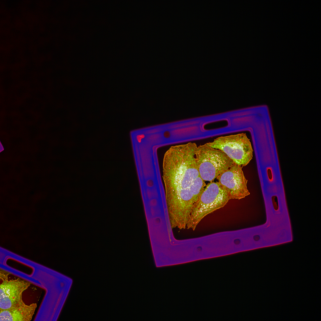
A549 cells on a Semarion SemaCyte microcarrier imaged on the Opera Phenix Plus high-content screening system.
Neurons imaged on the Operetta CLS high-content analysis system
×
さらに研究を加速させる検証済みのソリューション
ハイコンテントアナリシス用マイクロプレート
PhenoPlate™ 384ウェルマイクロプレートは、ハイコンテントアナリシスに最適化された高性能マイクロプレートです。3Dモデル撮像にはCellCarrier Spheroid ULAプレートもご利用いただけます。

オートメーションとワークステーション
スループットと生産性を向上し、変動性と試薬コストの削減を実現します。セルペインティング、3D細胞培養、または表現型スクリーニングのワークフローを改善するexplorer™ G3ワークステーションをぜひご活用ください。
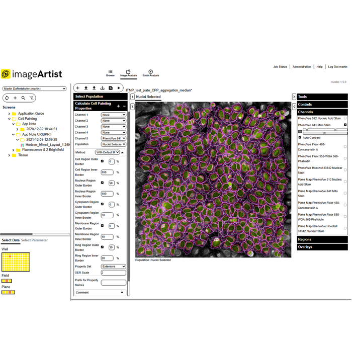
さらに効率的なデータ管理
Image Artist™プラットフォームは、スケーラブルで複数ユーザー対応の画像解析・マネジメント環境を提供します。Harmonyから自動エクスポートされた画像を、高性能コンピューティングと業界標準のオブジェクトストアによる最新の環境下で解析、保存、管理ができます。

Product information
Overview
Opera Phenix Plusで、細胞の理解をさらに深める
さらなる発見: ハイコンテントイメージングは、他のアプローチよりも多様な情報を提供し、細胞の状態を詳細に調べることができます。
詳細な定量化: シングルセルレベルから細胞集団全体まで、表現型の変化を正確に捉えられます。ハイコンテントイメージングは表現型の詳細な変化を定量します。
3Dイメージングの強化: 水浸対物レンズは、3D画像の質を向上させ、複雑な生物学的構造内の複雑な詳細を明らかにします。
スループットの向上: カメラを増設することで、スピードと効率が向上します。ハイコンテントイメージングは研究パイプラインをスピードアップします。
Revvityの最先端のハイコンテントイメージングソリューションで、研究を加速し新たな現象の発見につなげてください。
Specifications
| Dimensions | 134.0 cm (W) x 65.0 cm (D) x 47.0 cm (H) |
|---|
| Automation Compatible |
Yes
|
|---|---|
| Brand |
Opera Phenix Plus
|
| Imaging Modality |
明視野
共焦点
デジタル位相差
蛍光
|
| Unit Size |
1 unit
|
Video gallery
Opera Phenix Plus ハイコンテントスクリーニングシステム
Citations
Resources
Are you looking for resources, click on the resource type to explore further.
Brochure
3D cell culture workflow solutions
More than ever, researchers are turning to 3D cell cultures, microtissues and organoids to bridge the gap between 2D cell cultures...
Technical Note
3D volumetric analysis of luminal spaces inside cysts or organoids
High-content assays using 3D objects such as cysts or organoids can be challenging from the perspectives of both image acquisition...
Technical Note
3D volumetric and zonal analysis of solid spheroids
Multicellular 3D “oids” (tumoroids, spheroids, organoids) have the potential to better predict the effects of drug candidates...
Whitepaper
A brand-new modality on the horizon: how targeted protein degradation can address the unmet need in drug discovery and development
Targeted protein degradation (TPD) is an emerging drug discovery modality that offers the potential to probe biological pathways...
Technical Note
A scalable and reproducible workflow for high-content analysis of cytotoxic effects in RASTRUM 3D cell cultures
Explore our Technical Note unveiling a robust workflow for advanced cytotoxicity analysis in 3D cell cultures. This method...
Case Study
A scalable method to monitor protein levels and localizations in cells
In this case study, we present a novel method developed by researchers at the CeMM Research Center for studying protein levels and...
Loading...


How can we help you?
We are here to answer your questions.

