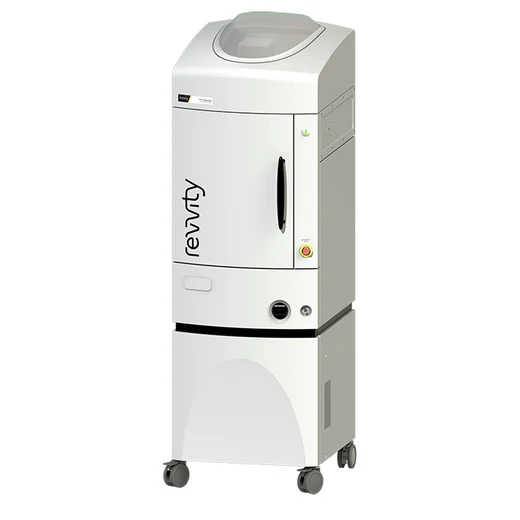
IVIS Spectrum In Vivo Imaging System


The IVIS® Spectrum in vivo imaging system combines 2D optical and 3D optical tomography in one platform. The system uses leading optical technology for preclinical imaging research and development ideal for non-invasive longitudinal monitoring of disease progression, cell trafficking and gene expression patterns in living animals.
Product information
Overview
An optimized set of high efficiency filters and spectral un-mixing algorithms lets you take full advantage of bioluminescent and fluorescent reporters across the blue to near infrared wavelength region. It also offers single-view 3D tomography for both fluorescent and bioluminescent reporters that can be analyzed in an anatomical context using our Digital Mouse Atlas or registered with our multimodality module to other tomographic technologies such as MR, CT or PET.
For advanced fluorescence pre-clinical imaging, the IVIS Spectrum has the capability to use either trans-illumination (from the bottom) or epi-illumination (from the top) to illuminate in vivo fluorescent sources. 3D diffuse fluorescence tomography can be performed to determine source localization and concentration using the combination of structured light and trans illumination fluorescent images. The instrument is equipped with 10 narrow band excitation filters (30nm bandwidth) and 18 narrow band emission filters (20nm bandwidth) that assist in significantly reducing autofluorescence by the spectral scanning of filters and the use of spectral unmixing algorithms. In addition, the spectral unmixing tools allow the researcher to separate signals from multiple fluorescent reporters within the same animal.
Features and Benefits
- High-sensitivity in vivo imaging of fluorescence and bioluminescence
- High throughput (5 mice) with 23 cm field of view
- High resolution (to 20 microns) with 3.9 cm field of view
- Twenty eight high efficiency filters spanning 430 – 850 nm
- Supports spectral unmixing applications
- Ideal for distinguishing multiple bioluminescent and fluorescent reporters
- Optical switch in the fluorescence illumination path allows reflection-mode or transmission-mode illumination
- 3D diffuse tomographic reconstruction for both fluorescence and bioluminescence
- Ability import and automatically co-register CT or MRI images yielding a functional and anatomical context for your scientific data.
- NIST traceable absolute calibrations
- Gas anesthesia inlet and outlet ports
- Class I Laser Product
Specifications
| Height |
206.0 cm
|
|---|---|
| Portable |
No
|
| Width |
65.0 cm
|
| Brand |
IVIS
|
|---|---|
| Imaging Modality |
Optical Imaging
|
| Optical Imaging Classification |
Bioluminescence Imaging
Fluorescence Imaging
|
| Quantity in a Package Amount |
1.0 each
|
| Unit Size |
1 Unit
|
Selected Publications / Citations / References
- Guccini et al (2021). Senescence Reprogramming by TIMP1 Deficiency Promotes Prostate Cancer Metastasis. Cancer Cell. 39, 1–15. https://doi.org/10.1016/j.ccell.2020.10.012
- Ullah et al (2021). Live imaging of SARS-CoV-2 infection in mice reveals neutralizing antibodies require Fc function for optimal efficacy. bioRxiv. https://doi.org/10.1101/2021.03.22.436337
- Leslie et al (2021). Non-invasive synchronous monitoring of neutrophil migration using whole body near-infrared fluorescence-based imaging. Sci Reports. https://doi.org/10.1038/s41598-021-81097-8
- Castellarin et al (2020). A rational mouse model to detect on-target, off-tumor CAR T cell toxicity. JCI Insight. https://doi.org/10.1172/jci.insight.136012
- Wang et al (2020). CRISPR-engineered human brown-like adipocytes prevent diet- induced obesity and ameliorate metabolic syndrome in mice. Sci Transl Med. 12. https://doi.org/10.1126/scitranslmed.aaz8664
- Zhang et al (2020). A Thermostable mRNA Vaccine against COVID-19. Cell. 182 (5): 1271-1283. https://doi.org/10.1016/j.cell.2020.07.024
- Hsu et el (2020). Transplantation of viable mitochondria improves right ventricular performance and pulmonary artery remodeling in rats with pulmonary arterial hypertension. J Thor & Cardiovasc Surgery. https://doi.org/10.1016/j.jtcvs.2020.08.014
- Metman et al (2020). Detection of Papillary Thyroid Cancer Nodal Metastasis after Intravenous Administration of a Fluorescent Tracer Targeting MET. EJSO. 46(2): E12-E13. https://doi.org/10.1016/j.ejso.2019.11.036
- Gu et al (2020). Rg1 in combination with mannitol protects neurons against glutamate-induced ER stress via the PERK-eIF2 α-ATF4 signaling pathway. Life Sci. 263: 118559. https://doi.org/10.1016/j.lfs.2020.118559
- Eckley et al (2019). Short-Term Environmental Conditioning Enhances Tumorigenic Potential of Triple-Negative Breast Cancer Cells. Tomography. 5(4): 346–357. https://dx.doi.org/10.18383%2Fj.tom.2019.00019
Resources
This product note highlights the features, benefits, and specs for the IVIS Spectrum system
Read how non-invasive optical imaging enables intricate host-pathogen interactions to be visualized and monitored in disease...
There’s a need to propel and reinvigorate the current state of drug discovery pipeline in tackling pressing neurodegenerative...
As the global population ages and life expectancy increases, neurodegenerative disorders like Alzheimer’s disease (AD) have the...
The primary goal of preclinical imaging is to improve the odds of clinical success and reduce drug discovery and development time...
Amyotrophic lateral sclerosis (ALS) is a devastating neurological disease for which there is no cure. Another lethal brain disease...


How can we help you?
We are here to answer your questions.


