


JP
Revvity Sites Globally
Select your location.
*e-commerce not available for this region.

AlphaLISA SureFire Ultra Human and Mouse Total MEK1 Detection Kit, 500 Assay Points


 View All
AlphaLISA SureFire Ultra Human and Mouse Total MEK1 Detection Kit, 500 Assay Points


AlphaLISA Surefire Ultra Total Protein


The AlphaLISA™ SureFire® Ultra™ Total MEK1 assay is a sandwich immunoassay for quantitative detection of MEK1 (both phosphorylated and non-phosphorylated) in cellular lysates using Alpha Technology. This assay is intended to be used as a normalization for phosphorylation studies.
| Feature | Specification |
|---|---|
| Application | 細胞シグナル伝達 |
| Sample Volume | 10 µL |
The AlphaLISA™ SureFire® Ultra™ Total MEK1 assay is a sandwich immunoassay for quantitative detection of MEK1 (both phosphorylated and non-phosphorylated) in cellular lysates using Alpha Technology. This assay is intended to be used as a normalization for phosphorylation studies.
Product variants
Unit Size: 500 assay points
Part #:
ALSU-TMEK1-A500
Unit Size: 10,000 assay points
Part #:
ALSU-TMEK1-A10K
Unit Size: 50,000 assay points
Part #:
ALSU-TMEK1-A50K
Unit Size: 100 assay points
Part #:
ALSU-TMEK1-A-HV
For research use only. Not for use in diagnostic procedures. All products to be used in accordance with applicable laws and regulations including without limitation, consumption, and disposal requirements under European REACH regulations (EC 1907/2006).
AlphaLISA SureFire Ultra Human and Mouse Total MEK1 Detection Kit, 500 Assay Points
AlphaLISA Surefire Ultra Total Protein
AlphaLISA SureFire Ultra Human and Mouse Total MEK1 Detection Kit, 500 Assay Points
Loading...
Product information
Overview
The AlphaLISA™ SureFire® Ultra™ Total MEK1 assay is a sandwich immunoassay for quantitative detection of MEK1 (both phosphorylated and non-phosphorylated) in cellular lysates using Alpha Technology. This assay is intended to be used as a normalization for phosphorylation studies.
The HV (high volume) kit contains reagents to run 100 wells in 96-well format, using a 60 µL reaction volume. The 500 point kit contains reagents to run 500 wells in 384-well format, using a 20 µL reaction volume. The 10,000 point kit contains reagents to run 10,000 wells in 384-well format, using a 20 µL reaction volume. The 50,000 point kit contains reagents to run 500 wells in 384-well format, using a 20 µL reaction volume.
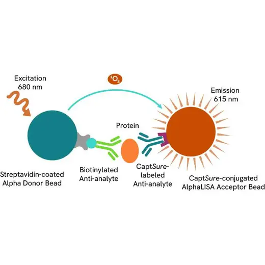
In the AlphaLISA™ SureFire® Ultra™ assay, Donor beads are coated with streptavidin to capture one of the antibodies, which is biotinylated. Acceptor beads are coated with a proprietary CaptSure™ agent that immobilizes the other antibody, labeled with a CaptSure™ tag. In the presence of target protein, the two antibodies bring the Donor and Acceptor beads close together, generating signal. The amount of light emission is directly proportional to the amount of protein present in the sample.
AlphaLISA™ SureFire® Ultra™ kits are compatible with:
- Cell and tissue lysates
- Antibody modulators
- Biotherapeutic antibodies
Alpha SureFire® kits can be used for:
- Cellular kinase assays
- Receptor activation studies
- Screening
How it works
Total-AlphaLISA SureFire Ultra assay principle
The Total-AlphaLISA SureFire Ultra assay measures the expression level of a protein target in a cell lysate.
The Total-AlphaLISA SureFire Ultra assay uses two antibodies which recognize two different distal epitopes on the targeted protein. AlphaLISA assays require two bead types: Acceptor and Donor beads. Acceptor beads are coated with a proprietary CaptSure™ agent to specifically immobilize the assay specific antibody, labeled with a CaptSure tag. Donor beads are coated with streptavidin to capture one of the detection antibodies, which is biotinylated. In the presence of targeted protein, the two antibodies bring the Donor and Acceptor beads in close proximity whereby the singlet oxygen transfers energy to excite the Acceptor bead, allowing the generation of a luminescent Alpha signal. The amount of light emission is directly proportional to the quantity of protein present in the sample.
Total-AlphaLISA SureFire Ultra two-plate assay protocol
The two-plate protocol involves culturing and treating the cells in a 96-well plate before lysis, then transferring lysates into a 384-well OptiPlate™ plate before the addition of Total-AlphaLISA SureFire Ultra detection reagents. This protocol permits the cells viability and confluence to be monitored. In addition, lysates from a single well can be used to measure multiple targets.

Total-AlphaLISA SureFire Ultra one-plate assay protocol
Detection of Total target protein with AlphaLISA SureFire Ultra reagents can be performed in a single plate used for culturing, treatment, and lysis. No washing steps are required. This HTS designed protocol allows for miniaturization while maintaining AlphaLISA SureFire Ultra quality.

Assay validation
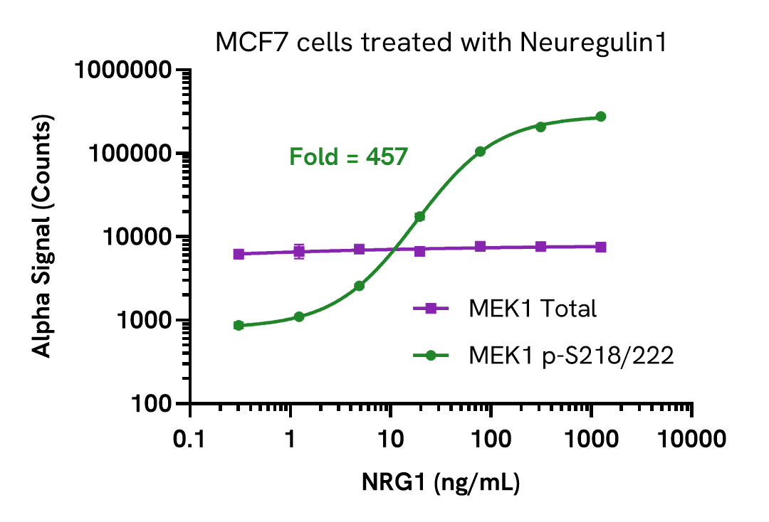
Validation of MEK1 Total in Neuregulin1 treated cells
MCF7 cells were seeded in a 96-well plate (40,000 cells/well) in complete medium, and incubated overnight at 37°C, 5% CO2. The cells were treated with increasing concentrations of Neuregulin1 (NRG1) for 10 minutes.
After treatment, the cells were lysed with 100 µL of Lysis Buffer for 10 minutes at RT with shaking (350 rpm). Phospho (Ser218/222) and Total MEK1 levels were evaluated using respective AlphaLISA SureFire Ultra assays. For the detection step, 10 µL of cell lysate (approximately 4,000 cells) was transferred into a 384-well white OptiPlate, followed by 5 µL of Acceptor mix and incubated for 1 hour at RT. Finally, 5 µL of Donor mix was then added to each well and incubated for 1 hour at RT in the dark. The plate was read on an Envision using standard AlphaLISA settings.
As expected, NRG1 triggered a dose-dependent increase in the levels of Phospho (Ser218/222) MEK1 while Total levels remained unchanged.
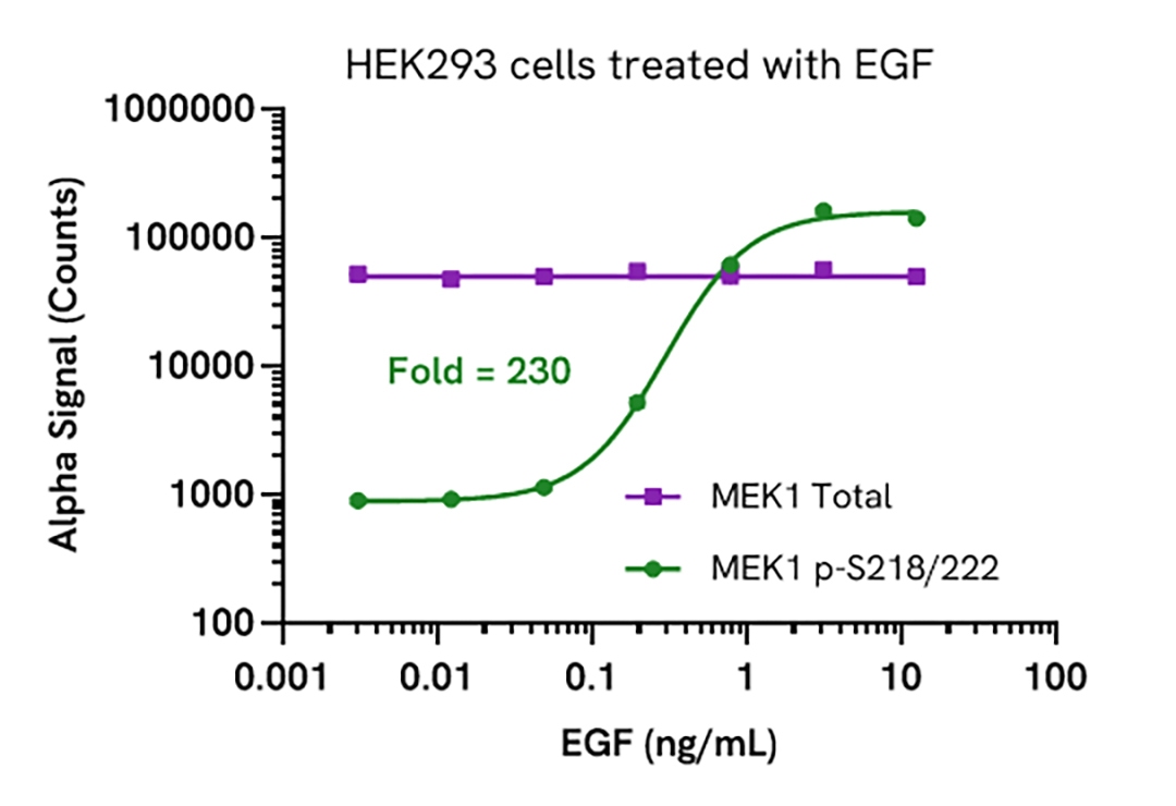
Validation of MEK1 Total in EGF treated cells
HEK293 cells were seeded in a 96-well plate (30,000 cells/well) in complete medium, and incubated for 48 hours at 37°C, 5% CO2. The cells were starved for 2 hours and treated with increasing concentrations of EGF for 10 minutes.
After treatment, the cells were lysed with 100 µL of Lysis Buffer for 10 minutes at RT with shaking (350 rpm). Phospho (Ser218/222) and Total MEK1 levels were evaluated using respective AlphaLISA SureFire Ultra assays. For the detection step, 10 µL of cell lysate (approximately 6,000 cells) was transferred into a 384-well white OptiPlate, followed by 5 µL of Acceptor mix and incubated for 1 hour at RT. Finally, 5 µL of Donor mix was then added to each well and incubated for 1 hour at RT in the dark. The plate was read on an Envision using standard AlphaLISA settings.
As expected, EGF triggered a dose-dependent increase in the levels of Phospho (Ser218/222) MEK1 while Total levels remained unchanged.
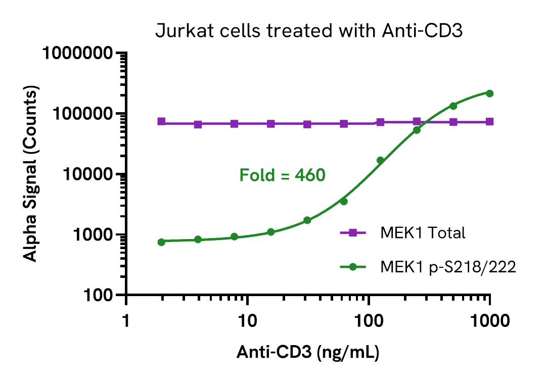
Validation of MEK1 Total in Anti-CD3 treated cells
Jurkat cells were seeded in a 96-well plate (50,000 cells/well) in HBSS + 0.1% BSA. The cells were starved for 60 minutes and then treated with increasing concentrations of Anti-CD3 antibody for 5 minutes.
After treatment, the cells were lysed with the addition of 50 µL of 5X Lysis Buffer for 10 minutes at RT with shaking (350 rpm). Phospho (Ser218/222) and Total MEK1 levels were evaluated using respective AlphaLISA SureFire Ultra assays. For the detection step, 10 µL of cell lysate (approximately 2,000 cells) was transferred into a 384-well white OptiPlate, followed by 5 µL of Acceptor mix and incubated for 1 hour at RT. Finally, 5 µL of Donor mix was then added to each well and incubated for 1 hour at RT in the dark. The plate was read on an Envision using standard AlphaLISA settings.
As expected, Anti-CD3 triggered a dose-dependent increase in the levels of Phospho (Ser218/222) MEK1 while Total levels remained unchanged.
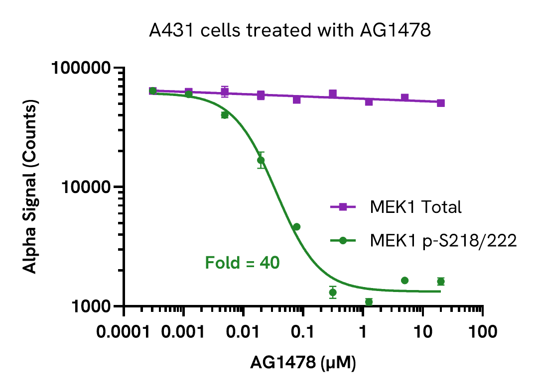
Validation of MEK1 Total in AG1478 treated cells
A431 cells were seeded in a 96-well plate (20,000 cells/well) in complete medium and incubated for 48 hours at 37°C, 5% CO2. The cells were treated with increasing concentrations of AG1478 for 2 hours and then treated with 1 ng/mL EGF for 30 minutes.
After treatment, the cells were lysed with 100 µL of Lysis Buffer for 10 minutes at RT with shaking (350 rpm). Phospho (Ser218/222) and Total MEK1 levels were evaluated using respective AlphaLISA SureFire Ultra assays. For the detection step, 10 µL of cell lysate (approximately 4,000 cells) was transferred into a 384-well white OptiPlate, followed by 5 µL of Acceptor mix and incubated for 1 hour at RT. Finally, 5 µL of Donor mix was then added to each well and incubated for 1 hour at RT in the dark. The plate was read on an Envision using standard AlphaLISA settings.
As expected, AG1478 triggered a dose-dependent decrease in the levels of Phospho (Ser218/222) MEK1 stimulated by EGF, while Total levels remained unchanged.
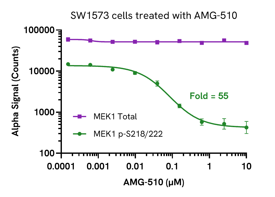
Validation of MEK1 Total in AMG-510 treated cells
SW1573 cells were seeded in a 96-well plate (40,000 cells/well) in complete medium, and incubated overnight at 37°C, 5% CO2. The cells were treated with increasing concentrations of AMG-510 for 2 hours.
After treatment, the cells were lysed with 100 µL of Lysis Buffer for 10 minutes at RT with shaking (350 rpm). Phospho (Ser218/222) and Total MEK1 levels were evaluated using respective AlphaLISA SureFire Ultra assays. For the detection step, 10 µL of cell lysate (approximately 4,000 cells) was transferred into a 384-well white OptiPlate, followed by 5 µL of Acceptor mix and incubated for 1 hour at RT. Finally, 5 µL of Donor mix was then added to each well and incubated for 1 hour at RT in the dark. The plate was read on an Envision using standard AlphaLISA settings.
As expected, AMG-510, a KRASG12C inhibitor, triggered a dose-dependent decrease in the levels of Phospho (Ser218/222) MEK1 while Total levels remained unchanged.
Assay specificity/selectivity
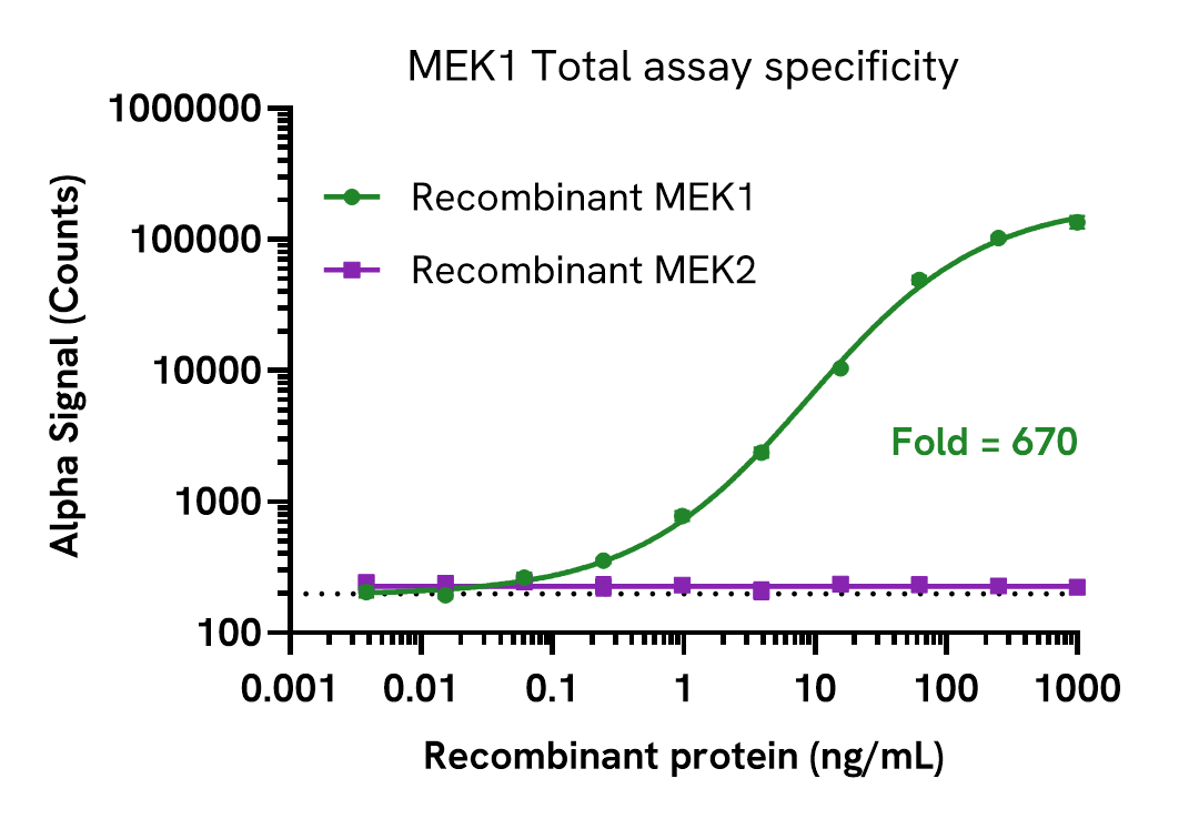
MEK1 Total assay specificity
Specificity of the MEK1 Total assay was assessed by using active MEK1 and MEK2 recombinant proteins.
Dilutions of recombinant MEK1 (Abcam, ab63209) and MEK2 (Merck, SLBN1818V) proteins were prepared in Lysis Buffer and evaluated using the AlphaLISA SureFire Ultra assay.
For the detection step, 10 µL of diluted protein was transferred into a 384-well white OptiPlate, followed by 5 µL of Acceptor mix and incubated for 1 hour at RT. Finally, 5 µL of Donor mix was then added to each well and incubated for 1 hour at RT in the dark. The plate was read on an Envision using standard AlphaLISA settings.
The MEK1 Total assay showed reactivity only to MEK1 protein, no cross reactivity to MEK2 protein was observed. The dotted line represents assay background.
Assay versatility
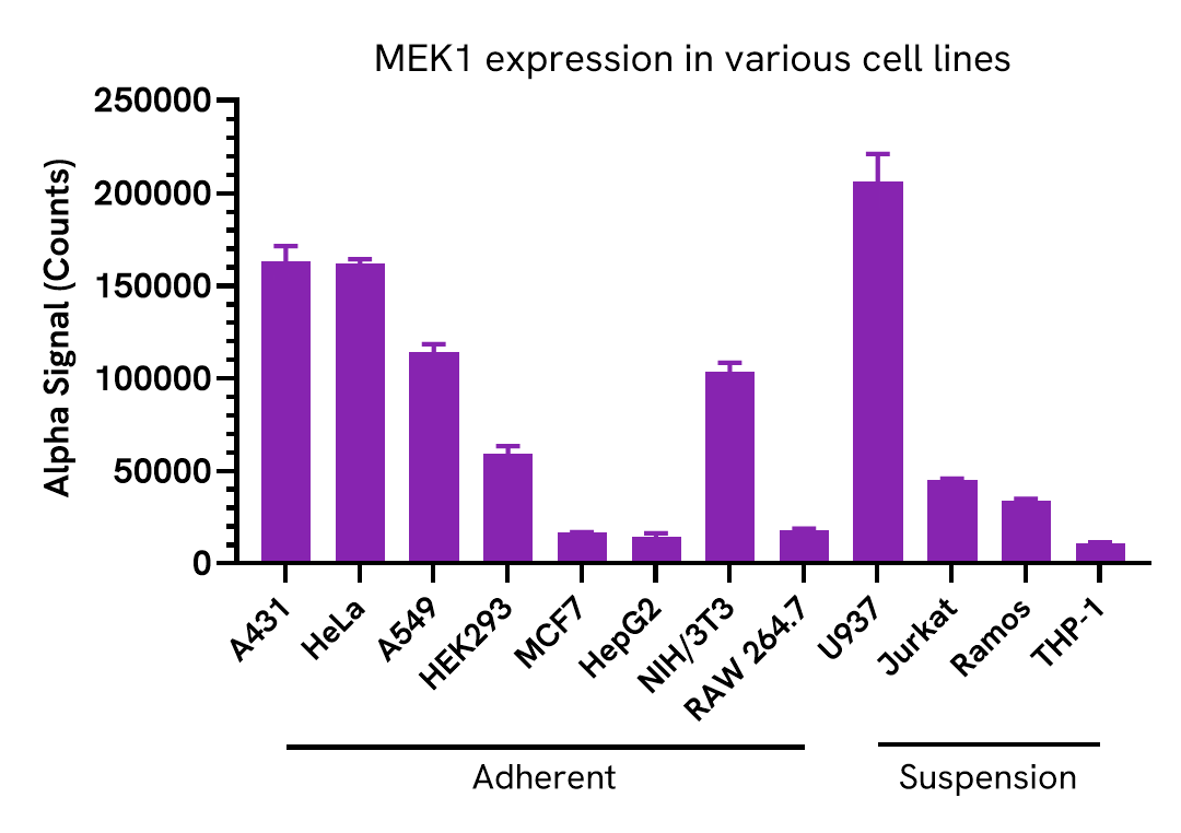
Versatility of MEK1 Total assay in various cell lines
Adherent cells were grown to confluency in a T175 flask at 37°C, 5% CO2, and were lysed with Lysis Buffer at a density of 0.5 x 10⁶ cells/mL. Suspension cells were harvested, washed in HBSS and lysed with Lysis Buffer at 1.6 x 10⁶ cells/mL.
Total MEK1 levels were evaluated using the AlphaLISA SureFire Ultra assay. For the detection step, 10 µL of cell lysate (5,000 adherent and 16,000 suspension cells) were transferred into a 384-well white OptiPlate, followed by 5 µL of Acceptor Mix and incubated for 1 hour at RT. Finally, 5 µL of Donor Mix was then added to each well and incubated for 1 hour at RT in the dark. The plate was read on an Envision using standard AlphaLISA settings.
Total MEK1 expression was detected in a wide range of human and mouse cell lines.
Assay sensitivity
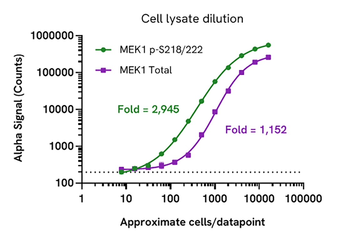
MEK1 assay sensitivity - cell lysate dilution
Cell lysate was prepared from A431 cells cultured to confluence in T175 flasks in complete medium. Cells were treated with 200 ng/mL EGF for 10 minutes and then lysed in 10 mL of Lysis Buffer.
Lysate was serially diluted in Lysis Buffer and assayed for Phospho (Ser218/222) and Total MEK1 using respective AlphaLISA SureFire Ultra kits. For the detection step, 10 µL of lysate was transferred into a 384-well white OptiPlate, followed by 5 µL of Acceptor mix and incubated for 1 hour at room temperature. Finally, 5 µL of Donor mix was then added to each well and incubated for 1 hour at RT in the dark. The plate was read on an Envision using standard AlphaLISA settings.
Approximate number of cells/datapoint is indicated on the graph. The dotted line represents assay background. This assay can detect MEK1 expression in less than 200 cells/datapoint.
Specifications
| Application |
Cell Signaling
|
|---|---|
| Automation Compatible |
Yes
|
| Brand |
AlphaLISA SureFire Ultra
|
| Cellular or Signaling Pathway |
MAPK
|
| Detection Modality |
Alpha
|
| Host Species |
Human
|
| Lysis Buffer Compatibility |
Lysis Buffer
|
| Molecular Modification |
Total
|
| Product Group |
Kit
|
| Sample Volume |
10 µL
|
| Shipping Conditions |
Shipped in Blue Ice
|
| Target |
MEK1
|
| Target Class |
Phosphoproteins
|
| Target Species |
Human
Mouse
|
| Technology |
Alpha
|
| Unit Size |
500 assay points
|
Video gallery
AlphaLISA SureFire Ultra Human and Mouse Total MEK1 Detection Kit, 500 Assay Points
AlphaLISA SureFire Ultra Human and Mouse Total MEK1 Detection Kit, 500 Assay Points
Resources
Are you looking for resources, click on the resource type to explore further.
Guide
AlphaLISA SureFire Ultra assay optimization
This guide outlines further possible optimization of cellular and immunoassay parameters to ensure the best possible results are...
Guide
AlphaLISA SureFire Ultra: the ultimate guide for successful experiments
The definitive guide for setting up a successful AlphaLISA SureFire Ultra assay
Several biological processes are regulated by...
Brochure
Alpha SureFire Ultra no-wash immunoassay catalog
Discover Alpha SureFire® Ultra™ assays, the no-wash cellular kinase assays leveraging Revvity's exclusive bead-based technology...
Application Note
Characterizing chemokine receptor inhibitors with AlphaLISA SureFire Ultra, Alpha SureFire Ultra Multiplex and LANCE Ultra cAMP assays
The measurement of protein phosphorylation is a useful tool for measuring the modulation of receptor activation by both antibodies...
Brochure
Species compatibility for HTRF, AlphaLISA SureFire Ultra and Alpha SureFire Ultra Multiplex assays
This document includes detailed tables listing HTRF™, AlphaLISA™ SureFire® Ultra™, and Alpha SureFire® Ultra™ Multiplex assays...
Guide
Understanding obesity: exploring cellular pathways and mechanisms
Obesity is a complex condition characterized by excessive fat accumulation, posing significant health and socioeconomic challenges...
Loading...


How can we help you?
We are here to answer your questions.

