


JP
Revvity Sites Globally
Select your location.
*e-commerce not available for this region.

AlphaLISA SureFire Ultra Human and Mouse Total Myogenin Detection Kit, 100 Assay Points
AlphaLISA SureFire Ultra Human and Mouse Total Myogenin Detection Kit, 100 Assay Points

AlphaLISA Surefire Ultra Total Protein
The AlphaLISA™ SureFire® Ultra™ Human and Mouse Total Myogenin assay is a sandwich immunoassay for quantitative detection of total myogenin in cellular lysates using Alpha Technology.
| Feature | Specification |
|---|---|
| Application | 細胞シグナル伝達 |
| Protocol Time | 2h at RT |
| Sample Volume | 30 µL |
The AlphaLISA™ SureFire® Ultra™ Human and Mouse Total Myogenin assay is a sandwich immunoassay for quantitative detection of total myogenin in cellular lysates using Alpha Technology.
Product variants
Unit Size: 100 assay points
Part #:
ALSU-TMYOG-A-HV
Unit Size: 500 assay points
Part #:
ALSU-TMYOG-A500
Unit Size: 10,000 assay points
Part #:
ALSU-TMYOG-A10K
Unit Size: 50,000 assay points
Part #:
ALSU-TMYOG-A50K
For research use only. Not for use in diagnostic procedures. All products to be used in accordance with applicable laws and regulations including without limitation, consumption and disposal requirements under European REACH regulations (EC 1907/2006).
AlphaLISA SureFire Ultra Human and Mouse Total Myogenin Detection Kit, 100 Assay Points
AlphaLISA Surefire Ultra Total Protein
Loading...
Product information
Overview
Myogenin (MYOG) is a muscle-specific transcription factor belonging to the MyoD family of basic helix-loop-helix proteins that regulates skeletal muscle differentiation and regeneration. Myogenin is expressed during the terminal differentiation phase of myogenesis, where it forms heterodimers with E-proteins to bind E-box sequences in muscle-specific gene promoters. It activates expression of structural muscle proteins including myosin heavy chain, troponin, and creatine kinase, driving the transition from proliferating myoblasts to multinucleated myotubes. Myogenin is essential for proper muscle fiber formation and is reactivated during muscle regeneration following injury. Dysregulation of myogenin is implicated in muscle wasting diseases, muscular dystrophies, and rhabdomyosarcoma, where it serves as both a diagnostic marker and potential therapeutic target for restoring muscle function and combating muscle-related pathologies.
The AlphaLISA SureFire Ultra Human and Mouse Total Myogenin is a sandwich immunoassay for the quantitative detection of total Myogenin in cellular lysates, using Alpha Technology.
Formats:
- The HV (high volume) kit contains reagents to run 100 wells in 96-well format, using a 60 μL reaction volume.
- The 500-point kit contains enough reagents to run 500 wells in 384-well format, using a 20 μL reaction volume.
- The 10,000-point kit contains enough reagents to run 10,000 wells in 384-well format, using a 20 μL reaction volume.
- The 50,000-point kit contains enough reagents to run 50,000 wells in 384-well format, using a 20 μL reaction volume.
AlphaLISA SureFire Ultra kits are compatible with:
- Cell and tissue lysates
- Antibody modulators
- Biotherapeutic antibodies
AlphaLISA SureFire Ultra kits can be used for:
- Cellular kinase assays
- Receptor activation studies
- High-throughput screening for preclinical studies
How it works
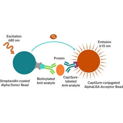
Total-AlphaLISA SureFire Ultra assay principle
The Total-AlphaLISA SureFire Ultra assay measures the expression level of a target protein in a biological sample (e.g. cell lysate).
The Total-AlphaLISA SureFire Ultra assay uses two antibodies which recognize two different distal epitopes on the target protein. AlphaLISA assays require two bead types: Acceptor and Donor Beads. Acceptor Beads are coated with a proprietary CaptSure™ agent to specifically immobilize the assay specific antibody, labeled with a CaptSure tag. Donor Beads are coated with streptavidin to capture one of the detection antibodies, which is biotinylated. In the presence of target protein, the two antibodies bring the Donor and Acceptor Beads in close proximity whereby the singlet oxygen transfers energy to excite the Acceptor Bead, allowing for the generation of a luminescent Alpha signal. The amount of light emission is directly proportional to the quantity of protein present in the sample.
Total-AlphaLISA SureFire Ultra two-plate assay protocol
The two-plate protocol involves culturing and treating the cells in a 96-well plate before lysis, then transferring lysates into a 384-well OptiPlate™ plate before the addition of Total-AlphaLISA SureFire Ultra detection reagents. This protocol enables cell viability and confluence to be monitored. In addition, lysates from a single well can be used to measure multiple targets.

Total-AlphaLISA SureFire Ultra one-plate assay protocol
Detection of Total target protein with AlphaLISA SureFire Ultra reagents can be performed in a single plate used for culturing, treatment, and lysis. No washing steps are required. This HTS designed protocol allows for miniaturization while maintaining robust AlphaLISA SureFire Ultra quality.

Assay validation
Decrease in Myogenin expression in response to BMP-2 treatment
C2C12 cells were seeded in a 96-well plate (20,000 cells/well) in complete medium and incubated overnight at 37°C, 5% CO2. The cells were treated with 50 ng/mL BMP-2 in serum-free medium for the indicated times.
After treatment, the cells were lysed with 100 µL of Lysis Buffer for 10 minutes at RT with shaking (350 rpm). Myogenin and Total ERK levels were evaluated using respective AlphaLISA SureFire Ultra assays. For the detection step, 10 µL of cell lysate (approximately 2,000 cells) was transferred into a 384-well white OptiPlate, followed by 5 µL of Acceptor mix and incubated for 1 hour at RT. Finally, 5 µL of Donor mix was then added to each well and incubated for 1 hour at RT in the dark. The plate was read on an Envision using standard AlphaLISA settings.
As expected, BMP-2 treatment decreased the levels of Myogenin in C2C12 cells while Total ERK levels were unchanged (data not shown).

Inhibition of Myogenin expression in C2C12 cells
C2C12 cells were seeded in a 96-well plate (40,000 cells/well) in complete medium and incubated overnight at 37°C, 5% CO2. The cells were treated with increasing concentrations of cycloheximide for 2 hours.
After treatment, the cells were lysed with 100 µL of Lysis Buffer for 10 minutes at RT with shaking (350 rpm). Myogenin and Total ERK levels were evaluated using respective AlphaLISA SureFire Ultra assays. For the detection step, 10 µL of cell lysate (approximately 4,000 cells) was transferred into a 384-well white OptiPlate, followed by 5 µL of Acceptor mix and incubated for 1 hour at RT. Finally, 5 µL of Donor mix was then added to each well and incubated for 1 hour at RT in the dark. The plate was read on an Envision using standard AlphaLISA settings.
The inhibition of protein synthesis by cycloheximide resulted in a decrease in the levels of Myogenin.

C2C12 cells were seeded in a 96-well plate (40,000 cells/well) in complete medium and incubated overnight at 37°C, 5% CO2. The cells were treated with increasing concentrations of H2O2 for 2 hours.
After treatment, the cells were lysed with 50 µL of Lysis Buffer for 10 minutes at RT with shaking (350 rpm). Myogenin and Total ERK levels were evaluated using respective AlphaLISA SureFire Ultra assays. For the detection step, 10 µL of cell lysate (approximately 8,000 cells) was transferred into a 384-well white OptiPlate, followed by 5 µL of Acceptor mix and incubated for 1 hour at RT. Finally, 5 µL of Donor mix was then added to each well and incubated for 1 hour at RT in the dark. The plate was read on an Envision using standard AlphaLISA settings.
As expected, H2O2 treatment causes a suppression of Myogenin expression, Total ERK levels are unchanged.

Assay versatility
Basal Myogenin expression in response to culturing conditions
RD and C2C12 cells were seeded in a 96-well plate at various cell density (10,000 to 80,000 cells/well) in complete medium, and incubated overnight at 37°C, 5% CO2. The cells were then lysed with 25 µL to 200 µL/well of Lysis Buffer to achieve the same cells/datapoint.
Myogenin and Total ERK levels were evaluated using respective AlphaLISA SureFire Ultra assays. For the detection step, 10 µL of cell lysate (approximately 4,000 cells) was transferred into a 384-well white OptiPlate, followed by 5 µL of Acceptor mix and incubated for 1 hour at RT. Finally, 5 µL of Donor mix was then added to each well and incubated for 1 hour at RT in the dark. The plate was read on an Envision using standard AlphaLISA settings.
High cell density acts as a signal for the onset of muscle differentiation. As expected, Myogenin expression is increased when cells are seeded at high density. Total ERK levels were unchanged (data not shown).

C2C12 cells were seeded in a 96-well plate (5,000 cells/well) in complete medium and incubated overnight at 37°C, 5% CO2. The cells were either left in complete DMEM (high serum) or DMEM + 2% horse serum (low serum) for 24 hours.
After treatment, the cells were lysed with 25 µL of Lysis Buffer for 10 minutes at RT with shaking (350 rpm). Myogenin and Total ERK levels were evaluated using respective AlphaLISA SureFire Ultra assays. For the detection step, 10 µL of cell lysate (approximately 4,000 cells) was transferred into a 384-well white OptiPlate, followed by 5 µL of Acceptor mix and incubated for 1 hour at RT. Finally, 5 µL of Donor mix was then added to each well and incubated for 1 hour at RT in the dark. The plate was read on an Envision using standard AlphaLISA settings.
Low serum conditions induces the cells to undergo differentiation from myoblasts into myotubes. Myogenin expression is activated in myoblasts as they begin the differentiation process. Total ERK levels were unchanged (data not shown).

Myogenin expression in various cell lines
Cells were seeded at 40,000 cells/well in a 96-well culture plate in complete medium and incubated overnight at 37°C, 5% CO2. Cells were lysed with 100 µL of Lysis Buffer.
Myogenin levels were evaluated by AlphaLISA SureFire Ultra. For the detection step, 10 µL of cell lysate (4,000 adherent cells) was transferred into a 384-well white OptiPlate, followed by 5 µL of Acceptor Mix and incubated for 1 hour at RT. Finally, 5 µL of Donor Mix was then added to each well and incubated for 1 hour at RT in the dark. The plate was read on an Envision using standard AlphaLISA settings.
As expected, Myogenin was only expressed in RD and C2C12 cells, both of which are derived from muscle tissue.

Specifications
| Application |
Cell Signaling
|
|---|---|
| Automation Compatible |
Yes
|
| Brand |
AlphaLISA SureFire Ultra
|
| Detection Modality |
Alpha
|
| Molecular Modification |
Total
|
| Product Group |
Kit
|
| Protocol Time |
2h at RT
|
| Sample Volume |
30 µL
|
| Shipping Conditions |
Shipped in Blue Ice
|
| Target |
Myogenin
|
| Target Class |
Phosphoproteins
|
| Target Species |
Human
Mouse
|
| Technology |
Alpha
|
| Therapeutic Area |
Rare Diseases
|
| Unit Size |
100 assay points
|
Resources
Are you looking for resources, click on the resource type to explore further.
Brochure
Alpha assays and reagents catalog
Alpha technolgy enables the rapid and straightforward mesaure of virtually any target. This includes enzymes, receptor-ligand...
Guide
AlphaLISA SureFire Ultra: the ultimate guide for successful experiments
The definitive guide for setting up a successful AlphaLISA SureFire Ultra assay
Several biological processes are regulated by...
Brochure
Alpha SureFire Ultra no-wash immunoassay catalog
Discover Alpha SureFire® Ultra™ assays, the no-wash cellular kinase assays leveraging Revvity's exclusive bead-based technology...
Brochure
Species compatibility for HTRF, AlphaLISA SureFire Ultra and Alpha SureFire Ultra Multiplex assays
This document includes detailed tables listing HTRF™, AlphaLISA™ SureFire® Ultra™, and Alpha SureFire® Ultra™ Multiplex assays...
Loading...


How can we help you?
We are here to answer your questions.

