


JP
Revvity Sites Globally
Select your location.
*e-commerce not available for this region.

AlphaLISA SureFire Ultra Human and Mouse Total STAT3 High Performance Detection Kit, 100 Assay Points
AlphaLISA SureFire Ultra Human and Mouse Total STAT3 High Performance Detection Kit, 100 Assay Points
AlphaLISA Surefire Ultra Total Protein
The AlphaLISA™ SureFire® Ultra™ High Performance Human and Mouse Total STAT3 assay is a sandwich immunoassay for quantitative detection of total STAT3 in cellular lysates using Alpha Technology.
| Feature | Specification |
|---|---|
| Application | 細胞シグナル伝達 |
| Protocol Time | 2h at RT |
| Sample Volume | 30 µL |
The AlphaLISA™ SureFire® Ultra™ High Performance Human and Mouse Total STAT3 assay is a sandwich immunoassay for quantitative detection of total STAT3 in cellular lysates using Alpha Technology.
Product variants
Unit Size: 100 assay points
Part #:
ALSU-TST3-B-HV
Unit Size: 500 assay points
Part #:
ALSU-TST3-B500
Unit Size: 10,000 assay points
Part #:
ALSU-TST3-B10K
Unit Size: 50,000 assay points
Part #:
ALSU-TST3-B50K
For research use only. Not for use in diagnostic procedures. All products to be used in accordance with applicable laws and regulations including without limitation, consumption and disposal requirements under European REACH regulations (EC 1907/2006).
AlphaLISA SureFire Ultra Human and Mouse Total STAT3 High Performance Detection Kit, 100 Assay Points
AlphaLISA Surefire Ultra Total Protein
Loading...
Product information
Overview
Signal Transducer and Activator of Transcription 3 (STAT3) is a transcription factor that mediates cellular responses to cytokines and growth factors, regulating genes involved in proliferation, survival, and immune responses. STAT3 is activated by phosphorylation at Tyr705, leading to dimerization, nuclear translocation, and DNA binding. STAT3 regulates expression of genes including c-MYC, cyclin D1, BCL-XL, and VEGF, promoting cell cycle progression and survival. Constitutive STAT3 activation is found in approximately 70% of tumors, where it drives oncogenesis and creates an immunosuppressive microenvironment. STAT3 inhibitors are being developed as cancer therapeutics to target tumor cells and restore anti-tumor immunity.
The AlphaLISA SureFire Ultra High Performance Human and Mouse Total STAT3 is a sandwich immunoassay for the quantitative detection of total STAT3 in cellular lysates, using Alpha Technology.
Formats:
- The HV (high volume) kit contains reagents to run 100 wells in 96-well format, using a 60 μL reaction volume.
- The 500-point kit contains enough reagents to run 500 wells in 384-well format, using a 20 μL reaction volume.
- The 10,000-point kit contains enough reagents to run 10,000 wells in 384-well format, using a 20 μL reaction volume.
- The 50,000-point kit contains enough reagents to run 50,000 wells in 384-well format, using a 20 μL reaction volume.
AlphaLISA SureFire Ultra kits are compatible with:
- Cell and tissue lysates
- Antibody modulators
- Biotherapeutic antibodies
AlphaLISA SureFire Ultra kits can be used for:
- Cellular kinase assays
- Receptor activation studies
- High-throughput screening for preclinical studies
How it works
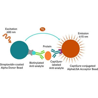
Total-AlphaLISA SureFire Ultra assay principle
The Total-AlphaLISA SureFire Ultra assay measures the expression level of a target protein in a biological sample (e.g. cell lysate).
The Total-AlphaLISA SureFire Ultra assay uses two antibodies which recognize two different distal epitopes on the target protein. AlphaLISA assays require two bead types: Acceptor and Donor Beads. Acceptor Beads are coated with a proprietary CaptSure™ agent to specifically immobilize the assay specific antibody, labeled with a CaptSure tag. Donor Beads are coated with streptavidin to capture one of the detection antibodies, which is biotinylated. In the presence of target protein, the two antibodies bring the Donor and Acceptor Beads in close proximity whereby the singlet oxygen transfers energy to excite the Acceptor Bead, allowing for the generation of a luminescent Alpha signal. The amount of light emission is directly proportional to the quantity of protein present in the sample.
Total-AlphaLISA SureFire Ultra two-plate assay protocol
The two-plate protocol involves culturing and treating the cells in a 96-well plate before lysis, then transferring lysates into a 384-well OptiPlate™ plate before the addition of Total-AlphaLISA SureFire Ultra detection reagents. This protocol enables cell viability and confluence to be monitored. In addition, lysates from a single well can be used to measure multiple targets.

Total-AlphaLISA SureFire Ultra one-plate assay protocol
Detection of Total target protein with AlphaLISA SureFire Ultra reagents can be performed in a single plate used for culturing, treatment, and lysis. No washing steps are required. This HTS designed protocol allows for miniaturization while maintaining robust AlphaLISA SureFire Ultra quality.

Assay validation
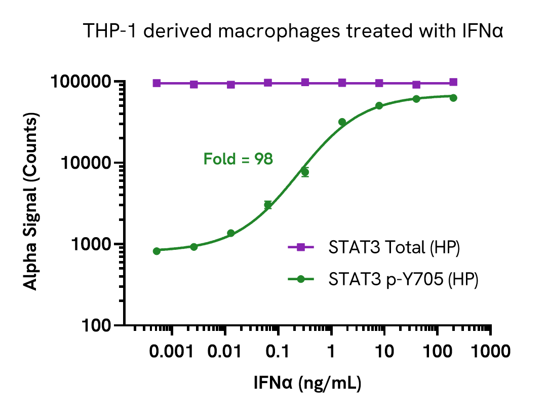
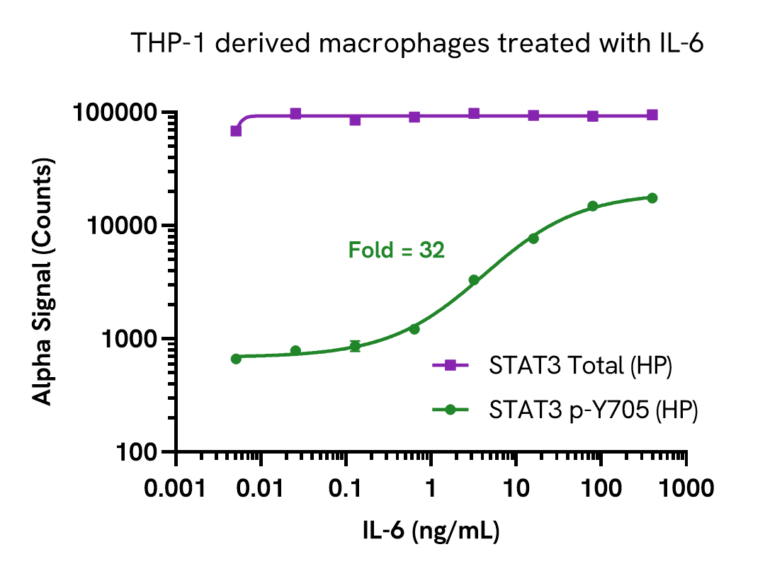
Induction of STAT3 (Tyr705) phosphorylation in THP-1 derived macrophages
THP-1 cells were seeded in a 96-well plate (50,000 cells/well) in complete medium containing 100 nM PMA and incubated for 24 hours at 37°C, 5% CO2. The THP-1 derived macrophages were starved in HBSS + 0.1% BSA for 2 hours and then stimulated with increasing concentrations of either IFNα or IL-6 for 20 minutes.
After treatment, the cells were lysed with 100 µL of Lysis Buffer for 10 minutes at RT with shaking (350 rpm). STAT3 Phospho (Tyr705) and Total levels were evaluated using respective AlphaLISA SureFire Ultra High Performance STAT3 assays. For the detection step, 10 µL of cell lysate (approximately 5,000 cells) was transferred into a 384-well white OptiPlate, followed by 5 µL of Acceptor mix and incubated for 1 hour at RT. Finally, 5 µL of Donor mix was then added to each well and incubated for 1 hour at RT in the dark. The plate was read on an Envision using standard AlphaLISA settings.
As expected, IFNα and IL-6 treatment resulted in a dose dependent increase in STAT3 (Tyr705) phosphorylation, while Total levels remained unchanged.
Induction of STAT3 phosphorylation in various cell models
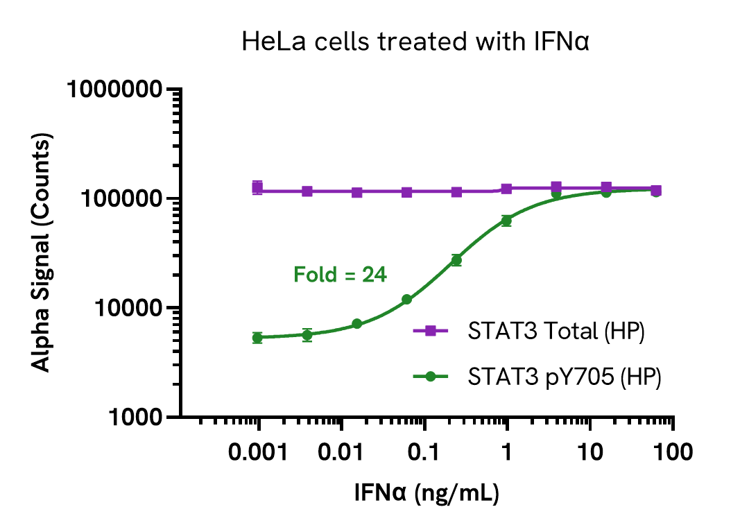
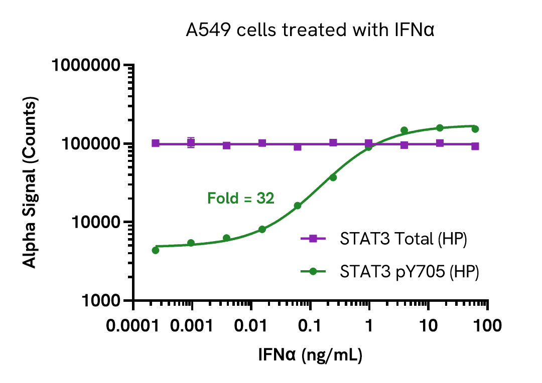
HeLa and A549 cells were seeded in a 96-well plate (20,000 cells/well) in complete medium and incubated overnight at 37°C, 5% CO2. Cells were starved for 20 hours and then stimulated with increasing concentrations of IFNα for 30 minutes.
After treatment, the cells were lysed with 100 µL of Lysis Buffer for 10 minutes at RT with shaking (350 rpm). STAT3 Phospho (Tyr705) and Total levels were evaluated using respective AlphaLISA SureFire Ultra High Performance STAT3 assays. For the detection step, 10 µL of cell lysate (approximately 4,000 cells) was transferred into a 384-well white OptiPlate, followed by 5 µL of Acceptor mix and incubated for 1 hour at RT. Finally, 5 µL of Donor mix was then added to each well and incubated for 1 hour at RT in the dark. The plate was read on an Envision using standard AlphaLISA settings.
As expected, IFNα treatment resulted in a dose dependent increase in STAT3 (Tyr705) phosphorylation, while Total levels remained unchanged.
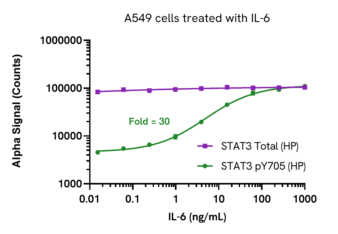
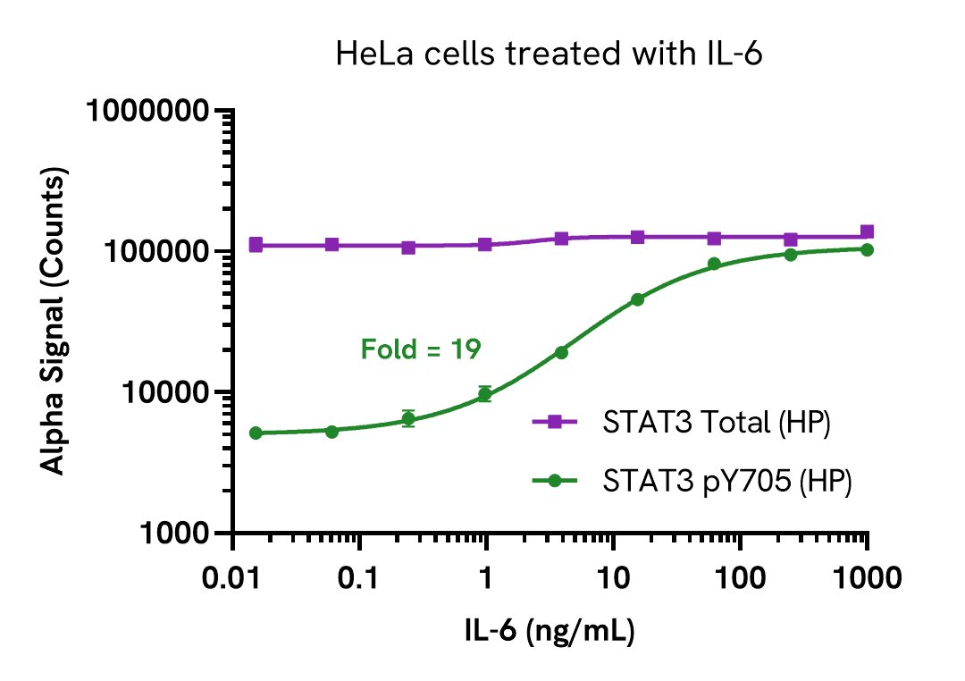
A549 and HeLa cells were seeded in a 96-well plate (20,000 cells/well) in complete medium and incubated overnight at 37°C, 5% CO2. Cells were starved for 20 hours and then stimulated with increasing concentrations of IL-6 for 15 minutes.
After treatment, the cells were lysed with 100 µL of Lysis Buffer for 10 minutes at RT with shaking (350 rpm). STAT3 Phospho (Tyr705) and Total levels were evaluated using respective AlphaLISA SureFire Ultra High Performance STAT3 assays. For the detection step, 10 µL of cell lysate (approximately 4,000 cells) was transferred into a 384-well white OptiPlate, followed by 5 µL of Acceptor mix and incubated for 1 hour at RT. Finally, 5 µL of Donor mix was then added to each well and incubated for 1 hour at RT in the dark. The plate was read on an Envision using standard AlphaLISA settings.
As expected, IL-6 treatment resulted in a dose dependent increase in STAT3 (Tyr705) phosphorylation, while Total levels remained unchanged.
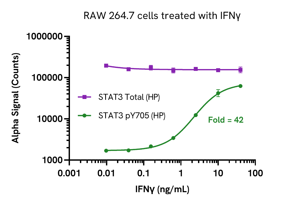
RAW 264.7 cells were seeded in a 96-well plate (20,000 cells/well) in complete medium and incubated overnight at 37°C, 5% CO2. Cells were starved for 2 hours and then stimulated with increasing concentrations of IFNγ for 20 minutes.
After treatment, the cells were lysed with 100 µL of Lysis Buffer for 10 minutes at RT with shaking (350 rpm). STAT3 Phospho (Tyr705) and Total levels were evaluated using respective AlphaLISA SureFire Ultra High Performance STAT3 assays. For the detection step, 10 µL of cell lysate (approximately 4,000 cells) was transferred into a 384-well white OptiPlate, followed by 5 µL of Acceptor mix and incubated for 1 hour at RT. Finally, 5 µL of Donor mix was then added to each well and incubated for 1 hour at RT in the dark. The plate was read on an Envision using standard AlphaLISA settings.
As expected, IFNγ treatment resulted in a dose dependent increase in STAT3 (Tyr705) phosphorylation, while Total levels remained unchanged.
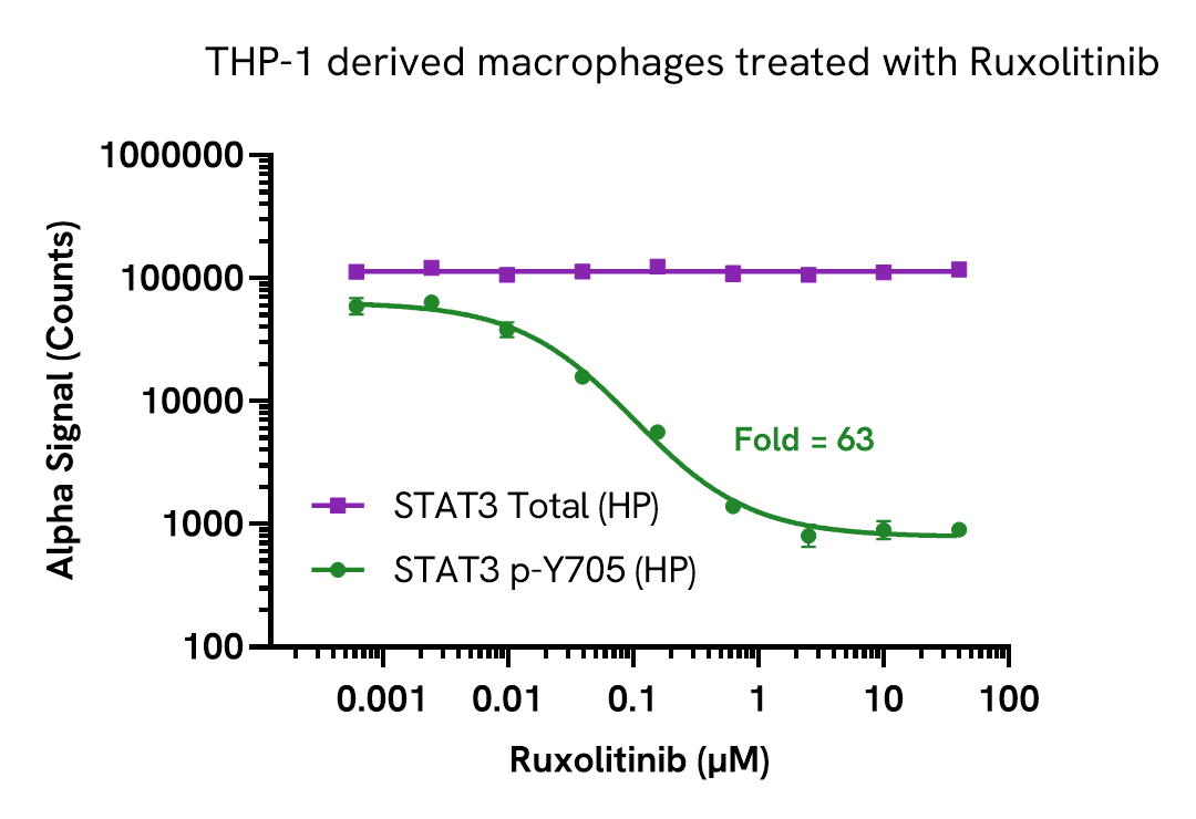
Inhibition of STAT3 (Tyr705) phosphorylation in THP-1 derived macrophages
THP-1 cells were seeded in a 96-well plate (50,000 cells/well) in complete medium containing 100 nM PMA and incubated for 24 hours at 37°C, 5% CO2. The THP-1 derived macrophages were treated with increasing concentrations of Ruxolitinib for 2 hours and then stimulated with 10 ng/mL IFNγ for 20 minutes.
After treatment, the cells were lysed with 100 µL of Lysis Buffer for 10 minutes at RT with shaking (350 rpm). STAT3 Phospho (Tyr705) and Total levels were evaluated using respective AlphaLISA SureFire Ultra High Performance STAT3 assays. For the detection step, 10 µL of cell lysate (approximately 5,000 cells) was transferred into a 384-well white OptiPlate, followed by 5 µL of Acceptor mix and incubated for 1 hour at RT. Finally, 5 µL of Donor mix was then added to each well and incubated for 1 hour at RT in the dark. The plate was read on an Envision using standard AlphaLISA settings.
As expected, treatment with Ruxolitinib resulted in a dose dependent decrease in the levels of Phospho STAT3 (Tyr705), while Total levels remained unchanged.
Assay versatility
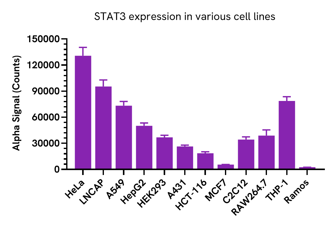
STAT3 expression in various cell lines
Adherent cells were grown to confluency in a T175 flask at 37°C, 5% CO2, and were lysed with Lysis Buffer at a density of 100,000 cells/mL. Suspension cells were washed and lysed with 5X Lysis Buffer at a density of 300,000 cells/mL.
STAT3 levels were evaluated using AlphaLISA SureFire Ultra High Performance assay. For the detection step, 10 µL of cell lysate (approximately 1,000 adherent cells and 3,000 suspension cells) were transferred into a 384-well white OptiPlate, followed by 5 µL of Acceptor Mix and incubated for 1 hour at RT. Finally, 5 µL of Donor Mix was then added to each well and incubated for 1 hour at RT in the dark. The plate was read on an Envision using standard AlphaLISA settings.
Assay sensitivity
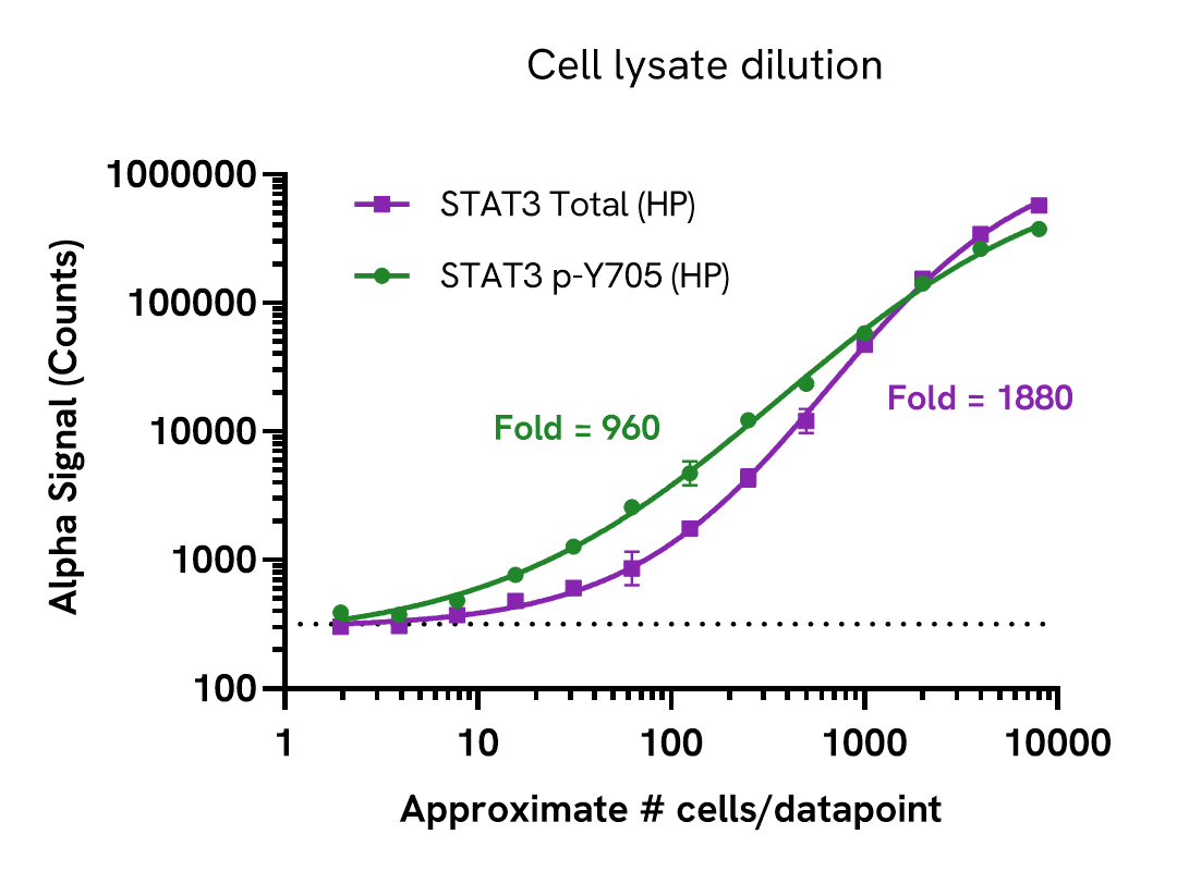
Total STAT3 High Performance assay sensitivity
THP-1 cells were seeded in a 96-well plate (50,000 cells/well) in complete medium containing 100 nM PMA and incubated for 24 hours at 37°C, 5% CO2. The THP-1 derived macrophages were starved in HBSS + 0.1% BSA for 2 hours and then stimulated with increasing concentrations of IFNα for 20 minutes.
After treatment, the cells were lysed with 100 µL of Lysis Buffer for 10 minutes at RT with shaking (350 rpm). STAT3 Phospho (Tyr705) and Total levels were evaluated using respective AlphaLISA SureFire Ultra STAT3 assays. For the detection step, 10 µL of cell lysate (approximately 5,000 cells) was transferred into a 384-well white OptiPlate, followed by 5 µL of Acceptor mix and incubated for 1 hour at RT. Finally, 5 µL of Donor mix was then added to each well and incubated for 1 hour at RT in the dark. The plate was read on an Envision using standard AlphaLISA settings.
Cell lysate was prepared from A431 cells cultured to confluency in a T175 flask treated with 2 µg/mL EGF for 10 minutes and lysed in 10 mL of Lysis Buffer for 10 minutes at RT with shaking.
Lysate was serially diluted in Lysis Buffer and STAT3 Phospho (Tyr705) and Total levels were evaluated using respective AlphaLISA SureFire Ultra High Performance STAT3 assays. For the detection step, 10 µL of cell lysate was transferred into a 384-well white OptiPlate, followed by 5 µL of Acceptor Mix and incubated for 1 hour at RT. Finally, 5 µL of Donor Mix was then added to each well and incubated for 1 hour at RT in the dark. The plate was read on an Envision using standard AlphaLISA settings.
Approximate number of cells is indicated. The dotted line represents assay background. The STAT3 Total High Performance assay has a broad dynamic range and can detect STAT3 expression in less than 50 cells/datapoint.
Specifications
| Application |
Cell Signaling
|
|---|---|
| Automation Compatible |
Yes
|
| Brand |
AlphaLISA SureFire Ultra
|
| Detection Modality |
Alpha
|
| Molecular Modification |
Total
|
| Product Group |
Kit
|
| Protocol Time |
2h at RT
|
| Sample Volume |
30 µL
|
| Shipping Conditions |
Shipped in Blue Ice
|
| Target |
STAT3
|
| Target Class |
Phosphoproteins
|
| Target Species |
Human
Mouse
|
| Technology |
Alpha
|
| Therapeutic Area |
Inflammation
Neuroscience
Oncology
|
| Unit Size |
100 assay points
|
Resources
Are you looking for resources, click on the resource type to explore further.
Brochure
Alpha assays and reagents catalog
Alpha technolgy enables the rapid and straightforward mesaure of virtually any target. This includes enzymes, receptor-ligand...
Guide
AlphaLISA SureFire Ultra: the ultimate guide for successful experiments
The definitive guide for setting up a successful AlphaLISA SureFire Ultra assay
Several biological processes are regulated by...
Brochure
Alpha SureFire Ultra no-wash immunoassay catalog
Discover Alpha SureFire® Ultra™ assays, the no-wash cellular kinase assays leveraging Revvity's exclusive bead-based technology...
Brochure
Species compatibility for HTRF, AlphaLISA SureFire Ultra and Alpha SureFire Ultra Multiplex assays
This document includes detailed tables listing HTRF™, AlphaLISA™ SureFire® Ultra™, and Alpha SureFire® Ultra™ Multiplex assays...
Loading...


How can we help you?
We are here to answer your questions.

