


JP
Revvity Sites Globally
Select your location.
*e-commerce not available for this region.

AlphaLISA SureFire Biotin-Free Human and Mouse Total EGR1 Detection Kit, 500 Assay Points

AlphaLISA SureFire Biotin-Free Human and Mouse Total EGR1 Detection Kit, 500 Assay Points
AlphaLISA SureFire Biotin Free Total Kit Schematic
The AlphaLISA™ SureFire® Biotin-Free Human and Mouse Total EGR1 assay is a sandwich immunoassay for quantitative detection of total EGR1 in cellular lysates using Alpha Technology.
| Feature | Specification |
|---|---|
| Application | 細胞シグナル伝達 |
| Protocol Time | 2h at RT |
| Sample Volume | 10 µL |
The AlphaLISA™ SureFire® Biotin-Free Human and Mouse Total EGR1 assay is a sandwich immunoassay for quantitative detection of total EGR1 in cellular lysates using Alpha Technology.
Product variants
Unit Size: 100 Assay Points
Part #:
ASBF-TEGR1-A-HV
Unit Size: 500 Assay Points
Part #:
ASBF-TEGR1-A500
Unit Size: 10,000 Assay Points
Part #:
ASBF-TEGR1-A10K
Unit Size: 50,000 Assay Points
Part #:
ASBF-TEGR1-A50K
For research use only. Not for use in diagnostic procedures. All products to be used in accordance with applicable laws and regulations including without limitation, consumption and disposal requirements under European REACH regulations (EC 1907/2006).
AlphaLISA SureFire Biotin-Free Human and Mouse Total EGR1 Detection Kit, 500 Assay Points
AlphaLISA SureFire Biotin Free Total Kit Schematic
Loading...
Product information
Overview
Early Growth Response 1 (EGR1) is an immediate-early transcription factor that rapidly responds to diverse stimuli including growth factors, cytokines, and stress signals to regulate cell proliferation, differentiation, and apoptosis. EGR1 is induced within minutes of stimulation through MAPK and calcium signaling pathways, binding to GC-rich sequences in target gene promoters. EGR1 regulates expression of genes involved in cell cycle control (p53, PTEN), differentiation (fibronectin, PDGF), and vascular responses (VEGF, tissue factor). EGR1 functions as both a tumor suppressor and oncogene depending on cellular context, with loss of expression in prostate cancer and glioblastoma but overexpression in breast cancer. EGR1 plays critical roles in vascular injury responses, wound healing, and synaptic plasticity, making it relevant to cardiovascular disease, fibrosis, and neurological disorders.
The AlphaLISA SureFire Biotin-Free Human and Mouse Total EGR1 Detection Kit is a sandwich immunoassay for the quantitative detection of total EGR1 in cellular lysates, using Alpha Technology.
Formats:
- The HV (high volume) kit contains reagents to run 100 wells in 96-well format, using a 60 μL reaction volume.
- The 500-point kit contains enough reagents to run 500 wells in 384-well format, using a 20 μL reaction volume.
- The 10,000-point kit contains enough reagents to run 10,000 wells in 384-well format, using a 20 μL reaction volume.
- The 50,000-point kit contains enough reagents to run 50,000 wells in 384-well format, using a 20 μL reaction volume.
AlphaLISA SureFire Biotin Free kits are compatible with:
- Cell and tissue lysates
- Antibody modulators
- Biotherapeutic antibodies
- Biotin rich samples
AlphaLISA SureFire Biotin Free kits can be used for:
- Cellular kinase assays
- Receptor activation studies
- High-throughput screening for preclinical studies
How it works
Total-AlphaLISA SureFire Biotin Free assay principle
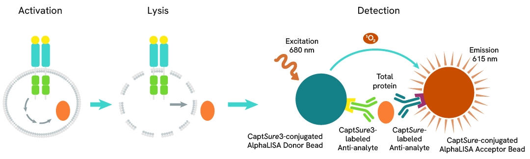
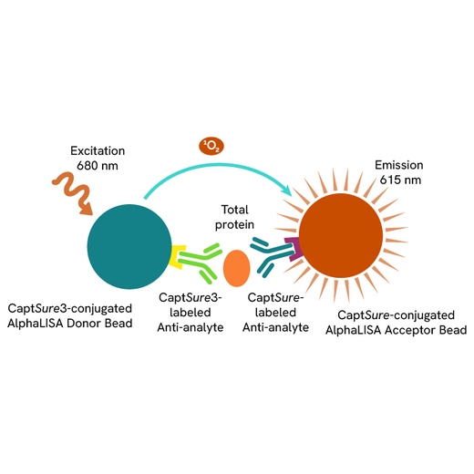
The Total-AlphaLISA Surefire Biotin Free assay measures the expression level of a protein target in a cell lysate.
The Total-AlphaLISA Surefire assay uses two antibodies which recognize two different distal epitopes on the targeted protein. AlphaLISA assays require two bead types: Acceptor and Donor beads. Acceptor beads are coated with a proprietary CaptSure™ agent to specifically immobilize the assay specific antibody, labeled with a CaptSure tag. Donor beads are coated with a proprietary CaptSure 3 agent to capture one of the detection antibodies, which is labeled with CaptSure 3 tag. In the presence of targeted protein, the two antibodies bring the Donor and Acceptor beads in close proximity whereby the singlet oxygen transfers energy to excite the Acceptor bead, allowing the generation of a luminescent Alpha signal. The amount of light emission is directly proportional to the quantity of protein present in the sample.
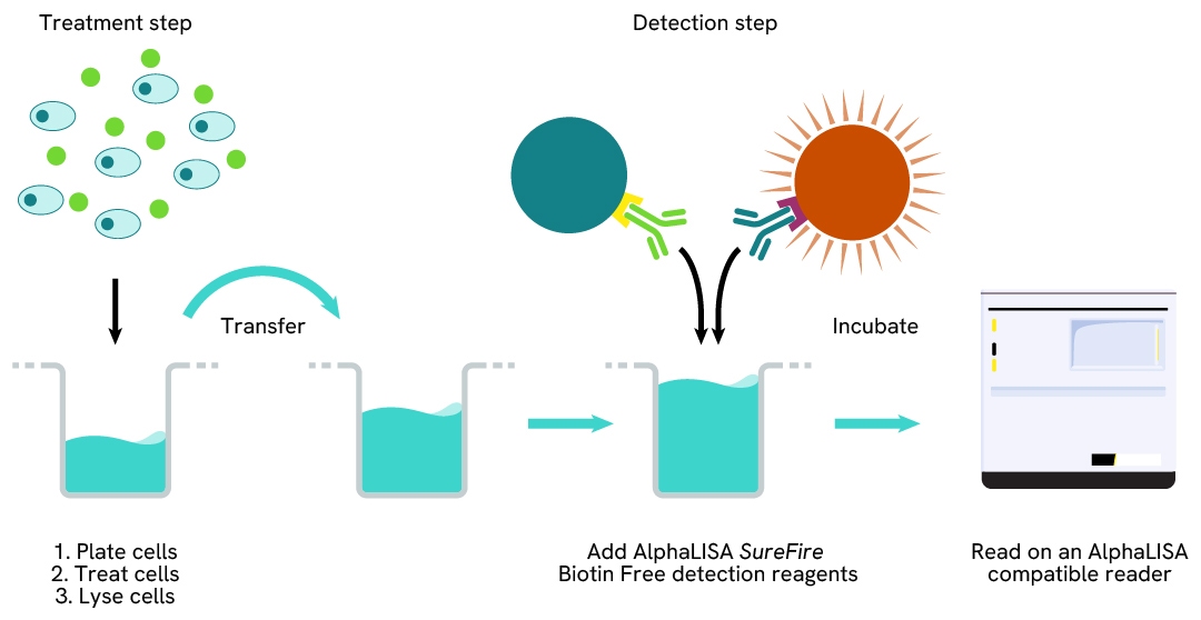
Total-AlphaLISA SureFire Biotin Free two-plate assay protocol
The two-plate protocol involves culturing and treating the cells in a 96-well plate before lysis, then transferring lysates into a 384-well Optiplate™ plate before the addition of Total-AlphaLISA SureFire Biotin Free detection reagents. This protocol permits the cells viability and confluence to be monitored. In addition, lysates from a single well can be used to measure multiple targets.
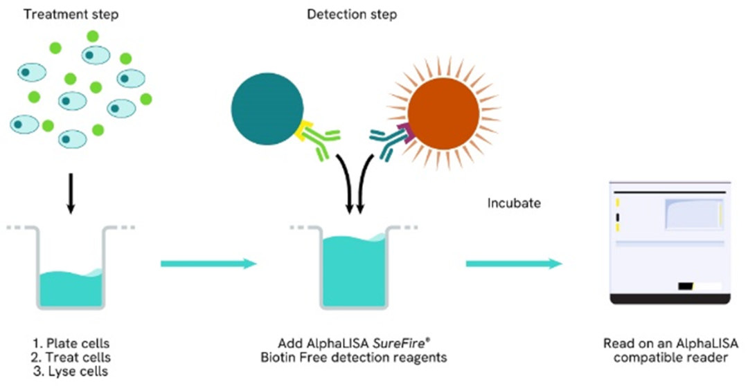
Total-AlphaLISA SureFire one-plate assay protocol
Detection of Total target protein with AlphaLISA SureFire Biotin Free reagents can be performed in a single plate used for culturing, treatment, and lysis. No washing steps are required. This HTS designed protocol allows for miniaturization while maintaining robust AlphaLISA SureFire quality.
Assay validation
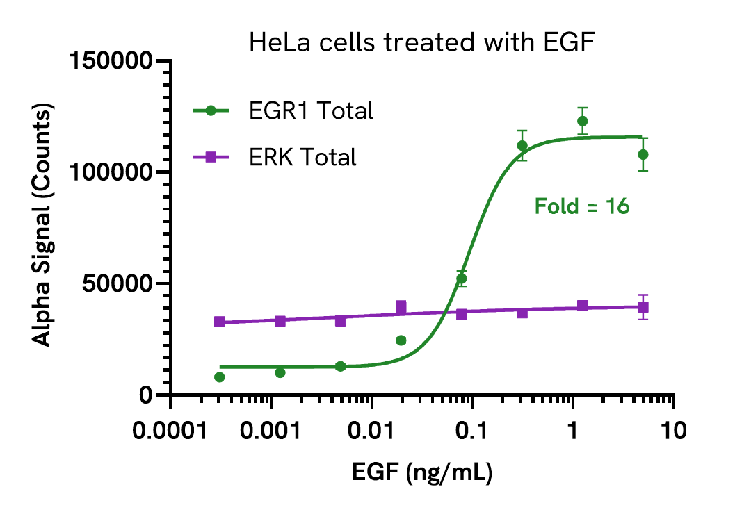
Induction of EGR1 in cells treated with EGF
HeLa cells were seeded in a 96-well plate (40,000 cells/well) in complete medium and incubated overnight at 37°C, 5% CO2. Cells were treated with increasing concentrations of EGF for 2 hours.
After treatment, the cells were lysed with 100 µL of Lysis Buffer for 10 minutes at RT with shaking (350 rpm). EGR1 and ERK Total levels were evaluated using respective AlphaLISA SureFire Biotin Free assays. For the detection step, 10 µL of cell lysate (approximately 4,000 cells) was transferred into a 384-well white OptiPlate, followed by 5 µL of Acceptor mix and incubated for 1 hour at RT. Finally, 5 µL of Donor mix was then added to each well and incubated for 1 hour at RT in the dark. The plate was read on an Envision using standard AlphaLISA settings.
As expected, EGF triggered a dose-dependent increase in the levels of Total EGR1 while ERK Total levels remained unchanged.
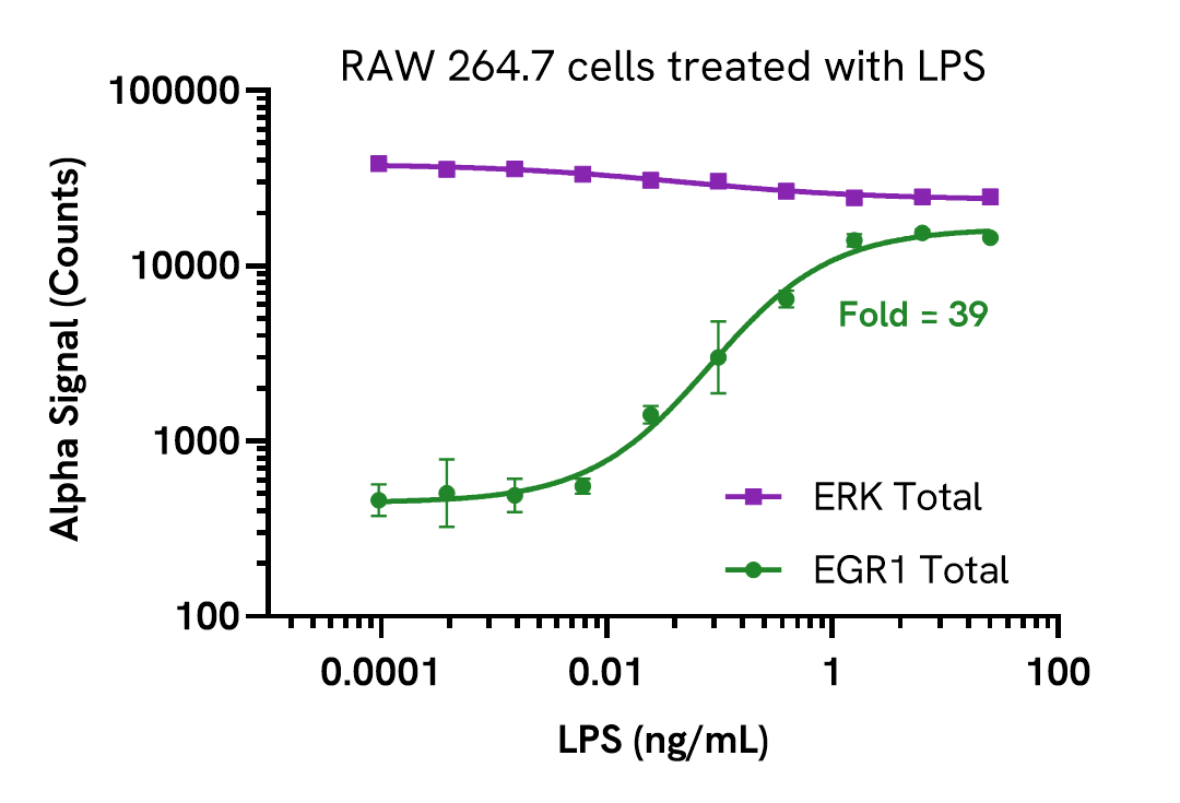
Induction of EGR1 in cells treated with LPS
RAW 264.7 cells were seeded in a 96-well plate (40,000 cells/well) in complete medium and incubated overnight at 37°C, 5% CO2. Cells were treated with increasing concentrations of LPS for 2 hours.
After treatment, the cells were lysed with 100 µL of Lysis Buffer for 10 minutes at RT with shaking (350 rpm). EGR1 and ERK Total levels were evaluated using respective AlphaLISA SureFire Biotin Free assays. For the detection step, 10 µL of cell lysate (approximately 4,000 cells) was transferred into a 384-well white OptiPlate, followed by 5 µL of Acceptor mix and incubated for 1 hour at RT. Finally, 5 µL of Donor mix was then added to each well and incubated for 1 hour at RT in the dark. The plate was read on an Envision using standard AlphaLISA settings.
As expected, LPS triggered a dose-dependent increase in the levels of Total EGR1 while ERK Total remained unchanged.
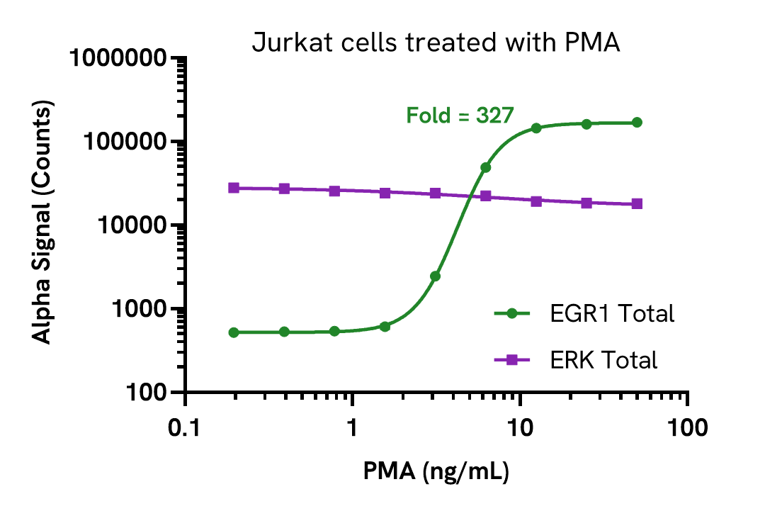
Induction of EGR1 in cells treated with PMA
Jurkat cells were seeded in a 96-well plate (400,000 cells/well) in complete RPMI 1640 medium and treated with increasing concentrations of PMA for 3 hours.
After treatment, the cells were lysed with the addition of 50 µL of 5X Lysis Buffer for 10 minutes at RT with shaking (350 rpm). EGR1 and ERK Total levels were evaluated using respective AlphaLISA SureFire Biotin Free assays. For the detection step, 10 µL of cell lysate (approximately 16,000 cells) was transferred into a 384-well white OptiPlate, followed by 5 µL of Acceptor mix and incubated for 1 hour at RT. Finally, 5 µL of Donor mix was then added to each well and incubated for 1 hour at RT in the dark. The plate was read on an Envision using standard AlphaLISA settings.
As expected, PMA triggered a dose-dependent increase in the levels of Total EGR1 while Total ERK remained unchanged.
Assay specificity/selectivity
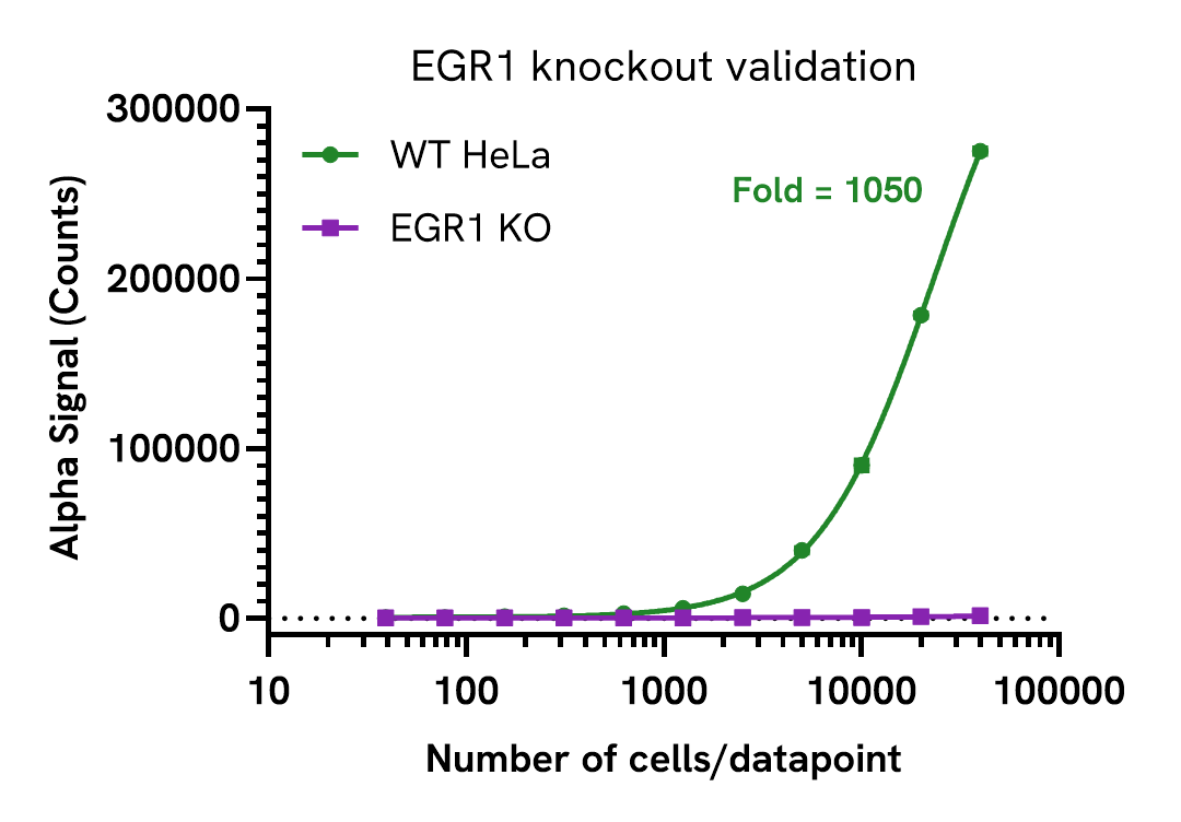
Knockout validation of EGR1 Total Assay
EGR1 levels were assessed in HeLa wild type (WT) and EGR1 KO (Abcam, ab274929) cell lines cultured to confluency in T175 flasks and treated with 2 ng/mL EGF for 2 hours. Each flask was lysed in 4 mL of Lysis Buffer for 10 minutes at RT with shaking. Lysates were serially diluted in Lysis Buffer and levels of EGR1 Total were evaluated using the AlphaLISA SureFire Biotin Free assay. For the detection step, 10 µL of cell lysate was transferred into a 384-well white OptiPlate, followed by 5 µL Acceptor Mix and incubated for 1 hour at RT. Finally, 5 µL of Donor Mix was added to each well and incubated for 1 hour at RT in the dark. The plate was read on an Envision using standard AlphaLISA settings.
EGR1 was only detected in WT cells, demonstrating assay specificity. Dotted line represents assay background.
Assay versatility
EGR1 expression in various cell lines
Adherent cells were grown to confluency in a T175 flask at 37°C, 5% CO2 and were lysed with Lysis Buffer at a density of 0.5 x 10⁶ cells/mL. Suspension cells were harvested, washed in HBSS and lysed with Lysis Buffer at 1.6 x 10⁶ cells/mL.
EGR1 levels were evaluated using the AlphaLISA SureFire Biotin Free assay. For the detection step, 10 µL of cell lysate (5,000 adherent and 16,000 suspension cells) were transferred into a 384-well white OptiPlate, followed by 5 µL of Acceptor Mix and incubated for 1 hour at RT. Finally, 5 µL of Donor Mix was then added to each well and incubated for 1 hour at RT in the dark. The plate was read on an Envision using standard AlphaLISA settings.

Specifications
| Application |
Cell Signaling
|
|---|---|
| Automation Compatible |
Yes
|
| Brand |
AlphaLISA SureFire Biotin-Free
|
| Detection Modality |
Alpha
|
| Product Group |
Kit
|
| Protocol Time |
2h at RT
|
| Sample Volume |
10 µL
|
| Shipping Conditions |
Shipped in Blue Ice
|
| Target |
EGR1
|
| Target Class |
Phosphoproteins
|
| Target Species |
Human
Mouse
|
| Technology |
Alpha
|
| Therapeutic Area |
NASH/Fibrosis
Oncology
|
| Unit Size |
500 Assay Points
|
Resources
Are you looking for resources, click on the resource type to explore further.
Guide
AlphaLISA SureFire Ultra: the ultimate guide for successful experiments
The definitive guide for setting up a successful AlphaLISA SureFire Ultra assay
Several biological processes are regulated by...
Loading...


How can we help you?
We are here to answer your questions.

