Revvity Sites Globally
Select your location.
*e-commerce not available for this region.
Select your location.
*e-commerce not available for this region.
High-content screening can be used to capture fine sub-cellular detail with very high resolution images, but there is always some level of compromise to the speed when you increase the resolution and therefore the amount of image data you capture. In certain applications, such as rare event studies or assays using microtissues, only part of the well or particular wells, are of interest. That means you want to acquire high resolution data from that region, and not waste time capturing the rest of the well or the entire plate.
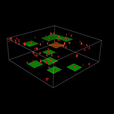
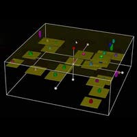
Intelligent acquisition technology for HCS takes these factors into account so that you can more accurately target your object of interest for significantly reduced acquisition and analysis times. This capability is available via the optional PreciScan plug-in for Harmony high-content analysis software. It enables a fully automated and integrated workflow of low magnification pre-scan, image analysis and higher magnification re-scan.
For example, this workflow can be used to locate 3D microtissues in all wells of a 96-well plate using a 5x brightfield scan, followed by acquiring a multicolour fluorescent z-stack at 40x magnification in the center of the tissue. It could also be used for locating stem cell colonies in co-cultures or identifying rare events in a large cell population. You could identify mitotic cells with a 10x pre-scan and follow up with 63x imaging of the nucleus. PreciScan also enables efficient tiled imaging of larger objects such as e.g. zebrafish or tissue slices.

Left image: Pre-scan at low magnification (10x) using PreciScan software to identify wells where microtissues have grown. Right image: Re-scan at higher magnification (20x) of only the wells in which the microtissues have grown and with microtissues centred in the image. 3D InSight Microtissue courtesy of InSphero AG.
For research use only. Not for use in diagnostic procedures.


