メニュー
Revvity logo
Contact us
JP
Revvity Sites Globally

Select your location.

*e-commerce not available for this region.

australia.webp Australia
austria.webp Austria
belgium.webp Belgium
brazil.webp Brazil *
canada.webp Canada
china.webp China *
denmark.webp Denmark
finland.webp Finland
france.webp France
germany.webp Germany
hong-kong.webp Hong Kong (China) *
india.webp India *
ireland.webp Ireland
italy.webp Italy
japan.webp Japan *
luxembourg.webp Luxembourg
mexico.webp Mexico *
netherlands.webp Netherlands
norway.webp Norway
philippines.webp Philippines *
republic of korea.webp Republic of Korea *
singapore.webp Singapore *
spain.webp Spain
sweden.webp Sweden
switzerland.webp Switzerland
thailand.webp Thailand *
uk.webp United Kingdom
usa.webp United States
Breadcrumb
...
  • ホーム
  • Products
  • 研究・開発
  • 細胞分析
  • Cell Counting and Image Cytometry
    • Cell Isolation
    • Cell Lines & Stem Cells
    • Cell Counting and Image Cytometry
    • セルヘルスと細胞生存率
    • Cellular Imaging & Analysis
    • Immunoassays
    • In Vivo Imaging
    • Microplates
  • Cell Counting & Image Cytometry Reagents
    • Cell Counting & Image Cytometry Slides, Plates, and Beads
    • Cell Counting & Image Cytometry Instruments
    • Cell Counting & Image Cytometry Reagents
  • Cell Cycle Assay Kits
    • Antibodies for Immunophenotyping
    • Apoptosis Assay Kits and Reagents
    • Cell Counting and Viability Assay Kits and Reagents
    • Cell Cycle Assay Kits
    • Cell Health and Fitness Assay Kits and Reagents
    • Proliferation and Tracer Reagents
    • Yeast Viability and Vitality Assay Kits and Reagents

Cell Cycle Assay Kits

View products & services View resource library

Cell Cycle Assay Kits for Cellometer™ cell counters and Celigo™ image cytometer

Cell cycle analysis is used to determine the proportion of cells in each stage of the cell cycle for a given cell population based on variations in DNA content. In our ViaStain cell cycle assays, each cell is stained with propidium iodide (PI), a fluorescent nuclear staining dye that intercalates with DNA. Because the dye cannot enter live cells, the cells are first fixed (with ethanol or methanol), and then stained with the cell cycle reagent. Cells preparing for division will contain increasing amounts of DNA and display proportionally increased fluorescence. Differences in fluorescence intensity are used to determine the percentage of cells in each phase of the cell cycle. We provide instrument-specific cell cycle assay kits for Cellometer cell counters and the Celigo image cytometer.


For research use only. Not for use in diagnostic procedures.
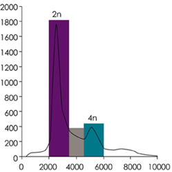
Histogram showing fluorescent intensity of Jurkat cells at various stages of the cell cycle. The purple bar (2n) represents G1-phase , the gray bar S-phase, and teal bar (4n) the G2/M phase.
The histogram shows fluorescent intensity of Jurkat cells at various stages of the cell cycle. The purple bar (2n) represents G1-phase , the gray bar S-phase, and teal bar (4n) the G2/M phase.

Request more information
Cell cycle
bg-element-listing-page bg-element-listing-page-mobile
Products & services
Resource library
Revvity Logo

©2025 Revvity - All rights reserved

Revvity is a trademark of Revvity, Inc. All other trademarks are the property of their respective owners.