


Single-cell sequencing raises the stakes on sample preparation
Single-cell and single-nucleus RNA sequencing (scRNA-seq, snRNA-seq) have transformed how researchers study gene expression, enabling the profiling of individual cells at a higher resolution than bulk methods. 10x Genomics has developed platforms such as the Chromium™, Visium™, and Xenium™ that have contributed to making this technology more accessible and affordable than ever. However, the sample preparation that feeds into a sequencing run is still extremely complex and sensitive to a variety of factors.
Key takeaways:
- Accurate cell counting can help protect sequencing investments
- Brightfield methods may have limitations for complex samples
- AOPI fluorescence-based counting offers enhanced accuracy
- Revvity cell counting instruments support single-cell sequencing workflows with AOPI technology
One of the most important and easily improved variables within a sequencing workflow is accurate sample counting. Before a single sample goes onto the chip, an accurate measurement of concentration and viability is required at multiple steps to ensure proper sample dilution and loading. Getting an inaccurate count is not a minor inconvenience.
A full 8-sample chip run requires a significant investment of resources. Each failed sample that requires a repeat run will incur not only the significant additional costs associated with reagents and consumables but also duplicate the person-hours required to run the sample, add sequencing queue delays, necessitate batch-to-batch corrections, and potentially result in the loss of precious and irreplaceable patient samples.
Cell counting is recommended at multiple points throughout the workflow: (i) after initial cell or nuclei isolation, (ii) after any enrichment or depletion steps, and (iii) immediately before loading the chip (CG000126). Loading too many cells or nuclei increases the probability of doublet formation, while loading too few cells can result in a higher rate of empty droplets. An inaccurate count at any step can propagate through the sample preparation process and lead to run failure.
Where brightfield counting methods fall short
Brightfield-based methods can have difficulty distinguishing subcellular debris from intact dead cells. This can present a challenge when samples contain extracellular debris, as is common with dissociated primary tissue, erythrocyte-depleted monocyte preps, and nuclei suspensions. Debris may be counted alongside the desired objects, producing an inflated concentration estimate that leads to underloading the chip and may increase the likelihood of run failure rates.
In single-nuclei workflows, Trypan blue has been shown to overcount nuclei by as much as 400% compared to fluorescence-based methods, depending on tissue type (Figure 1). For fixed samples, 10x Genomics explicitly notes that Trypan blue is not recommended.
Figure 1. Nuclei count comparison between propidium iodide (PI) fluorescence staining and Trypan blue across brain, lung, and kidney tissue preparations. Data sourced from 10x Genomics.
For a detailed overview of the limitations of brightfield counting methods such as Trypan blue, see Revvity's recent blog post: Got the cell counting blues? Why Trypan Blue may no longer be the gold standard for cell counting and viability measurements.
The case for fluorescence-based counting with AOPI
Acridine orange (AO) and propidium iodide (PI) staining is a fluorescence-based method that intercalates with double-stranded DNA (dsDNA). AO is cell membrane permeable and can stain all nucleated cells, both live and dead. PI enters only cells with compromised membranes, indicating that they are dead or dying. Critically, debris without nuclear dsDNA does not fluoresce in either channel and is therefore invisible to the counting algorithm.
AOPI is particularly well-suited to sample types common in high-resolution NGS workflows:
- Primary tissue dissociates
- Nuclei preparations
- Fixed cells and nuclei used in 10x Flex workflows
AOPI
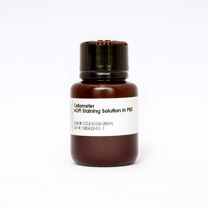 ViaStain AOPI Staining Solution, 25 mL
is also instantaneous with no wash steps, incubation times, or cytotoxicity effects.
ViaStain AOPI Staining Solution, 25 mL
is also instantaneous with no wash steps, incubation times, or cytotoxicity effects.
Revvity cell counters for single-object sequencing workflows
Revvity’s Cellometer™ Ascend™
 Cellometer Ascend Automated Cell Counter
, Cellaca MX™
Cellometer Ascend Automated Cell Counter
, Cellaca MX™
 Cellaca MX High-throughput Cell Counter
, and Cellaca PLX™
Cellaca MX High-throughput Cell Counter
, and Cellaca PLX™
 Cellaca PLX High-throughput Image Cytometer
cell counters are well-suited for these applications. Both instruments use AOPI fluorescence as their primary counting modality and are compatible with the cell, nuclei, and fixed-sample protocols used in single-cell sequencing workflows. The Cellaca is specifically highlighted in 10x Genomics protocol documentation (CG000478) as a compatible instrument for counting both live and fixed cells and nuclei ahead of Chromium™ RNA Profiling runs.
Cellaca PLX High-throughput Image Cytometer
cell counters are well-suited for these applications. Both instruments use AOPI fluorescence as their primary counting modality and are compatible with the cell, nuclei, and fixed-sample protocols used in single-cell sequencing workflows. The Cellaca is specifically highlighted in 10x Genomics protocol documentation (CG000478) as a compatible instrument for counting both live and fixed cells and nuclei ahead of Chromium™ RNA Profiling runs.
The Cellometer Ascend is suited to lower-throughput labs, supporting up to eight samples per run with as little as 5 µL of sample per count. The Cellaca uses the same underlying AOPI technology but scales to 24 samples in under six minutes and may be integrated into automation workflows for core facilities and high-throughput environments.
Accurate cell counting is one of the simplest and most cost-effective steps a researcher can take to protect the quality of a sequencing run.
To learn more about Revvity instrumentation, please visit the Revvity cell counting solutions page.
For research use only. Not for use in diagnostic procedures.


