
Despite decades of progress in cancer research, over 90% of cancer drugs entering clinical trials fail to reach the market.1,2 A major reason for this high failure rate is the gap between traditional models and the complexity of human cancer biology. 2D cell cultures lack the architecture and heterogeneity of tumors, while animal models often fall short in recapitulating human-specific biology, take months to generate results, and raise ethical concerns about animal welfare.
Microphysiological systems (MPS), also known as complex in vitro models (CIVM), offer an attractive alternative to 2D cell cultures or animal models. These platforms enable living cells or tissues to be maintained in controlled microenvironments designed to mimic the physiological aspects of tissue and organ function.
Types of MPS include:
- Organ-on-chips: miniature, microfluidic devices containing engineered human tissues and cells within a polymer chip.
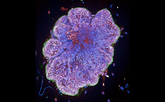
- Organoids: miniature, self-organized 3D tissue cultures derived from stem or cancer cells that maintain patient-specific tumor characteristics.
- Spheroids: 3D cell cultures that self-assemble into sphere-like formations and are well suited for scalable drug testing.
By recapitulating key features of human biology, MPS provide more predictive insights into disease mechanisms, drug responses, and personalized treatment strategies. They also align with the ethical goals of the 3R framework—Replacement, Reduction, and Refinement of animal use—that has guided biomedical research for over 50 years.
Key applications of MPS in cancer research
Given these advantages, MPS are being adopted across cancer research to model critical cancer processes that traditional systems cannot capture. For example, metastasis cannot be properly studied in flat 2D cultures that lack the physiologically relevant tissue architecture.
Disease modeling: Patient-derived organoids and tumor-on-chip platforms recreate tumor heterogeneity and allow researchers to study cancer progression in controlled environments.
Drug testing: MPS enable high-throughput screening of candidate compounds under conditions that more closely mimic human tissue.
Tumor microenvironment studies: These systems allow the inclusion of stromal and immune cells, helping researchers explore complex interactions between tumors and their surrounding microenvironment.
Metastasis modeling: Organ-on-chips and 3D cultures provide insights into the metastatic cascade which involves multiple processes that are challenging to replicate in vitro.
Advancing cancer drug discovery with screening-ready microphysiological systems
Despite their promise, large-scale and high-throughput integration of MPS into drug discovery pipelines has remained limited. Key barriers include the complexity of handling 3D models, sample heterogeneity, and the time- and labor-intensive processes required for automation and scaling. In addition, 3D image analysis presents challenges, requiring advanced algorithms to analyze the complex morphology and spatial organization of these cultures.
In our recent literature review, we explore how researchers are overcoming these challenges and using MPS for high-throughput cancer drug screening. For example:
- Ozer et al. developed a microfluidic organ-on-chip assay that captures the early stages of cancer metastasis and applied it for high-throughput drug profiling.3
- Bozal et al. designed a fully automated high-content screening platform for multi-modal analysis of 3D organoid cultures.4
- Mysior and Simpson created a scalable approach to generate large numbers of uniform spheroids and extract single-cell and subcellular measurements following drug perturbations.5
References:
- Jentzsch V, Osipenko L, Scannell JW, Hickman JA. Costs and causes of oncology drug attrition with the example of Insulin-Like growth Factor-1 receptor inhibitors. JAMA Network Open. 2023;6(7):e2324977. doi:10.1001/jamanetworkopen.2023.24977
- Kuo Y, Barrett JS. Consideration of the root causes in candidate attrition during oncology drug development. Clinical Pharmacology in Drug Development. 2024;13(9):952-960. doi:10.1002/cpdd.1464
- Ozer LY, Fayed HS, Ericsson J, Zen AAH. Development of a cancer metastasis-on-chip assay for high throughput drug screening. Frontiers in Oncology. 2024;13. doi:10.3389/fonc.2023.1269376
- Bozal SB, Sjogren G, Costa AP, et al. Development of an automated 3D high content cell screening platform for organoid phenotyping. SLAS Discovery. 2024;29(7):100182. doi:10.1016/j.slasd.2024.100182
- Mysior MM, Simpson JC. An automated high-content screening and assay platform for the analysis of spheroids at subcellular resolution. PLoS ONE. 2024;19(11):e0311963. doi:10.1371/journal.pone.0311963
For research use only. Not for use in diagnostic procedures.