


The most overlooked step in cell counting
The impact of cell counting extends far beyond determining how many cells are present in a sample. Accurate measurement of cell concentration and viability influences many downstream workflows, from routine cell culture and genomics experiments to high-throughput drug screening, bioprocess development, and cell therapy research.
Over the past decade, automated cell counters have become increasingly common alternatives to manual hemocytometers in laboratories requiring greater throughput and reproducibility. These systems use imaging and automated analysis to deliver rapid, objective measurements that reduce much of the operator bias associated with manual counting.
Key takeaways:
- Standardizing sample preparation can help improve cell counting reproducibility. Automated liquid handling can reduce variation in concentration and viability measurements.
- Cell settling, pipetting shear, and staining inconsistency affect cell counts. Pre-analytical variables can distort concentration and viability before samples reach the cell counter.
- Automated liquid handling supports consistent, gentler sample preparation. Programmable pipetting, wide-bore tips, and controlled AO/PI staining help protect fragile cells.
- The AssayMate workstation and the Cellaca cell counter illustrate scalable cell counting automation. The workflow cuts 20 manual steps to four and hands-on preparation time by 70%.
However, the quality of the cell count depends in part on how the sample is prepared before it reaches the instrument. If cells settle before aspiration, experience shear stress during pipetting, or are inconsistently stained, the analyzer cannot recover the original biological state of the sample.1 Increasingly, researchers recognize that improving the consistency of cell counting requires attention to sample preparation, not just the instrument.
Why sample preparation is the hidden variable
Three pre-analytical variables can affect cell concentration and viability measurements:
- Cell settling and clumping: Suspension cells begin settling almost immediately after mixing. A technician drawing from the top of a tube may measure a different concentration than one aspirating from the bottom. Manual mixing helps, but every researcher mixes differently, with some pipetting three times, others ten. These differences can introduce variability that is unrelated to the biological sample itself.2
- Mechanical stress during pipetting: Not all cells tolerate pipetting equally. Primary cells, stem cells, suspension cultures, and engineered immune cells are particularly susceptible to mechanical stress. Rapid aspiration through narrow pipette tips can damage plasma membranes, lowering measured viability before the sample reaches the analyzer.3
- Inconsistent staining: Whether using trypan blue or fluorescent dyes such as Acridine Orange/Propidium Iodide (AO/PI), reproducible viability measurements require consistent reagent ratios and consistent mixing. Uneven staining, incorrect ratios, or prolonged dye exposure can all influence results, independent of changes in the underlying sample.1
A field application perspective
Tip: If cell count variability appears unexpectedly, verify the sample preparation workflow before troubleshooting the analyzer. Inconsistent mixing, prolonged delays before aspiration, or aggressive pipetting are common sources of variability that can affect the resulting measurement. Standardizing these steps often improves reproducibility without changing the analytical method.4
How automation solves these challenges
Automated liquid handling systems address some of the variability introduced during sample preparation.
| Challenge | Impact | Automated solution |
|---|---|---|
| Cell settling and clumping | Uneven sampling produces inaccurate concentration measurements | Automated tip mixing immediately before aspiration creates a homogeneous suspension |
| Shear stress during pipetting | Fragile cells rupture, lowering apparent viability | Programmable low aspiration/dispense speeds with wide-bore tips reduce mechanical stress |
| Viability stain variability | Premature mixing causes localized dye toxicity | Air-gap techniques separate cells and stain until final dispense for controlled mixing |
By preparing every sample under identical, reproducible conditions, the aim is to reduce the variation introduced by the preparation process but still retain any biological variability between samples.
Standardizing the workflow before analysis
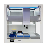
Revvity's AssayMate™ workstation can be used to automate defined sample preparation steps before cell counting.
The workflow includes four stages:
- Load: Cell stock, culture media, and AO/PI solution are loaded into a 12-column reservoir. Integrated temperature control keeps samples and reagents stable during setup.
- Dilute: Twelve biological replicates are prepared across four dilution fractions on a 96-well storage plate, with automated gentle mixing (10× per replicate).
- Stain: AO/PI solution is added at a 1:1 ratio with controlled pipetting to minimize cell disruption during staining.
- Transfer: Replicates are loaded into the 8×3 cell counting plate for scanning on the Cellaca™ MX or Cellaca PLX cell counters. Because handling is automated, every plate is processed under identical conditions.
In this workflow, automation replaces a series of repeated manual pipetting and mixing steps with a defined sequence that can be reproduced across runs. The reported workflow reduces 20 manual steps to four, and has demonstrated a 70% cut in hands-on preparation time while maintaining R² > 0.99 linearity. The primary benefit for most labs is the reduction in operator-to-operator variability.
The automation spectrum: choosing the right level
Cell counting automation is not one-size-fits-all. Laboratories can adopt technologies that match their throughput, budget, and operational requirements.
| Level | Approach | Throughput | Best for | Revvity solutions |
|---|---|---|---|---|
| 1) Manual | Hemocytometer + trypan blue | <10 samples/day | Small research labs, education, backup verification | Disposable Hemocytometers |
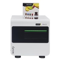
| 2) Benchtop automated | Standalone imaging counter, manual sample prep | 10–50 samples/day | Mid-sized research labs, QC, clinical diagnostics | Cellometer™ Ascend™ automated cell counter Cellaca™ cell counters Celigo™ image cytometer |
| 3) Semi-automated | Liquid handler integrated with cell counter | 50–500 samples/day | HTS labs, bioprocess development, CROs | AssayMate™ workstation + Cellaca™ cell counters |
| 4) Automated work cell | End-to-end robotic automation with LIMS integration | 500+ samples/day | Cell therapy manufacturing, pharma drug discovery | Integrated Lab Automation + Cellaca™ cell counters or Celigo™ image cytometer |
Many organizations follow a natural progression, starting manually to establish protocols, then adding benchtop automation for accuracy, integrating liquid handling to scale throughput, and ultimately deploying full work cells for commercial-scale manufacturing.
Looking beyond the cell counter
Advances in imaging algorithms and analysis software will continue to improve cell counting technology. However, improvements can be achieved now from better sample preparation, without waiting for the next instrument generation.
Automated liquid handling can standardize sample preparation, minimize mechanical stress, reduce operator variability, and integrate with imaging systems, incubators, robotic plate handlers, and LIMS.
For scientists working in drug discovery, bioprocess development, or cell-based research, this integrated approach provides a way to reduce the manual variation associated with sample preparation and frees researchers to focus on experimental design rather than repetitive manual preparation.
The most consistent cell count does not begin inside the cell counter. It begins with the quality of the sample delivered to it.
For research use only. Not for use in diagnostic procedures.
References
- Piccinini F, Tesei A, Arienti C, Bevilacqua A. Cell counting and viability assessment of 2D and 3D cell cultures: expected reliability of the trypan blue assay. Biotechnology Reports. 2015.
- Strober W. Trypan Blue Exclusion Test of Cell Viability. Current Protocols in Immunology. 2015;111:A3.B.1–A3.B.3.
- Freshney RI. Culture of Animal Cells: A Manual of Basic Technique and Specialized Applications. 7th Edition. Wiley. 2016.
- Riss TL, Moravec RA, Niles AL, et al. Cell Viability Assays. Assay Guidance Manual. Eli Lilly & National Center for Advancing Translational Sciences. 2016.
- Revvity. AssayMate™ Liquid Handling Applications: High-Throughput Cell Counting and Viability Automation.