


Cancer researchers need well-characterized models and quantitative tools to evaluate therapeutic approaches and study drug response. Cell culture or animal models for studying potential therapies may not always be reflective of actual human tumors. Phan et al. 1 demonstrate that patient-derived organoids (PDOs) can be used as an alternative model for preclinical evaluation of candidate cancer drugs.
About 90% of potential cancer therapies fail in clinical trials, and this is attributed to failures of the models to accurately represent human disease. 2 In recognition of this challenge, regulatory bodies have recently changed the requirements for animal model use in preclinical testing. 2 This has accelerated the adoption of New Approach Methodologies (NAMs) that take a human-centered approach. 2
Key takeaways:
- Patient-derived organoids support human-relevant cancer drug screening. PDOs can model key tumor features and enable preclinical evaluation of candidate therapies as a human-centered new approach methodology.
- High-throughput PDO workflows can reveal donor-specific drug sensitivities. Phan et al. tested 240 compounds at two concentrations across four ovarian cancer PDOs, showing heterogeneous treatment responses.
- Whole-well image cytometry strengthens organoid quantification. The Celigo™ image cytometer measures organoid number and size across the entire well, including edge-positioned objects important for accurate organoid counts.
- Automated brightfield and fluorescence imaging expands organoid assay readouts. The Celigo image cytometer supports plate imaging in minutes plus Caspase 3/7 and Calcein AM viability measurements for high-throughput NAM workflows.
Identifying drug sensitivities in patient-derived tumor organoids
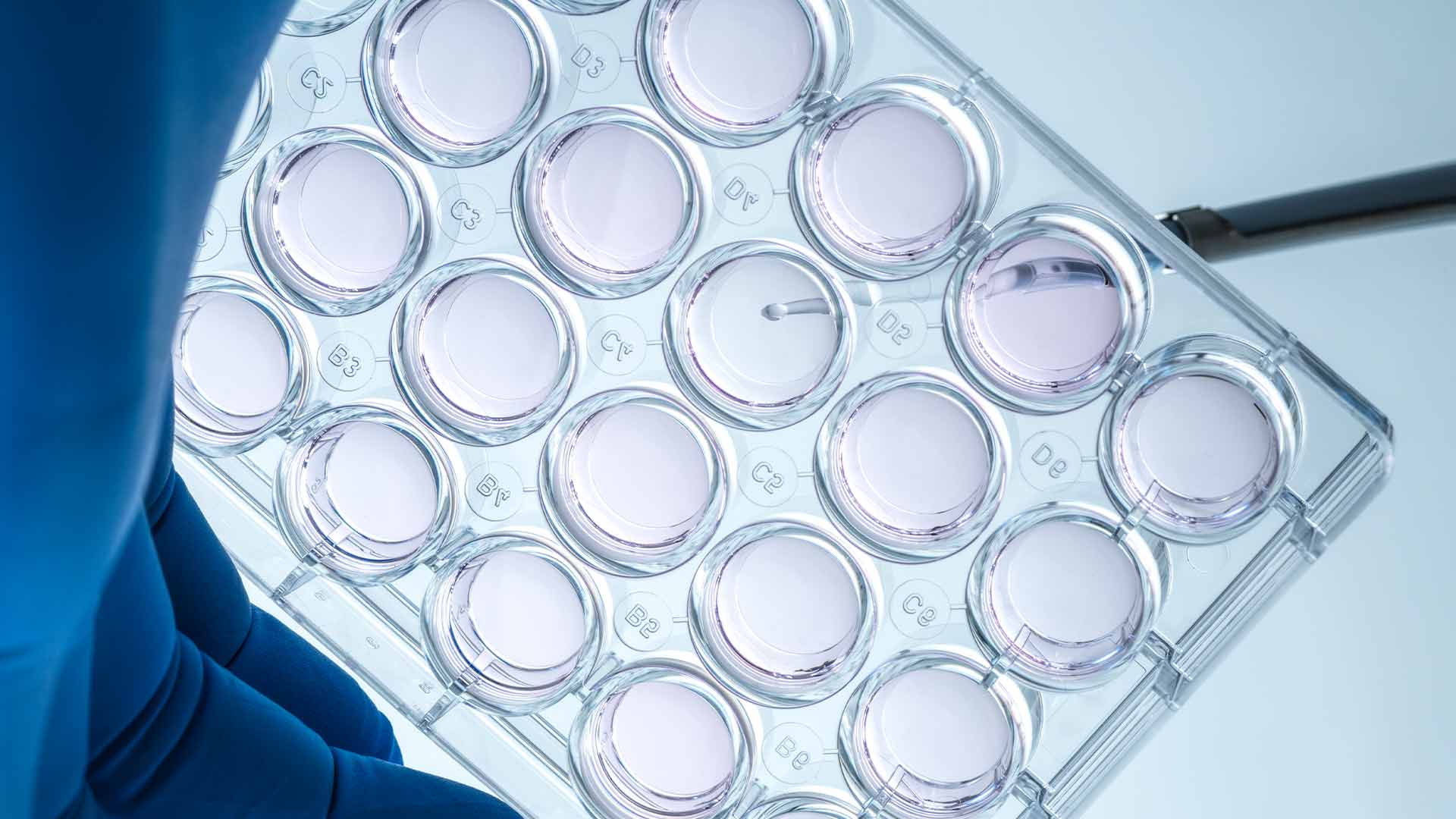
In their study, Phan et al. 1 focus on demonstrating the potential of PDOs as a NAM for evaluating cancer drug responses. Their method uses cells derived from surgically removed tumors and grows them into small clusters of cells that mimic key features of the original tumor. They grow these organoids in a ring along the edge of each well in a multi-well plate. This format facilitates exposure of organoids to a wide range of drug candidates in an automated, high-throughput manner.
To develop this model and method, Phan et al. 1 studied PDOs derived from four donors with different ovarian cancer tumor types. An automated system was used to culture the organoids, allowing them to test 240 compounds at two different concentrations for 480 different conditions.
The primary outputs of this study were:
A. the number and size of the organoids at the end of the compound exposure, and
B. the amount of ATP as a measure of cell viability.
Compounds demonstrating cytotoxic activity would be expected to result in fewer, smaller organoids and decreased ATP levels.
Drug response findings
Phan et al. 1 identified only one compound that all four PDOs responded to. This finding is of interest because response to the PI3K/mTOR cell growth and survival signaling pathway inhibitor BGT226 has not been consistently associated with typical cancer biomarkers.
Similarly, the PDO from donor 1 responded to several cyclin-dependent kinase inhibitors that prevent cell division. When this information was used to predict the effects of other cyclin-dependent kinase inhibitors, only some of their predictions were confirmed by experimental results. These findings demonstrate that testing a candidate compound on tumor-derived organoids may provide more representative data upon which to predict drug response.
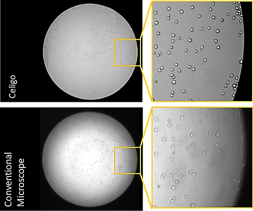
Throughout the development of their method, Phan et al. 1 relied on the Celigo™ image cytometer from Revvity for the analysis of PDO number and size. The Celigo image cytometer can image the cells or organoids in a well from edge to edge (Figure 1). This allows it to identify objects even when they are at the edge of a well, which can be important for studies where organoid number is a primary output.
The Celigo image cytometer can also be integrated into an automation system and can image a plate in minutes. These capabilities are compatible with high-throughput workflows such as those used by Phan et al. 1
Figure 1: Images of cells in two-dimensional culture demonstrate the Celigo image cytometer’s whole-well imaging capability.
The Celigo image cytometer also has fluorescent channels that can be used for whole-well fluorescence imaging. Though Phan et al.1 primarily relied on brightfield imaging to quantify the number and growth of their organoids, they also used the fluorescent channels to measure cell viability via Caspase 3/7 as well as Calcein AM as they developed their method.
Conclusion
The work described by Phan et al. 1 demonstrates the use of the Celigo image cytometer to support cancer research using NAMs such as PDOs. The whole-well imaging, speed via integration with automation, and fluorescent imaging capabilities can support organoid-based drug screening and other NAM applications.
[ Learn more ]. https://www.revvity.com/product/celigo-5c-config-200-bffl-5c
References
- Phan, N., Hong, J.J., Tofig, B. et al. A simple high-throughput approach identifies actionable drug sensitivities in patient-derived tumor organoids. Commun Biol 2, 78 (2019).
- Liu W, Pang PD, Wu CA, Tagle D, Wu JC. New approach methodologies for drug discovery. Cell. 2026;189(7):1877-1903.
For research use only. Not for use in diagnostic procedures.