


Automated imaging and analysis of 3D tumor spheroids, cancer stem cells
The Celigo™ image cytometer
 Celigo Image Cytometer
has been developed to fully automate imaging and analysis of tumorspheres, embryoid bodies and cancer stem colonies. The instrument provides a unique live cell analysis method for studying large multicellular structures and has been used in the development of 3D tumor spheroid-based functional assays for target validation and drug evaluation.
Celigo Image Cytometer
has been developed to fully automate imaging and analysis of tumorspheres, embryoid bodies and cancer stem colonies. The instrument provides a unique live cell analysis method for studying large multicellular structures and has been used in the development of 3D tumor spheroid-based functional assays for target validation and drug evaluation.
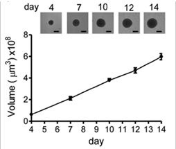
Suspension cultures of tumor spheres, including mammospheres and neurospheres, can be rapidly and accurately analyzed using the Celigo image cytometer. This automated morphometric analysis tool significantly reduces the time and effort needed to quantify key aspects of 3D spheres including size, growth, growth tracking over time and response to chemotherapeutics. Analysis of tumor spheres generated from different cancer cell lines and primary cancer cells can be used to evaluate sphere-forming efficiency, tumorigenicity and self-renewal of cancer stem/tumor-initiating cells.
Tumor sphere analysis benefits
- Non-destructive quantification of live spheroids for correlation with malignant cell behavior
- Whole-well imaging of suspension spheroids (96-well to 6-well)
- Rapid data analysis (scan and analyze an entire 12-well plate in <15 min)
- Analysis of sphere number, size, shape and growth kinetics over time
- Identify and count spheroids in flat or U-bottom wells
- Eliminate cumbersome manual morphology measurements and improve data reproducibility
- Monitor growth of spheroids over time
- Screen numerous spheroid plates per day, 96- and 384- well plates
- Scan an ultra-low adhesion 96-well plate in 8 minutes
- Measure viability using dual-fluorescence assays
- Multi-parameter analysis
Counts / size / short & long diameter / est. volume / perimeter /area
Spheroid growth curve
Tumor sphere analysis on Celigo image cytometer
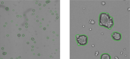
Live images of segmented primary mammospheres generated from MCF10DCIS cells (a premalignant clone of the MCF10A cell line), day 7. Images courtesy of S. Stecklein & R. Jensen, University of Kansas Medical Center.
Mammosphere analysis on Celigo image cytometer
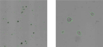
Live images of segmented primary mammospheres generated from HMLE cells (immortalized primary mammary epithelial cells), day 7. Images courtesy of S. Stecklein & R. Jensen, University of Kansas Medical Center.


")
")
For research use only. Not for use in diagnostic procedures.