


Measure the cell invasion from a spheroid into Matrigel®
- Directly image tumor spheroids in various microwell formats
- Non-invasive brightfield imaging allows the user to image the same plate over multiple days
- Perform a two-color fluorescent viability assay
Introduction
The Celigo™ image cytometer
 Celigo Image Cytometer
has been developed to fully automate live cell analysis of tumorspheres. This automated morphometric analysis tool significantly reduces the time and effort needed to quantify key aspects of 3D spheres including size, growth, growth tracking over time and response to chemotherapeutics.
Celigo Image Cytometer
has been developed to fully automate live cell analysis of tumorspheres. This automated morphometric analysis tool significantly reduces the time and effort needed to quantify key aspects of 3D spheres including size, growth, growth tracking over time and response to chemotherapeutics.
Reduced cell invasion from a U-87 MG 3D spheroid into Matrigel® in the presence of 17-AAG
Experimental setup
- Day 0-4: form spheroids
- Day 4: add Matrigel® to provide a semi-solid gel-like matrix
- Day 4-7: use The Celigo™ image cytometer to determine the area occupied by individual cells or cell clusters
Brightfield spheroid images

U-87 MG + 17-AAG
Measure cell invasion from 3D spheroid into Matrigel®
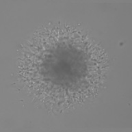
Pre-Matrigel®
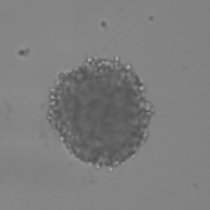
Brightfield spheroid
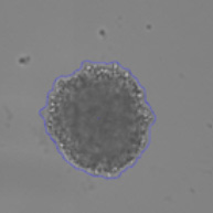
Brightfield identified spheroid
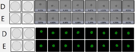
Percent confluence and represented fill view as seen in the Celigo™ software
Measure cell invasion from 3D spheroid into Matrigel®
48 hr after the addition of Matrigel®
Brightfield spheroid
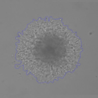
Brightfield identified spheroid
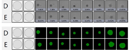
Percent confluence and represented fill view as seen in the Celigo™ software


")
")
For research use only. Not for use in diagnostic procedures.