


Identify, characterize and monitor cell populations based on morphology
A mixture of adherent and suspension cells were seeded into a 96-well plate. Using the Celigo™ image cytometer
 Celigo Image Cytometer
gating interface, we provide a live cell analysis method to identify two distinct populations using morphology. Designed to produce enhanced brightfield cell images, in combination with the advanced image analysis software, the Celigo has been used to identify, characterize and monitor specific cell sub-populations based on morphological features.
Celigo Image Cytometer
gating interface, we provide a live cell analysis method to identify two distinct populations using morphology. Designed to produce enhanced brightfield cell images, in combination with the advanced image analysis software, the Celigo has been used to identify, characterize and monitor specific cell sub-populations based on morphological features.
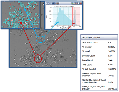
Population analysis based on cell roundness
The Celigo image cytometer records cell images, displays gated cell images, calculates cell morphological parameters and plots the data for population gating.
Camptothecin-induced morphological changes
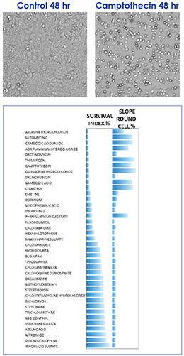
Alteration in cellular morphology was used as an indicator of compound toxicity. We can observe an example (top) of the rounding up of an adherent cell after 48 hrs treatment with Camptothecin. Using this alteration in morphology in a label-free assay, we screened a number of compounds. The percentage survival index calculated from this assay is shown.
Brightfield Images of Colo-205 Cells on Celigo image cytometer.
Monitor drug-induced morphological changes
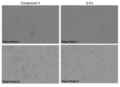
Cells were treated with Compound X (left) and 5-FU (right) and images were acquired at multiple time points. After 5-FU treatment, a morphological change is observed at time point 3.


")
")
For research use only. Not for use in diagnostic procedures.