メニュー
Revvity logo
Contact us
JP
Revvity Sites Globally

Select your location.

*e-commerce not available for this region.

australia.webp Australia
austria.webp Austria
belgium.webp Belgium
brazil.webp Brazil *
canada.webp Canada
china.webp China *
denmark.webp Denmark
finland.webp Finland
france.webp France
germany.webp Germany
hong-kong.webp Hong Kong (China) *
india.webp India *
ireland.webp Ireland
italy.webp Italy
japan.webp Japan *
luxembourg.webp Luxembourg
mexico.webp Mexico *
netherlands.webp Netherlands
norway.webp Norway
philippines.webp Philippines *
republic of korea.webp Republic of Korea *
singapore.webp Singapore *
spain.webp Spain
sweden.webp Sweden
switzerland.webp Switzerland
thailand.webp Thailand *
uk.webp United Kingdom
usa.webp United States
Breadcrumb
...
  • ホーム
  • Resources
  • Knowledge Base
  • CellCountingSub Section
  • Measure Apoptosis
Cell Counting and Image Cytometry

Measure Apoptosis

Section
Celigo Applications
Celigo Applications
Cell Counting Method Selection
Cell Counting and Image Cytometry FAQs
Cell-based Assays for Bioprocessing
Cell-based Assays for Gene Therapy Development
Cellometer Applications
Modern Virology Assays
Sub Section
3D Cell Models
3D Cell Models
Assays for Virology Research
Brightfield Applications
Celigo Cell Counting Applications
Celigo Fluorescent Assays
Celigo Image Cytometer Direct Cell Counting Assays for Immunotherapy
Cell Line Development
Migration and Invasion Assays
iPSC Reprogramming
Topic
Measure Apoptosis
Embryoid Bodies and Patient-Derived Organoids
3D Confrontation Assays
Growth Inhibition
Measure Apoptosis
Measure Migration onto extracellular matrix
Quantify Invasion into Matrigel
Tumor Spheroid Viability
Tumorsphere Formation & Clonogenic Survival
  1. Directly image tumor spheroids in various microwell formats
  2. Non-invasive brightfield imaging allows the user to image the same plate over multiple days
  3. Perform a two-color fluorescent viability assay

Introduction

The Celigo™ image cytometer has been developed to fully automate live cell analysis of tumorspheres. This automated morphometric analysis tool significantly reduces the time and effort needed to quantify key aspects of 3D spheres including size, growth, growth tracking over time and response to chemotherapeutics.

growth inhibition celigo image cytometer

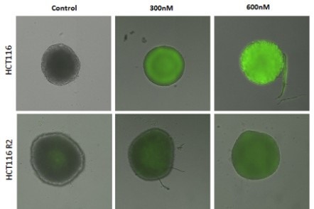
Measure an increase in caspase 3/7 activity in HCT116 3D tumor spheroids
 

measure an increase in caspase

HCT116R2 and HCT116 spheroids were stained with Caspase 3/7 reagent and imaged. The above images present a merge of brightfield and Caspase 3/7 (green) images.

For research use only. Not for use in diagnostic procedures.

ON THIS PAGE
Revvity Logo

©2025 Revvity - All rights reserved

Revvity is a trademark of Revvity, Inc. All other trademarks are the property of their respective owners.