Benefits of Celigo cell counting applications
- Rapid brightfield imaging and label-free in situ analysis of cells
- Whole-well image capture with a 1 µ/pixel resolution
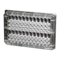
- Imaging of multi-well plates (1536-well to 6-well) and T-flasks (T-25 and T-75)
- Easy-to-use interface to image and identify a wide variety of cell types in brightfield
- Straightforward image segmentation and cell identification software
- Graphic and numeric reports of growth curves, cell counts, doubling time and doubling rates
Cell counting
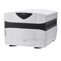
The Celigo™ image cytometer
 Celigo Image Cytometer
cell counting application a reliable live cell analysis method and features whole-well and label-free brightfield imaging. It has increased counting accuracy of cell cultures and eliminates the need for detachment enzymes and hematocytometers to determine cell culture density. The flexibility and ease-of-use of the Celigo cell counting application also allow cell growth to be monitored over time and facilitates the daily management of cultures. The standardization of this process leads to increased consistency of quantitative cell-based assays.
Proliferation assay and cytotoxicity assay for drug screening introduction
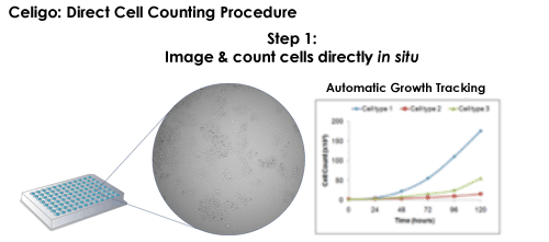
Celigo direct cell counting procedure

Celigo counting benefits
- Image and count cells directly in situ
- Label-free cell counting requires no reagents
- Whole-well brightfield imaging and segmentation counts the cells in every plate well
- Multiple reads of the same sample reduce cost and effort
- Normalization of wells using the actual number of cells in each well
- Allows for morphological inspection and analysis
For research use only. Not for use in diagnostic procedures.


