メニュー
Revvity logo
Contact us
JP
Revvity Sites Globally

Select your location.

*e-commerce not available for this region.

australia.webp Australia
austria.webp Austria
belgium.webp Belgium
brazil.webp Brazil *
canada.webp Canada
china.webp China *
denmark.webp Denmark
finland.webp Finland
france.webp France
germany.webp Germany
hong-kong.webp Hong Kong (China) *
india.webp India *
ireland.webp Ireland
italy.webp Italy
japan.webp Japan *
luxembourg.webp Luxembourg
mexico.webp Mexico *
netherlands.webp Netherlands
norway.webp Norway
philippines.webp Philippines *
republic of korea.webp Republic of Korea *
singapore.webp Singapore *
spain.webp Spain
sweden.webp Sweden
switzerland.webp Switzerland
thailand.webp Thailand *
uk.webp United Kingdom
usa.webp United States
Breadcrumb
...
  • ホーム
  • Resources
  • Knowledge Base
  • CellCountingSub Section
  • Transwell Migration
Cell Counting and Image Cytometry

Transwell Migration

Section
Celigo Applications
Celigo Applications
Cell Counting Method Selection
Cell Counting and Image Cytometry FAQs
Cell-based Assays for Bioprocessing
Cell-based Assays for Gene Therapy Development
Cellometer Applications
Modern Virology Assays
Sub Section
Migration and Invasion Assays
3D Cell Models
Assays for Virology Research
Brightfield Applications
Celigo Cell Counting Applications
Celigo Fluorescent Assays
Celigo Image Cytometer Direct Cell Counting Assays for Immunotherapy
Cell Line Development
Migration and Invasion Assays
iPSC Reprogramming
Topic
Transwell Migration
Chemotaxis
Invasion into Matrigel
Transwell Migration
Wound Healing

Image and quantify transwell invasion and migration assays using suspension and adherent cells

  1. Image transwell invasion of adherent cells without dissociation of cells from the transwell insert
  2. Directly count the number of invading/migrating cells
  3. Fluorescently label and image invading cells

Perform and quantify transwell invasion assay by direct cell count

Transwell diagram

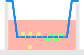
Suspension cells located inside the insert migrate through the porous membrane toward the chemoattractant from the bottom plate. Celigo image cytometer is used to automatically count migrated cells in the bottom plate.

Image and count the number of cells in a transwell Invasion assay

DAPI BF whole well

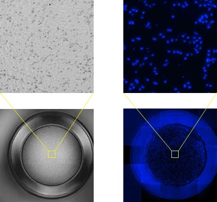
Brightfield and DAPI images of transwell invasion of adherent cancer cells

Transwell DAPI bright field overlay

Overlay of brightfield and DAPI

Bright field images of transwell membrane surface

High-resolution brightfield images of the transwell membrane

Thumbnail pictures and cell numbers are automatically displayed upon the completion of the cell counts

Acquisition of high-resolution DAPI and brightfield images of the transwell membrane surface in a 24 well plate format took ~ 3 minutes

For research use only. Not for use in diagnostic procedures.

ON THIS PAGE
Revvity Logo

©2025 Revvity - All rights reserved

Revvity is a trademark of Revvity, Inc. All other trademarks are the property of their respective owners.