


Rapid, label-free counting and characterization of live cell analysis of embryoid bodies
Embryonic stem (ES) and induced pluripotent stem (iPS) cell colonies spontaneously differentiate and form three dimensional multicellular aggregates. called embryoid bodies (EBs), when cultured in suspension without key stem cell growth factors. EB formation is a common intermediate during the in vitro differentiation of ES/iPS cells into specialized cell types. The size distribution of embryoid bodies plays a significant role in the efficiency of differentiation and in production yields (Dang et al. 2004: Ng et al. 2005: Folconnet et al. 2006).
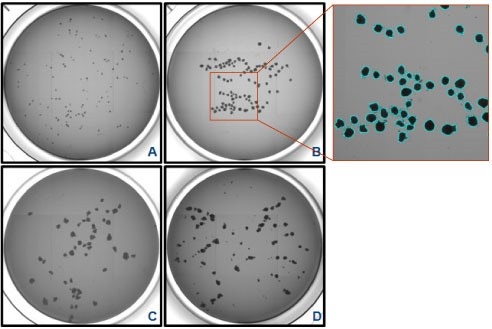
Cultures maintained by enzymatic passage result in the formation of a heterogeneous embryoid bodies population, varying in size and morphology, and in the ultimate yield of differentiated cells. Recent efforts to enrich EBs of preferred size to increase differentiation potential have included biocompatible coatings (Valamehr et al. 2008), microcontact printing (Bouwens et al. 2008), and forced aggregation systems (Burridge et al, 2007) with some success. The generation of a live cell analysis method and an embryoid body counting algorithm for the Celigo™ image cytometer
 Celigo Image Cytometer
has enabled automated imaging and size analysis of EBs. These developments for improved embryoid body generation have led to a need for routine quality assessment of the embryoid body number, size, and shape.
Celigo Image Cytometer
has enabled automated imaging and size analysis of EBs. These developments for improved embryoid body generation have led to a need for routine quality assessment of the embryoid body number, size, and shape.
The Celigo colony counting embryoid body application quickly analyzes embryoid body populations of all shapes and sizes and does so in a non-destructive manner so that the embryoid bodies analyzed can be subsequently used. Current methods typically require acquisition of hundreds of images on a standard microscope followed by manual analysis or loading into another software program which provides only basic measurements (e.g., EB diameter). Because these analyses are difficult and slow. thorough characterization of embryoid bodies prior to differentiation is often omitted. A fast, automated live cell analysis method for embryoid body analysis greatly reduces the time and effort needed and allows quality control of embryoid bodies prior to investing in long-term differentiation cultures. In addition, embryoid body characteristics which enhance differentiation pathways can be identified to improve differentiation efficiency and reduce culture costs.
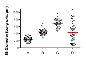
The colony counting embryoid body application uses the Celigo large area rapid scanning capability to count and determine the size,shape.,and morphology of embryoid bodies. The system records whole-well images of multi-well (384W to 6W) plates, enabling the tracking of live embryoid body characteristics for correlation with final differentiation patterns.
Colony counting features
- EB Count
- Diameter (um)
- Short Axis Diameter
- Long Axis Diameter
- Area
- Perimeter
- Form Factor
- Smoothness
- Aspect Ratio
- % Well Sampled


")
")
For research use only. Not for use in diagnostic procedures.