


JP
Revvity Sites Globally
Select your location.
*e-commerce not available for this region.

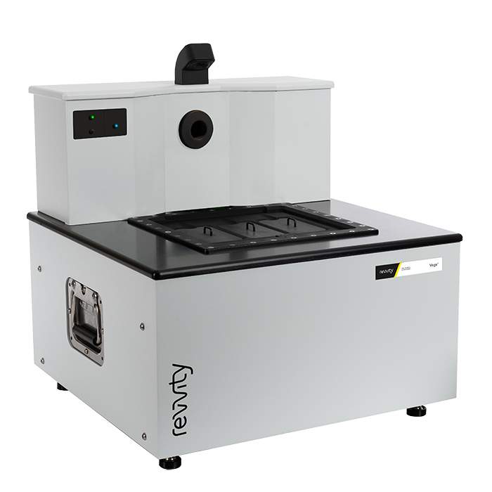
Vega Preclinical Ultrasound System
The Vega™ ultrasound system is Revvity's latest addition of leading preclinical in vivo imaging technology. With an innovative design, Vega is a hands-free automated ultrasound platform that delivers high-resolution 2D and 3D imaging in just a few minutes.
Hands-free, automated, high-throughput ultrasound
For research use only. Not for use in diagnostic procedures.
Vega Preclinical Ultrasound System
Vega® automated, hands-free, high throughput preclinical ultrasound system
Part #:
D5240000
Imaging Modality:
超音波
Loading...
Vega Ultrasound System
Easy to use
Requires minimal amount of training, no dedicated sonographer required.
More consistent results
Removes operator variability resulting in more consistent data over longitudinal studies.
Increased data accuracy
By removing physical contact between the transducer and the animal, tissues are not 'distorted' or 'warped' during 3D image acquisition.
3D widefield imaging
Allows whole subject imaging for visualizing effects of disease or therapies on specific organs or surrounding tissues.
High speed scanning
Automated hands-free transducers allow for fast and consistent scanning.
Streamlined imaging workflow
Users can prep subjects while scanner is operating.
Video overview
Key features
Hands free
Obtain more consistent results with a hands-free ultrasound imaging system that removes the challenges associated with conventional hand-held transducers.
Automated
Innovative bottom-up imaging approach through the use of automated transducers located under the imaging stage is easy-to-use and involves minimal training with no dedicated sonographer required.
High throughput
Sequential scanning of 3 mice, fast scan times with most taking less than one minute, and a streamlined workflow by prepping the next set of mice on the benchtop work together to achieve increased throughput.
Widefield scanning
Fast 3D whole subject imaging for visualizing effects of disease or therapies in a broader anatomical context reducing the risk of missing findings using conventional handheld transducers.
Multiple modes for multiple applications
Vega comes standard with 2 integrated transducers covering many scanning modes from high-resolution and vascular imaging to deep tissue imaging, elastography, non-linear contrast, cardiac imaging, and more.
Flexible
Through the use transducers located under the imaging stage, Vega eliminates the inconvenience of manually swapping out transducers providing ultimate ease and flexibility when switching to different modes.
Imaging modes
Standard
B- & M- modes
Brightness mode for common scanning applications and motion mode for measuring cardiac function.
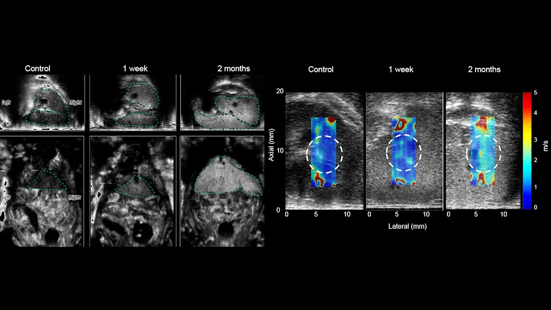
SWE
Elastography
Evaluate and measure tissue stiffness using shear wave elastography (SWE).
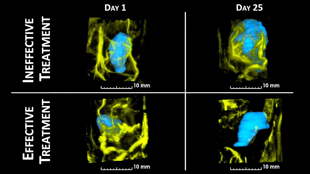
CEUS
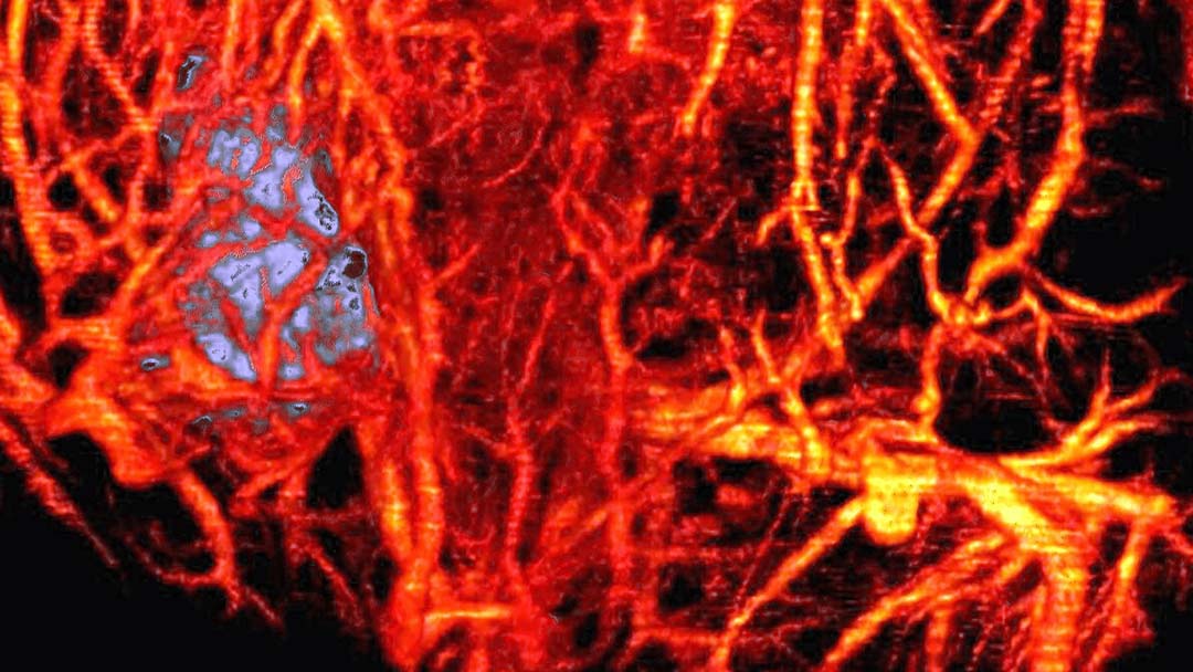
Acoustic angiography
Visualize and quantify tumor vessel network & density or reveal therapy response in tissue over time.
NCL
Non-linear contrast
Leverage VesselVue™ agents for high resolution imaging of tissue perfusion and blood flow.
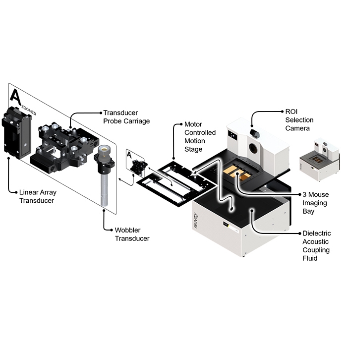
Inside the Vega system
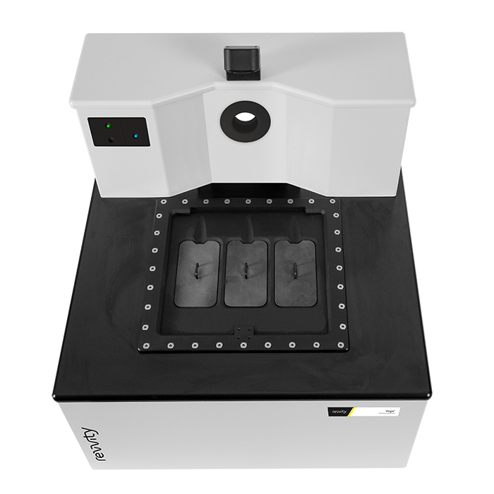
Three mouse imaging bay
Improves imaging throughput by combining fast scan times with a heated three-bay imaging stage, allowing for the sequential scanning of three mice. This capability, alongside its speed, dramatically increases efficiency.
Transducer probe carriage
Housing unit for both the wobbler and linear array transducers located under the imaging stage offering true hands-free ultrasound imaging.
Linear array transducer
Ideal for B-mode, M-mode, 4D-mode, shear wave elastography (SWE), and non-linear contrast (NLC) imaging.
Wobbler transducer
Single-element, high-frequency B-mode transducer ideal for tumor and organ imaging. Dual-element annular array, high and low frequency wobbler offering both B-mode and acoustic angiography for tumor, organ, and vascular imaging.
Highlighted applications
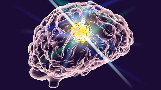
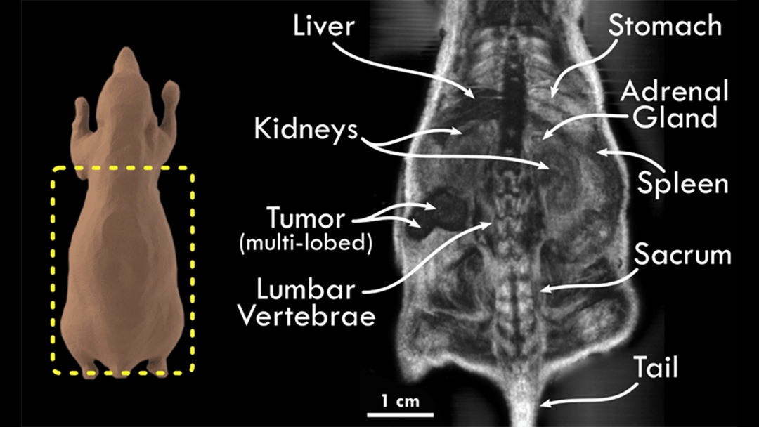
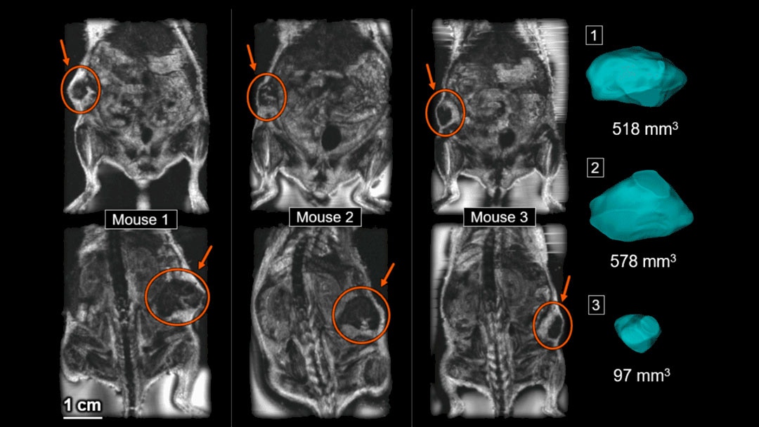
Ultrasound to study glioblastoma
Vega ultrasound as a complementary imaging tool to MRI for assessing glioblastoma in mice.
What our clients say about us

Vega accelerates drug discovery with rapid imaging and user-friendly design
The Vega is now an indispensable part of our preclinical ultrasound imaging core lab. The Vega's emphasis on rapid image acquisition allows us to conduct larger in vivo ultrasound studies to support our drug discovery pipeline, and its intuitive interface has enabled our collaborators to collect and analyze their own data.
Justin Elstrott, Ph.D.
Principal Scientist of Technology, Department of Biomedical Imaging, Genentech Inc.
Vega 3D ultrasound accelerates oncology research with high-throughput analysis
When we needed to expand our pre-clinical imaging modalities to support our numerous oncology researchers, the Vega 3D ultrasound system was at the top of our list. Getting rapid and accurate tumor volumes is critical for our work and the Vega provides a high throughput method to acquire these results from unlabeled samples without the need for a dedicated technician.
James Hayden
Managing Director, Imaging Shared Resource, Ellen and Ronald Caplan Cancer Center, The Wistar Institute
Image gallery
Widefield ultraound
Tumor volume
Evaluate liver stiffness using shear wave elastography
Tumor vasculature
Acoustic angiography
×
FAQs
-
What is the throughput on the Vega system?
The Vega system incorporates 3 mouse bays for hands free sequential imaging.
-
How fast can the Vega do whole body scans?
In B-mode, the Vega can perform a whole-body scan in less than one minute.
-
Do I need to use ultrasound gel to image mice on the Vega?
There is an option to use ultrasound gel or water as a coupling agent. Water is the most convenient due to ease of cleanup.
-
Does the Vega come with a heated mouse bed?
Yes. Each of the 3 bays is heated to 37°C, ensuring mice are kept at the optimum physiologic temperature.
-
Can I image vasculature?
Yes. The Vega has 2 modes, non-linear contrast (NLC) to visualize blood flow and perfusion and acoustic angiography for high resolution vascular imaging. Both modes use VesselVue™ microbubble contrast agents.
-
What is shear wave elastography (SWE)?
SWE is used to measure tissue stiffness which can be an indicator of disease progression. The speed of the shear waves can be directly correlated to Young's modulus, the engineering unit of stiffness.
-
Why is tissue stiffness measurement important?
As an example, liver stiffness can be an indicator of MASH/NASH, fibrosis, or cancer.
-
How can I see a demo on the Vega system?
Contact us for a virtual or onsite demo.
Product information
Overview
Designed with the researcher in mind, the Vega removes the challenges associated with traditional hand-held ultrasound, and uses a bottom-up imaging approach through the use of automated hands-free transducers located under the imaging stage. This unique design requires minimal training with no dedicated sonographer needed, enables high-throughput imaging, and produces more consistent results than conventional hand-held ultrasound systems.
This powerful ultrasound system gives you:
- Hands-free - Automated transducer positioning and movement
- Easy-to-use requiring minimal training
- High-speed, high-throughput performance with 3 mice scanning in just a few minutes
- 3D widefield acquisitions enabling whole subject imaging
- Standard B-Mode and M-Mode capability
- Shear Wave Elastography (SWE) mode for quantifying tissue stiffness
- Acoustic Angiography (AA) mode for visualization of microvasculature
- Flexible visualization and analysis software
- Fits on the benchtop
Specifications
| Dimensions | 54.0 cm (W) x 54.0 cm (D) x 61.0 cm (H) |
|---|---|
| Weight |
45.0 kg
|
| Brand |
Vega
|
|---|---|
| Imaging Modality |
Ultrasound
|
| Unit Size |
1 unit
|
Citations
Resources
Are you looking for resources, click on the resource type to explore further.
Loading...


How can we help you?
We are here to answer your questions.

