


JP
Revvity Sites Globally
Select your location.
*e-commerce not available for this region.

Quantum GX3 microCT System
This product replaces the Quantum™ GX2 (part # CLS149276)
The Quantum GX3 - advanced microCT system for ex vivo and in vivo imaging. The versatile Quantum GX3 microCT offers the flexibility researchers need by delivering the functionality to perform longitudinal studies, across a wide range of species, with fast, low-dose scanning and high spatial resolution.
For research use only. Not for use in diagnostic procedures.
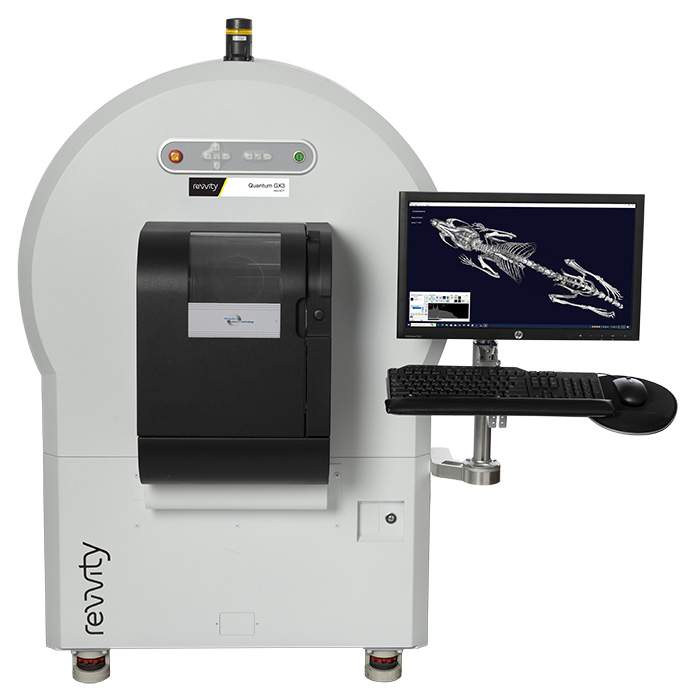

Quantum GX3 microCT System
Quantum GX3 microCT imaging system
Loading...
Product information
Overview
The Quantum GX3 system represents the latest advances in microCT imaging with high image resolution, high speed, low-dose, and flexibility.
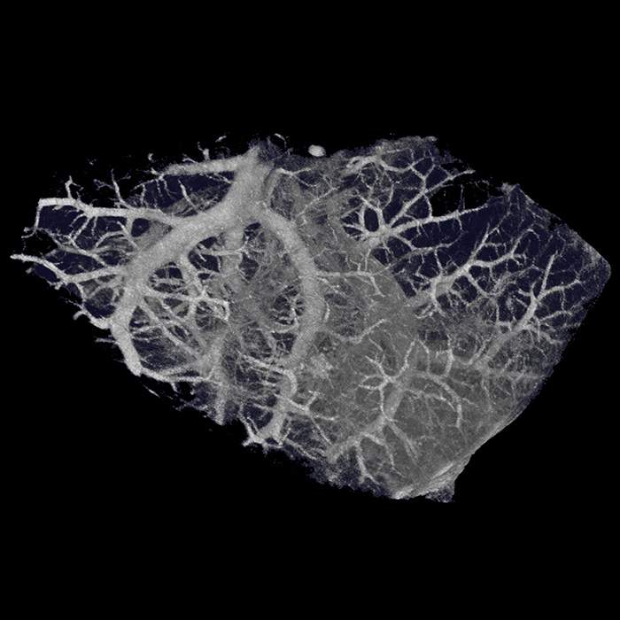
With the combination of higher resolution, increased field of view (FOV) range, and enhanced image-based respiratory and cardiac gating, the Quantum GX3 low-dose microCT system enables researchers to gain a better understanding of healthy and diseased tissue in a broad range of areas including bone, respiratory, cardiovascular, liver/kidney, brain, and oncology research.
Additional product information
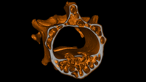
Superior Spatial Resolution
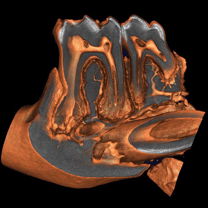
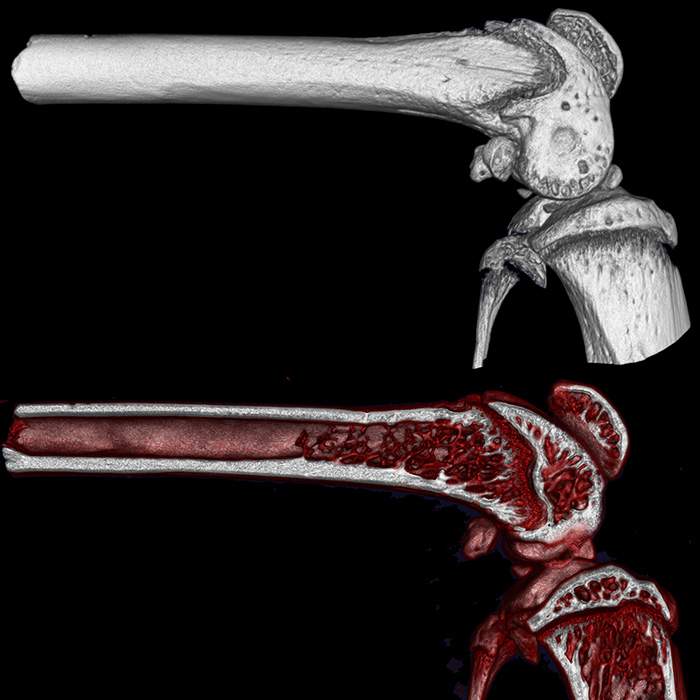
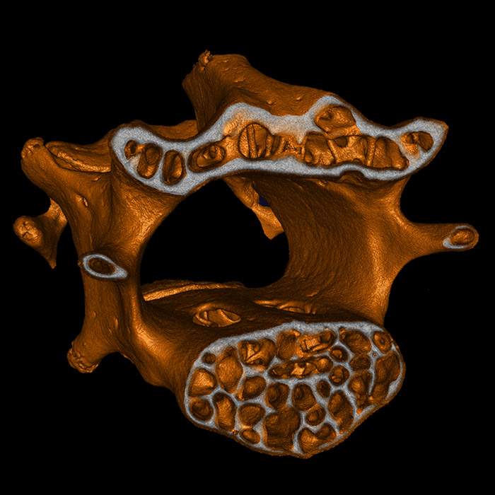
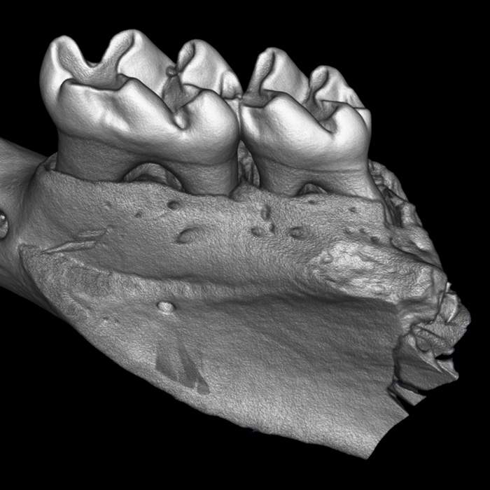
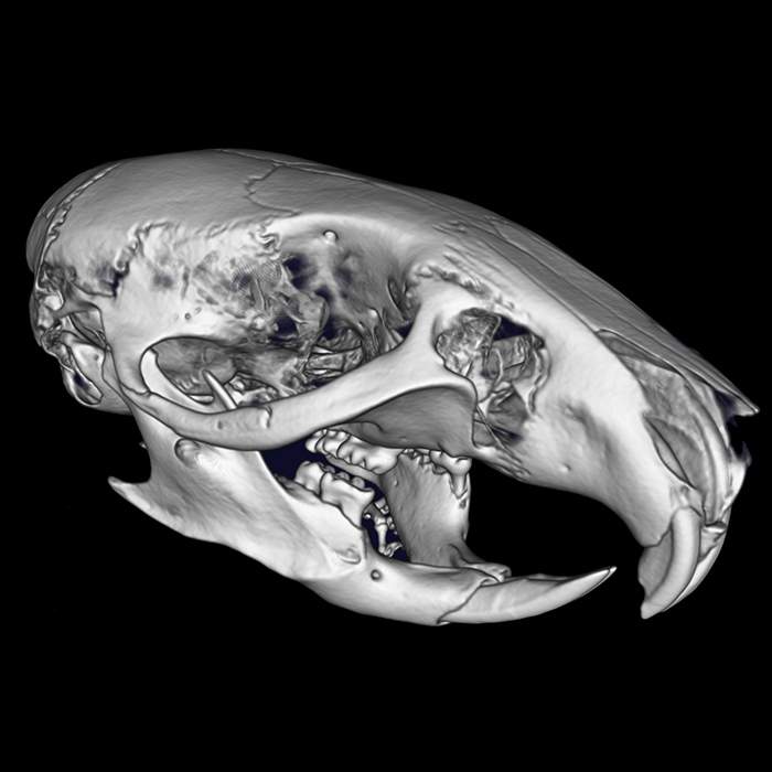
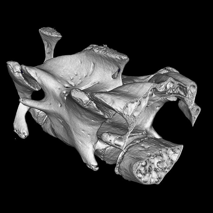
The Quantum GX3 microCT system offers exceptionally high spatial resolution down to 5 µm and pixel size of 2.86 µm (at 8 mm field of view) enabling high image quality of ex vivo biological samples, small bones and fine anatomical structures in vivo in greater detail.
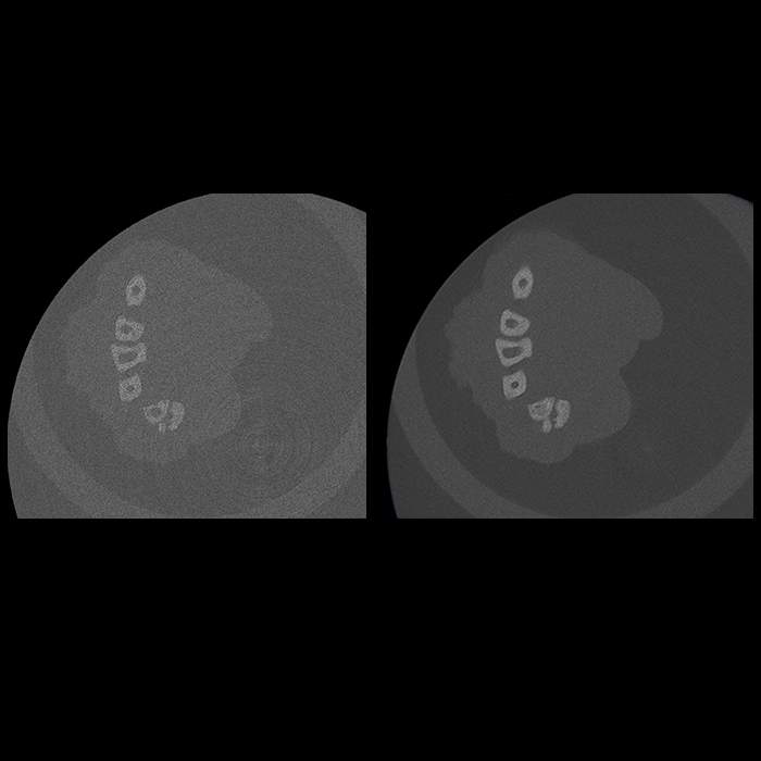
Wide FOV Range
With a wide range of field of view (FOV), the Quantum GX3 gives you the flexibility needed to choose the right balance of resolution and pixel size depending on the size of the specimen or part of the animal needing to be imaged.
The variable FOV ranging from 8 mm for imaging ex vivo biological samples, to the largest FOV of 86 mm, ideal for imaging lungs of larger animals in a single scan, the Quantum GX3 is the most versatile microCT on the market.


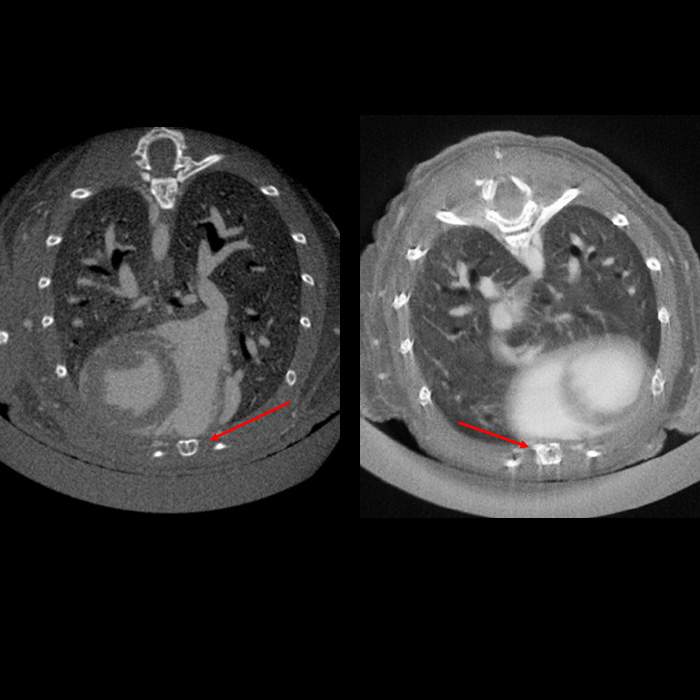
Improved Image-based Gating
Accurate microCT reconstructions often require reducing motion artifacts due to cardiac and respiratory motion. Using a retrospective two-phase image-based gating technique, the Quantum GX3 enables superior cardiac and lung function measurements for a range of species, including mice, rats, and ferrets.
Two Scanning Modes
With the Quantum GX3, researchers have the option of selecting either continuous scanning mode for high throughput fast image acquisition or step scanning mode for higher resolution imaging.


Easy Co-registration
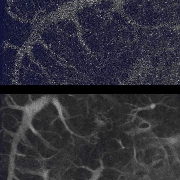
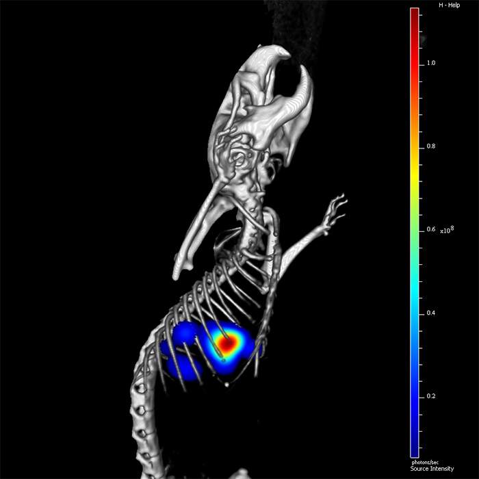
Seamless co-registration of data from the Quantum GX3 can be easily combined with readouts from Revvity’s IVIS® 3D optical system providing more comprehensive insights into the molecular and anatomical features of diseased and healthy biology in the same subject.
Analysis at Your Fingertips
With the convenience of the touchscreen monitor, data capture and analysis on the Quantum GX3 has never been easier. Reconstructions are automatically created within seconds of scanning and basic analysis tools allow you to visualize tissue densities, make quantitative measurements, and export for further analysis in DICOM format. Compliment your microCT data and take your analysis to the next level with Analyze 14.0 software for advanced imaging visualization and measurements.

| Features | Benefits |
|---|---|
| CMOS flat panel detector, 14-bit | Delivers robust, reliable, and high-quality images |
| X-ray source (20-100 kV, 200 uA, 20 W) | Increased x-ray penetration for imaging dense tissue resulting in better signal-to-noise and superior images |
| 5 Field of Views (8, 18, 36, 72, 86 mm) | Imaging of a wide range of animals and specimens |
| Spatial Resolution (5 µm | 2.86 µm smallest pixel size) | Superior resolution for visualizing anatomical features in greater detail |
| Scan times (3.9 sec – fastest) |
|
| Reconstruction times (6 sec – fastest) | Increases throughput accelerating research & development |
| 6 changeable filters with automatic sensing | Software verification of filter selection to ensure correct filter is being used for the imaging study |
| Respiratory and cardiac gating | TestImage-based retrospective cardiac and respiratory gating in mice, rats, and ferrets |
| Continuous and step scanning modes | Option of continuous mode for faster acquisition or step mode for higher resolution imaging |
| Active ring reduction | Automatically removes ring artifacts for increased image quality |
| Touchscreen monitor | Easy to use and interactive |
| Co-registration | Easily combine data from the Quantum GX3 with IVIS® 3D optical data to obtain both functional and anatomical readouts from the same animal |
Specifications
| Dimensions | 980.0 mm (W) x 1536.0 mm (H) |
|---|---|
| Weight |
530.0 kg
|
| Brand |
Quantum
|
|---|---|
| Imaging Modality |
microCT
|
| Unit Size |
1 unit
|
Image gallery
Quantum GX3 microCT System
Citations
Resources
Are you looking for resources, click on the resource type to explore further.
Technical Brief
3D optical and microCT data – The power of multimodality imaging
3D optical and microCT data – The power of multimodality imaging
Case Study
A novel non-Invasive in vivo tool for the assessment of NASH
Non-alcoholic fatty liver disease (NAFLD) describes a progressive pathology that affects the liver. Fat accumulation causes fatty...
Technical Note
Analysis of bone loss in two osteoporosis mouse models using high resolution microCT imaging
Technical Brief
Assessing osseointegration using the Quantum GX3 microCT system
Learn how the Quantum GX3 micro CT system enhances orthopedic and biomaterial studies by delivering high resolution, quantitative...
Brochure
Biologics workflow solutions
Precision biologics are playing an increasingly powerful role as part of therapeutic strategies such as monoclonal antibodies...
Loading...


How can we help you?
We are here to answer your questions.

