JP
Revvity Sites Globally
Select your location.
*e-commerce not available for this region.


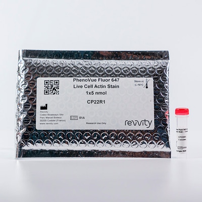
PhenoVue Fluor 647 Live Cell Actin Stain - 1x5nmol

 View All
PhenoVue Fluor 647 Live Cell Actin Stain - 1x5nmol
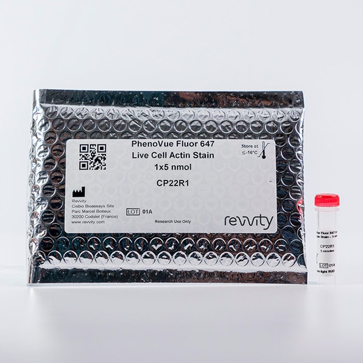
PhenoVue Fluor 647 Live Cell Actin Stain - 1x5nmol
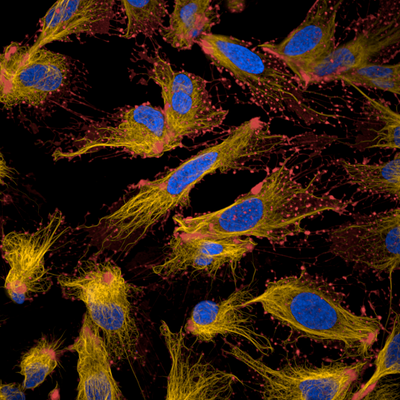
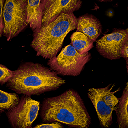
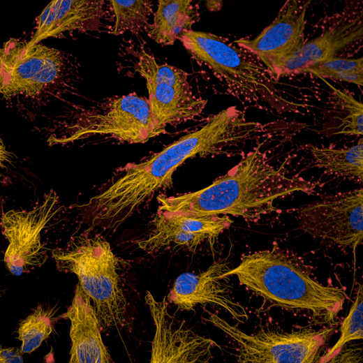
PhenoVue™ Fluor 647 live cell actin stain is a no wash, cell permeable fluorogenic dye which specifically bind to actin filaments and is part of Revvity’s portfolio of cellular imaging reagents.
PhenoVue Fluor 647 live cell actin stain can be used to visualize actin cytoskeleton in live cells, emiting a far-red fluorescence, and is validated for use in imaging microscopy and high-content screening applications. It exhibits a maximum excitation wavelength at 650 nm and a maximum emission wavelength of 670 nm.
View our extensive validation data in the Product Information Sheet within the Resources tab below.
| Feature | Specification |
|---|---|
| Color | Red |
| Filter | Cy5 |
| Fluorophore | PhenoVue™ Fluor 647 |
| Organelle and Cell Compartment | Actin |
PhenoVue™ Fluor 647 live cell actin stain is a no wash, cell permeable fluorogenic dye which specifically bind to actin filaments and is part of Revvity’s portfolio of cellular imaging reagents.
PhenoVue Fluor 647 live cell actin stain can be used to visualize actin cytoskeleton in live cells, emiting a far-red fluorescence, and is validated for use in imaging microscopy and high-content screening applications. It exhibits a maximum excitation wavelength at 650 nm and a maximum emission wavelength of 670 nm.
View our extensive validation data in the Product Information Sheet within the Resources tab below.
For research use only. Not for use in diagnostic procedures.
PhenoVue Fluor 647 Live Cell Actin Stain - 1x5nmol
PhenoVue Fluor 647 Live Cell Actin Stain - 1x5nmol
Loading...
Product information
Overview
PhenoVue™ Fluor 647 live cell actin stain is a no wash, cell permeable fluorogenic dye which specifically bind to actin filaments (F-actin) in live cells. Sensitive, rapid and photostable, PhenoVue Fluor 647 live cell actin stain exhibits far-red emission and can be multiplexed with blue, green and orange colors such as PhenoVue nuclear, lysosomal, mitochondrial or tubulin stains.
Like other actin stains derived from jasplakinolide, cytotoxicity can be observed with long exposure time (>24h) which can be significantly limited at concentrations comprised between 30 and 300nM while maintaining high brightness and image quality.
Depending on the cellular model, intracellular retention of PhenoVue Fluor 647 live cell actin stain can be further improved in the presence of efflux pump inhibitor such as PhenoVue Probenecid, Ready to Use Solution.
Typical working concentration: 100 nM
Equivalent number of microplates:
- 1 to 5 x 96-well microplates
- 1 to 5 x 384-well microplates
- 2 to 8 x 1536-well microplates
Specifications
| Color |
Red
|
|---|---|
| Form |
Solution in DMSO
|
| Maximum Emission Wavelength (Emmax) |
670 nm
|
| Maximum Excitation Wavelength (Exmax) |
650 nm
|
| Application |
High Content Imaging
Microscopy
|
|---|---|
| Brand |
PhenoVue™
|
| Detection Modality |
Fluorescence
|
| Filter |
Cy5
|
| Fluorophore |
PhenoVue™ Fluor 647
|
| Organelle and Cell Compartment |
Actin
|
| Quantity |
1 x 5 nmol
|
| Sample Type |
Live and fixed samples
|
| Shipping Conditions |
Shipped in Dry Ice
|
| Storage Conditions |
-16 °C or below, protected from light
|
| Type |
Individual reagent
|
Image gallery
PhenoVue Fluor 647 Live Cell Actin Stain - 1x5nmol
Spectra viewer
Resources
Are you looking for resources, click on the resource type to explore further.
Flyer
PhenoVue Cellular Imaging Reagents Flyer
This flyer describes Revvity's PhenoVue cellular imaging reagents.
Product Info
PhenoVue Fluor Live Cell Actin Stain
This is a product information sheet for PhenoVue Fluor live cell actin stain. View validation data, product information, protocols...
Flyer
PhenoVue reagents & imaging microplates product list
PhenoVue reagents & imaging microplates product list
Loading...


How can we help you?
We are here to answer your questions.

