JP
Revvity Sites Globally
Select your location.
*e-commerce not available for this region.


PhenoVue Fluor 594 - WGA

 View All
PhenoVue Fluor 594 - WGA
PhenoVue Fluor 594 - WGA
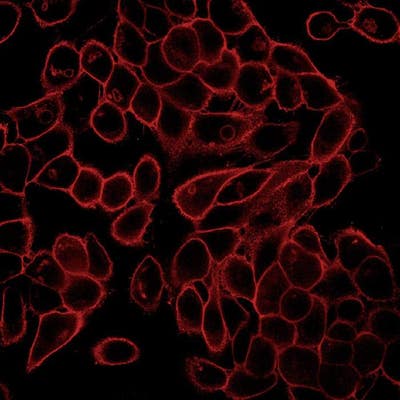
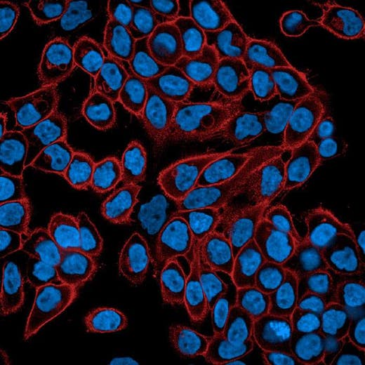
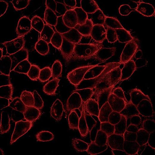
PhenoVue Fluor 594 - WGA is a fluorescent lectin which displays high affinity for sialic acid and N-acetylglucosamine residues of glycoproteins and glycolipids present at the cellular plasma membranes. It can be used for cellular membrane staining, particularly the Golgi apparatus.
PhenoVue Fluor 594 - WGA exhibits bright green fluorescence and is validated for use in imaging microscopy and high-content screening applications.
Part of Revvity's portfolio of cellular imaging reagents, PhenoVue Fluor 594 - WGA has a maximum excitation wavelength of 590 nm and a maximum emission wavelength of 617 nm.
View our extensive validation data in the Product Information Sheet within the Resources tab below.
| Feature | Specification |
|---|---|
| Color | Red/Orange |
| Filter | Texas-Red |
| Fluorophore | PhenoVue™ Fluor 594 |
| Organelle and Cell Compartment | ゴルジ体 |
PhenoVue Fluor 594 - WGA is a fluorescent lectin which displays high affinity for sialic acid and N-acetylglucosamine residues of glycoproteins and glycolipids present at the cellular plasma membranes. It can be used for cellular membrane staining, particularly the Golgi apparatus.
PhenoVue Fluor 594 - WGA exhibits bright green fluorescence and is validated for use in imaging microscopy and high-content screening applications.
Part of Revvity's portfolio of cellular imaging reagents, PhenoVue Fluor 594 - WGA has a maximum excitation wavelength of 590 nm and a maximum emission wavelength of 617 nm.
View our extensive validation data in the Product Information Sheet within the Resources tab below.
Product variant
Quantity: 5 x 1 mg
Part #:
CP15941
For research use only. Not for use in diagnostic procedures.
PhenoVue Fluor 594 - WGA
PhenoVue Fluor 594 - WGA
Loading...
Product information
Overview
WGA is a plant lectin that binds different carbohydrate motifs and displays high affinity for sialic acid and N-acetylglucosamine residues of glycoproteins and glycolipids present at the cellular plasma membranes.
Fluorescent WGA derivatives are commonly used for staining the cellular membranes of mammalian cells, particularly Golgi apparatus which is glycoprotein-enriched. In addition, fluorescent WGA derivatives are also routinely used for the staining of skeletal muscle cells and tissues, as well as in cardiac fibrosis.
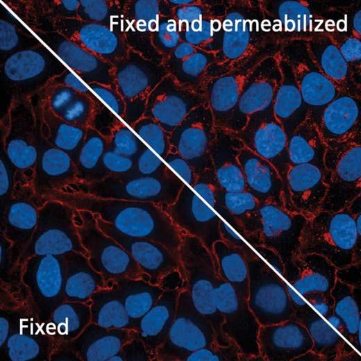
PhenoVue Fluor 594 - WGA can be used to visualize cellular membranes in immunofluorescence, immunohistochemistry and flow cytometry, as well as high-content analysis and screening applications.
See product information sheet for more information on fixed-cell staining.
Recommended working concentration: 5 µg/ml (146 nM)
Equivalent number of microplates:
- 36 - 100 x 96-well microplates
- 30 - 100 x 384-well microplates
- 55 - 160 x 1536-well microplates
Specifications
| Color |
Red/Orange
|
|---|---|
| Form |
Lyophilized
|
| Maximum Emission Wavelength (Emmax) |
617 nm
|
| Maximum Excitation Wavelength (Exmax) |
590 nm
|
| Application |
High Content Imaging
Microscopy
|
|---|---|
| Brand |
PhenoVue™
|
| Detection Modality |
Fluorescence
|
| Filter |
Texas-Red
|
| Fluorophore |
PhenoVue™ Fluor 594
|
| Organelle and Cell Compartment |
Golgi Apparatus
|
| Quantity |
5 x 1 mg
|
| Sample Type |
Live and fixed samples
|
| Shipping Conditions |
Shipped Ambient
|
| Storage Conditions |
2-8 °C, protected from light
|
| Type |
Individual reagent
|
Image gallery
PhenoVue Fluor 594 - WGA
Spectra viewer
Resources
Are you looking for resources, click on the resource type to explore further.
Loading...


How can we help you?
We are here to answer your questions.

