JP
Revvity Sites Globally
Select your location.
*e-commerce not available for this region.


PhenoVue Fluor 400LS - Concanavalin A
 View All
PhenoVue Fluor 400LS - Concanavalin A


PhenoVue Fluor 400LS - Concanavalin A
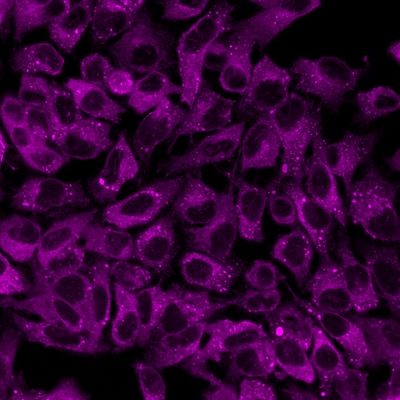
PhenoVue Fluor 400LS - Concanavalin A is a long Stokes shift fluorescent dye conjugated lectin which displays high affinity for glycoproteins and glycolipids present at the cellular membranes. It can be used for cellular membrane staining, particularly the endoplasmic reticulum.
Part of Revvity's portfolio of cellular imaging reagents, PhenoVue Fluor 400LS - Concanavalin A exhibits bright orange fluorescence and can be easily multiplexed for imaging and high-content screening applications. It has been validated on Revvity's HCS instruments such as Operetta CLS and Opera Phenix Plus, and has a maximum excitation wavelength of 395 nm and a maximum emission wavelength of 585 nm.
View our extensive validation data in the Product Information Sheet within the Resources tab below.
| Feature | Specification |
|---|---|
| Color | Orange |
| Filter | Ex: 375-440 nm, Em: 550-650 nm |
| Fluorophore | PhenoVue™ Fluor 400LS |
| Organelle and Cell Compartment | 小胞体 |
PhenoVue Fluor 400LS - Concanavalin A is a long Stokes shift fluorescent dye conjugated lectin which displays high affinity for glycoproteins and glycolipids present at the cellular membranes. It can be used for cellular membrane staining, particularly the endoplasmic reticulum.
Part of Revvity's portfolio of cellular imaging reagents, PhenoVue Fluor 400LS - Concanavalin A exhibits bright orange fluorescence and can be easily multiplexed for imaging and high-content screening applications. It has been validated on Revvity's HCS instruments such as Operetta CLS and Opera Phenix Plus, and has a maximum excitation wavelength of 395 nm and a maximum emission wavelength of 585 nm.
View our extensive validation data in the Product Information Sheet within the Resources tab below.
Product variant
Quantity: 2 x 1 mg
Part #:
CP94001
For research use only. Not for use in diagnostic procedures.
PhenoVue Fluor 400LS - Concanavalin A
PhenoVue Fluor 400LS - Concanavalin A
Loading...
Product information
Overview
Concanavalin A is a plant homotetrameric lectin known to activate the immune system or induce apoptosis and autophagy. Concanavalin A displays high affinity for α-mannopyranosyl and α-glucopyranosyl residues of glycoproteins and glycolipids present at the cellular membranes.
Fluorescent Concanavalin A derivatives are commonly used for staining the cellular membranes of mammalian cells, particularly the endoplasmic reticulum.
PhenoVue Fluor 400LS - Concanavalin A can be used to visualize cellular membranes in immunofluorescence as well as high-content analysis and screening applications.
Its unique photophysical property enables extended multiplexing capability by adding a fifth color while ensuring no spectral overlap, high brightness as well as photostability.
Quantity or Volume Per Vial: 1mg (9.62 nmoles)
Recommended Working Concentration: 50 µg/mL
Equivalent Number of Microplates:
- 1 - 4 x 96-well microplates
- 1 - 4 x 384-well microplates
- 2 - 6 x 1536-well microplates
Specifications
| Color |
Orange
|
|---|---|
| Form |
Lyophilized
|
| Maximum Emission Wavelength (Emmax) |
585 nm
|
| Maximum Excitation Wavelength (Exmax) |
395 nm
|
| Application |
High Content Imaging
Microscopy
|
|---|---|
| Brand |
PhenoVue™
|
| Detection Modality |
Fluorescence
|
| Filter |
Ex: 375-440 nm, Em: 550-650 nm
|
| Fluorophore |
PhenoVue™ Fluor 400LS
|
| Organelle and Cell Compartment |
Endoplasmic Reticulum
|
| Quantity |
2 x 1 mg
|
| Sample Type |
Live and fixed samples
|
| Shipping Conditions |
Shipped Ambient
|
| Storage Conditions |
2-8 °C, protected from light
|
| Type |
Individual reagent
|
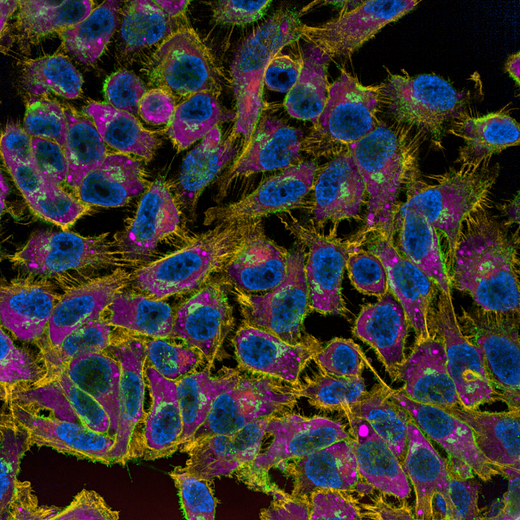
Image gallery
PhenoVue Fluor 400LS - Concanavalin A
Spectra viewer
Resources
Are you looking for resources, click on the resource type to explore further.
Technical Note
Expanding imaging multiplexing capabilities with PhenoVue Fluor 400LS - Phalloidin
This technical note offers tips and best practices for using PhenoVue® Fluor 400LS Phalloidin stain to optimize fluorescent signal...
Flyer
PhenoVue Cellular Imaging Reagents Flyer
This flyer describes Revvity's PhenoVue cellular imaging reagents.
Product Info
PhenoVue Fluor - Concanavalin A Conjugates Product Information Sheet
Fluorescent Concanavalin A conjugates represent a method of choice for labelling the cellular membranes of mammalian cells...
Flyer
PhenoVue reagents & imaging microplates product list
PhenoVue reagents & imaging microplates product list
Loading...


How can we help you?
We are here to answer your questions.

