JP
Revvity Sites Globally
Select your location.
*e-commerce not available for this region.


PhenoVue 641 Mitochondrial Stain

 View All
PhenoVue 641 Mitochondrial Stain


PhenoVue 641 Mitochondrial Stain
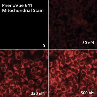
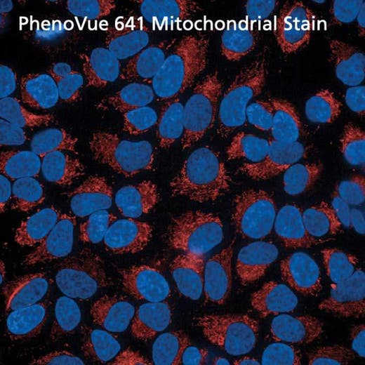
PhenoVue 641 Mitochondrial stain is a fluorescent dye which accumulates in heathly mitochondria of live cells.
PhenoVue 641 Mitochondria stain exhibits bright red fluorescence and is validated for use in imaging microscopy and high-content screening applications.
Part of Revvity's portfolio of cellular imaging reagents, PhenoVue 578 Mitochondrial stain has a maximum excitation wavelength of 641 nm and a maximum emission wavelength of 662 nm, which makes it an alternative to the similar MitoTracker™ Deep Red FM.
View our extensive validation data in the Product Information Sheet within the Resources tab below.
| Feature | Specification |
|---|---|
| Color | Red |
| Filter | Cy5 |
| Organelle and Cell Compartment | ミトコンドリア |
PhenoVue 641 Mitochondrial stain is a fluorescent dye which accumulates in heathly mitochondria of live cells.
PhenoVue 641 Mitochondria stain exhibits bright red fluorescence and is validated for use in imaging microscopy and high-content screening applications.
Part of Revvity's portfolio of cellular imaging reagents, PhenoVue 578 Mitochondrial stain has a maximum excitation wavelength of 641 nm and a maximum emission wavelength of 662 nm, which makes it an alternative to the similar MitoTracker™ Deep Red FM.
View our extensive validation data in the Product Information Sheet within the Resources tab below.
Product variant
Quantity: 20 x 50 µg (92 nmoles)
Part #:
CP3D1
For research use only. Not for use in diagnostic procedures.
PhenoVue 641 Mitochondrial Stain
PhenoVue 641 Mitochondrial Stain
Loading...
Product information
Overview
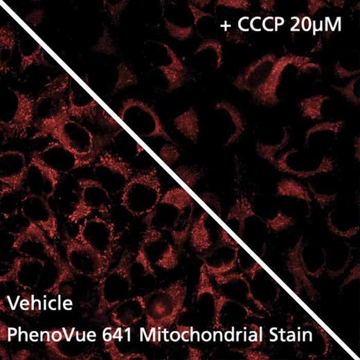
- PhenoVue 641 Mitochondrial stain is a cationic cyanine based structure. With mitochondrial membrane potential, it accumulates in mitochondria through electrostatic interactions and reacts with thiol moieties forming stable thioether bonds. Therefore PhenoVue 641 Mitochondrial stain is well retained after cell fixation.
Fluorescent mitochondrial stains are commonly used for visualizing and quantifying mitochondria whose dysfunctions, including impaired biogenesis, dynamics or trafficking, are a hallmark of some human diseases, such as neurodegenerative disorders.
PhenoVue 641 Mitochondrial stain can be used to detect mitochondria in indirect immunofluorescence, immunohistochemistry and flow cytometry, as well as high-content analysis and screening applications.
Typical working concentration: 100 nM (53 ng/mL)
Equivalent number of microplates:
- 650 - 1850 x 96-well microplates
- 550 - 1950 x 384-well microplates
- 1000 - 3050 x 1536-well microplates
Specifications
| Color |
Red
|
|---|---|
| Form |
Desiccated
|
| Maximum Emission Wavelength (Emmax) |
662 nm
|
| Maximum Excitation Wavelength (Exmax) |
641 nm
|
| Application |
High Content Imaging
Microscopy
|
|---|---|
| Brand |
PhenoVue™
|
| Detection Modality |
Fluorescence
|
| Filter |
Cy5
|
| Organelle and Cell Compartment |
Mitochondria
|
| Quantity |
20 x 50 µg (92 nmoles)
|
| Sample Type |
Live cells with fixation post staining
|
| Shipping Conditions |
Shipped in Dry Ice
|
| Storage Conditions |
-16 °C or below, protected from light
|
| Type |
Individual reagent
|
Image gallery
PhenoVue 641 Mitochondrial Stain
Spectra viewer
Resources
Are you looking for resources, click on the resource type to explore further.
Loading...


How can we help you?
We are here to answer your questions.

