JP
Revvity Sites Globally
Select your location.
*e-commerce not available for this region.


PhenoVue 577 Lysosomal stain
 View All
PhenoVue 577 Lysosomal stain
PhenoVue 577 Lysosomal Stain
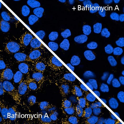
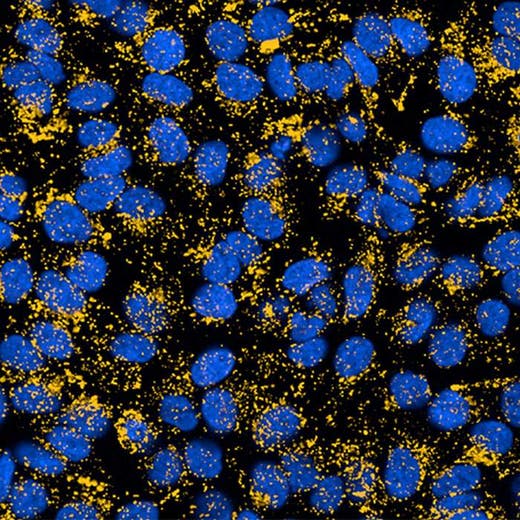
PhenoVue 577 Lysosomal stain is a fluorescent dye which accumulates in acidic vesicles such as the lysosomes. The bright red fluorescence associated with PhenoVue 577 Lysosomal stain are validated for use in imaging microscopy and High Content Screening Systems.
View our extensive validation data in the Product Information Sheet within the Resources tab below.
| Feature | Specification |
|---|---|
| Color | Red/Orange |
| Filter | RFP |
| Organelle and Cell Compartment | リソソーム |
PhenoVue 577 Lysosomal stain is a fluorescent dye which accumulates in acidic vesicles such as the lysosomes. The bright red fluorescence associated with PhenoVue 577 Lysosomal stain are validated for use in imaging microscopy and High Content Screening Systems.
View our extensive validation data in the Product Information Sheet within the Resources tab below.
Product variant
Quantity: 20 x 50 µL
Part #:
CP10R1
For research use only. Not for use in diagnostic procedures.
PhenoVue 577 Lysosomal stain
PhenoVue 577 Lysosomal Stain
Loading...
Product information
Overview
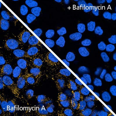
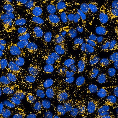
Lysosomes are abundant organelles which contain more than sixty acidic enzymes. Lysosomes play essential role in macromolecules and organelles break down through the autophagy-lysosomal system. Defective regulation of lysosomal function has been reported in several diseases such as lysosomal storage disorders and neurodegenerative diseases like Alzheimer’s and Parkinson’s. PhenoVue 577 lysosomal stain is partially protonated at neutral pH, and highly protonated at acidic pH. In living cells, PhenoVue 577 lysosomal stain accumulates in acidic vesicles such as lysosomes, resulting in bright and specific lysosomal fluorescent staining. PhenoVue 577 lysosomal stain can be used to lysosomes in immunofluorescence, as well as high-content analysis and screening applications.
Quantity per vial: 19.9 µg (50 nmoles)
Typical working concentration: 19.96 ng/mL
Equivalent number of microplates:
- 650 - 2050 x 96-well microplates
- 550 - 2050 x 384-well microplates
- 1050 - 3250 x 1536 microplates
Specifications
| Color |
Red/Orange
|
|---|---|
| Form |
1mM solution in DMSO
|
| Maximum Emission Wavelength (Emmax) |
590 nm
|
| Maximum Excitation Wavelength (Exmax) |
577 nm
|
| Application |
High Content Imaging
Microscopy
|
|---|---|
| Brand |
PhenoVue™
|
| Detection Modality |
Fluorescence
|
| Filter |
RFP
|
| Organelle and Cell Compartment |
Lysosomes
|
| Quantity |
20 x 50 µL
|
| Sample Type |
Live samples only
|
| Shipping Conditions |
Shipped in Dry Ice
|
| Storage Conditions |
-16 °C or below, protected from light
|
| Type |
Individual reagent
|
Image gallery
PhenoVue 577 Lysosomal stain
Spectra viewer
Resources
Are you looking for resources, click on the resource type to explore further.
Flyer
PhenoVue Cellular Imaging Reagents Flyer
This flyer describes Revvity's PhenoVue cellular imaging reagents.
Product Info
PhenoVue Lysosomal Stains Product Information Sheet
In living cells, PhenoVue lysosomal stains accumulate in acidic vesicles such as lysosomes, resulting in bright and specific...
Flyer
PhenoVue reagents & imaging microplates product list
PhenoVue reagents & imaging microplates product list
Loading...


How can we help you?
We are here to answer your questions.

