JP
Revvity Sites Globally
Select your location.
*e-commerce not available for this region.


PhenoVue 488 Lysosomal Stain

PhenoVue 488 Lysosomal Stain
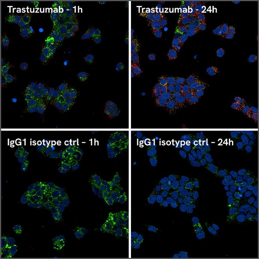
Representative images of BT-474 cells acquired with the Opera Phenix™ Plus high-content screening system. Cells were stained with PhenoVue 488 lysosomal stain (green) to visualize lysosomes and with the PhenoVue Fluor 647 pH-sensitive Fab anti-Human IgG (red) to detect Trastuzumab internalization.
PhenoVue™ 488 lysosomal stain is a fluorescent dye which accumulates in acidic vesicles, such as lysosomes. Bright green fluorescence is associated with the PhenoVue 488 lysosomal stain and it is validated for use in imaging microscopy and high-content screening systems.
Part of Revvity's portfolio of cellular imaging reagents, the PhenoVue 488 lysosomal stain, has a maximum excitation wavelength of 490 nm and a maximum emission wavelength of 519 nm. View our validation data in the Product Information Sheet within the Resources tab below.
| Feature | Specification |
|---|---|
| Color | Green |
| Filter | FITC |
| Fluorophore | PhenoVue™ Fluor 488 |
| Organelle and Cell Compartment | リソソーム |
| Quantity | 1 vial |
PhenoVue™ 488 lysosomal stain is a fluorescent dye which accumulates in acidic vesicles, such as lysosomes. Bright green fluorescence is associated with the PhenoVue 488 lysosomal stain and it is validated for use in imaging microscopy and high-content screening systems.
Part of Revvity's portfolio of cellular imaging reagents, the PhenoVue 488 lysosomal stain, has a maximum excitation wavelength of 490 nm and a maximum emission wavelength of 519 nm. View our validation data in the Product Information Sheet within the Resources tab below.
Product variant
Unit Size: 25 µL (1000x)
Part #:
CP104881
For research use only. Not for use in diagnostic procedures.
PhenoVue 488 Lysosomal Stain
Representative images of BT-474 cells acquired with the Opera Phenix™ Plus high-content screening system. Cells were stained with PhenoVue 488 lysosomal stain (green) to visualize lysosomes and with the PhenoVue Fluor 647 pH-sensitive Fab anti-Human IgG (red) to detect Trastuzumab internalization.
Loading...
Product information
Overview
Lysosomes are abundant organelles which contain more than sixty acidic enzymes. Lysosomes play an essential role in macromolecule and organelle break down through the autophagy-lysosomal system. Defective regulation of lysosomal function has been reported in several diseases, such as lysosomal storage disorders and neurodegenerative diseases, including Alzheimer’s and Parkinson’s. PhenoVue 488 lysosomal stain is a pH-sensitive probe which accumulates in acidic vesicles such as lysosomes, resulting in bright and specific lysosomal fluorescent staining. PhenoVue 488 lysosomal stain can be used to visualize lysosomes in immunofluorescence, as well as high-content analysis and screening applications.
Equivalent number of microplates:
- 0.8-2.5 x 96-well microplates
- 0.7-2.5 x 384-well microplates
- 1.3-4 x 1536-well microplates
Specifications
| Color |
Green
|
|---|---|
| Form |
DMSO solution
|
| Maximum Emission Wavelength (Emmax) |
526 nm
|
| Maximum Excitation Wavelength (Exmax) |
496 nm
|
| Application |
High Content Imaging
Microscopy
|
|---|---|
| Assay Points |
2 x 96-well or 1 x 384-well microplates
|
| Brand |
PhenoVue™
|
| Detection Modality |
Fluorescence
|
| Filter |
FITC
|
| Fluorophore |
PhenoVue™ Fluor 488
|
| Organelle and Cell Compartment |
Lysosomes
|
| Product Group |
Organelle and cell compartment
|
| Quantity |
1 vial
|
| Sample Type |
Live cells only
|
| Shipping Conditions |
Shipped in Dry Ice
|
| Storage Conditions |
-16° C or below, protected from light
|
| Type |
Individual reagent
|
| Unit Size |
25 µL (1000x)
|
Spectra viewer
Resources
Are you looking for resources, click on the resource type to explore further.
Product Info
PhenoVue Lysosomal Stains Product Information Sheet
In living cells, PhenoVue lysosomal stains accumulate in acidic vesicles such as lysosomes, resulting in bright and specific...
Loading...


How can we help you?
We are here to answer your questions.

