


JP
Revvity Sites Globally
Select your location.
*e-commerce not available for this region.

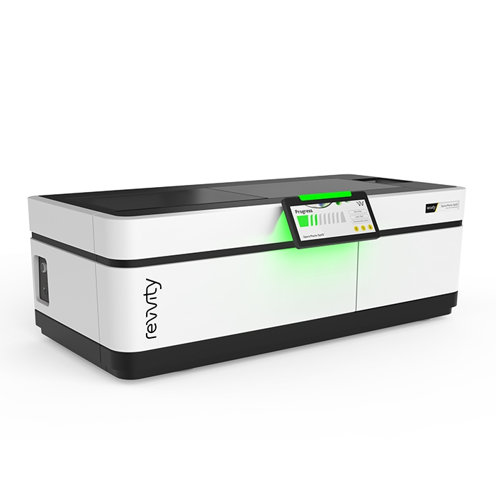
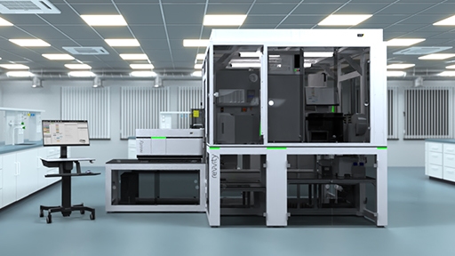
Opera Phenix OptIQ High-Content Imaging System - Single Camera
The single camera Opera Phenix OptIQ™ System is configured for the research and budget of biopharma start-ups and smaller academic labs. A blend of innovation and affordability, this single-camera imager is engineered to help you accelerate your research and discoveries.
What's new:
- >95% QE camera(s)
- Advanced laser-based autofocus
- Hypoxia applications
- 10x high NA lens
- Phenologic.AI classification tools
- Find Organoids building block
For research use only. Not for use in diagnostic procedures.

Opera Phenix OptIQ High-Content Imaging System - Single Camera
Opera Phenix OptIQ High-Content Screening System
Part #:
HH25002001
Imaging Modality:
AI-based phase contrast, 明視野, 共焦点, デジタル位相差, 蛍光
Loading...
Imaging just got smarter with the Opera Phenix OptIQ System
Discover more
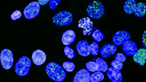
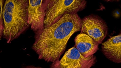
High content imaging provides a greater depth of information than other approaches, allowing you to explore cellular processes in detail.
Quantify in detail
Analyze phenotypic changes at scale, from single cells to entire populations. High-content imaging empowers detailed quantification.
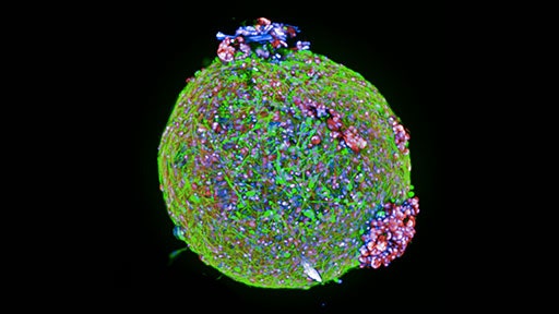
Enhance 3D imaging
Water immersion objectives elevate 3D image quality, revealing intricate details within complex biological structures.
Boost throughput
Add more cameras to increase speed and efficiency. High-content imaging accelerates your research pipeline.
Video overview
Key features
Improved camera
Increase imaging quality by using a high-quantum efficiency camera (>95% QE) for multi-color acquisition, especially for extensive stacks for 3D models.
Microlens-enhanced spinning disk confocality
A pinhole and a microlens disk spinning in tandem for increased light efficiency. Pinhole distances suited for 3D samples, reducing out-of-focus light.
Automated water-immersion objectives
Improve image quality and get better data by enhancing the signal and improving the z resolution while capturing more light.
Machine learning
Easily create algorithms without being an image analysis expert using our Phenologic proprietary machine-learning technology.
Intelligent image acquisition
Image only the objects you are interested in, centered 3D models at high magnification, thereby reducing imaging time and data size.
Powerfully simple analysis
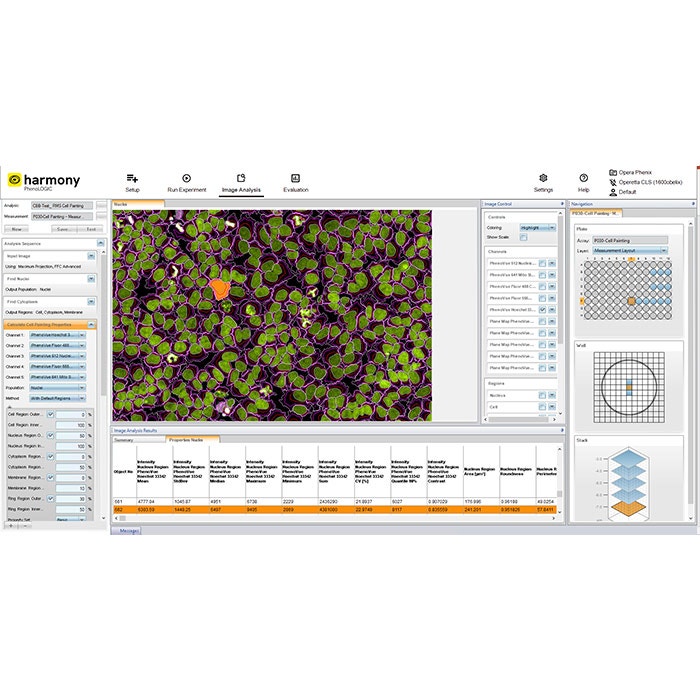
Harmony™ high-content imaging and analysis software can make you more productive faster with ready-made templates and simple steps to a custom analysis.
3D analysis
Explore your cell models by visualizing them in a 3D- and an XYZ-viewer and quantify volumetric and other 3D related phenotypic readouts.
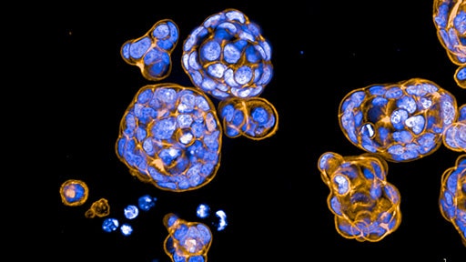
Find Organoids building block
The Find Organoids building block within Harmony software can detect organoids in brightfield stacks using 2D or 3D detection with improved object splitting.
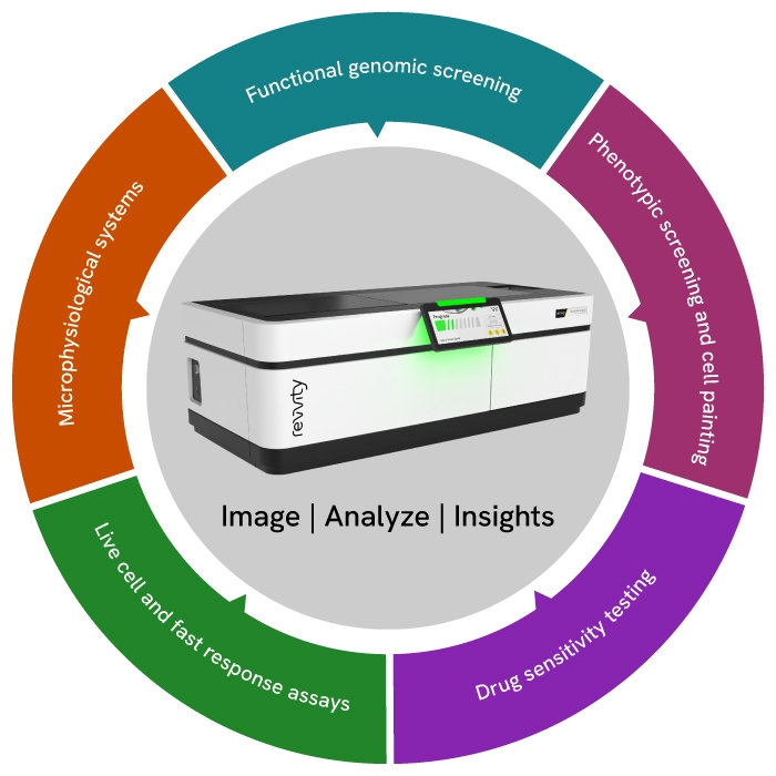
Applications
Microphysiological Systems (MPS)
Deep understanding of 3D cell models with clearer images that reveal critical features and processes.
Functional genomic screening
Create genetic evidence with ease by phenotyping cellular response to thousands of gene manipulations in a single experiment.
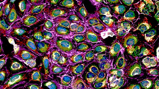
Cell painting
Simplify multiparametric screening with out of the box reagents and analysis to reduce complexity (and create more time for you to focus on the science).
Drug sensitivity testing
Determine rational treatment selections by rapidly characterizing cellular function in response to drug treatment.
Fast response assays
Capture rapid cellular events and dynamic processes with fast frame rates and on-board liquid dispensing.
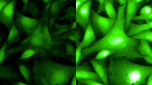
Hypoxic environment applications
Capture cellular images in low oxygen concentrations using the onboard environmental control chamber allowing cells to be imaged in a more realistic physiological condition.
Configuration details
| Single | ||
|---|---|---|
| Lasers | 375/425 nm | Unavailable |
| 405 nm | ✓ | |
| 488 nm | ✓ | |
| 561 nm | ✓ | |
| 640 nm | ✓ | |
| System options | Number of cameras (confocal) | 1 |
| Environmental control | ✓ | |
| Water immersion lenses | ✓ | |
| Transmitted light | ✓ | |
| On-board liquid handling | Optional | |
| Robotics / Automation compatible | ✓ | |
| Imaging modes | Minimal crosstalk imaging | ✓ |
| Ratiometric FRET | Suitable | |
| 3D imaging | ✓ | |
| Multi-color fluorescence imaging | ✓ | |
| Multi-color simultaneous confocal imaging | Unavailable | |
| Brightfield and digital phase contrast | ✓ | |
| Fluorescence widefield imaging | ✓ | |
| Fast frame rate imaging | ✓ |
Image acquisition and analysis simplified with Harmony software
Intuitive workflow
Harmony software offers an intuitive user interface that guides you from image acquisition to analysis and evaluation.
Templates for quick set-up
Templates allow you to set up acquisition channels and parameters efficiently.
Ready-made solutions
Choose from pre-built solutions for common image analysis tasks, simplifying your workflow.
Customizable building blocks
Create, configure, and customize your own high-content analysis applications using image analysis building blocks.
Advanced features
Harmony includes advanced analysis capabilities, such as texture and STAR morphology analysis, providing detailed descriptions of cellular morphology and robust differentiation of phenotypes.
Data management
The software automatically stores analysis results and metadata, including assay layout, instrument settings, and user-defined keywords and annotations.
AI-based image analysis
Using pre-trained deep-learning image-analysis models, Phenologic.AI allows for segmentation and classification of cells in brightfield and fluorescent images for easier analysis of live and fixed cell assays.
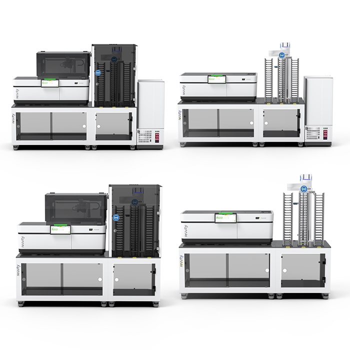
Scale smarter: Automate high-content imaging with the Opera Phenix OptIQ system
Peace-of-mind
plate:: handler™ FLEX automation and imaging options provided and serviced by Revvity, with a single point of contact.
Easy-to-use
Ready-to-use application templates covering major use cases.
Free-up resources, increase utilization and safeguard results
Supports autonomous 24x7 plate processing with plates to be imaged in sequence, in loops or following user-defined schedules Full barcode tracking is included.
Shared equipment use
Users can queue plates for imaging with user-specific methods and adjustable priorities within the software.
Grows with your needs
Seamless upgrade path, with plate::handler™ FLEX sharing the same hardware & software components as our larger full-workflow automation platforms.
Hypoxia options for incubator
Combine your automated set up with a hypoxia compatible incubator to enable applications under hypoxic conditions.
Progress faster with our verified solutions
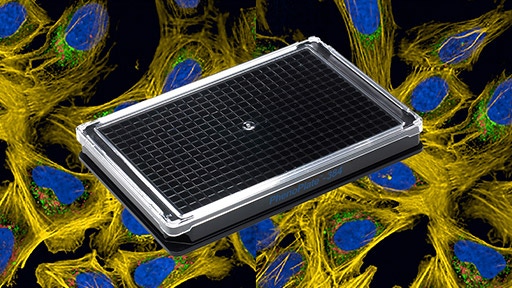
Microplates for high-content imaging
Utilize PhenoPlate™ 384-well microplates designed to support optimal performance in high-content imaging applications. Employ CellCarrier Spheroid ULA plates for imaging 3D models.

PhenoVue cellular imaging reagents
Ready-to-use kits and reagents with straightforward protocols. Extensively tested to provide optimal formulations and long-term stability.
Automation and workstations
Improve throughput, productivity, and reduce variability and reagent costs. Benefit from automated workflows using the explorer™ G3 workstations for cell painting, 3D cell culture, or phenotypic screening.
Efficient data management
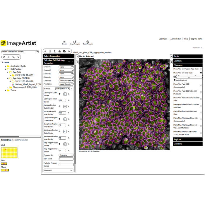
Export results automatically into the Image Artist™ platform that uses high performance computing and an industry standard object store to provide a scalable, multi-user solution for image analysis and management.

Product information
Overview
Opera Phenix OptIQ high-content imager (single camera) for your high-content applications.
Key features:
- Improved Camera
- Microlens-enhanced spinning disk confocality
- Automated water-immersion objectives
- Machine learning
- Intelligent image acquisition
- Powerfully simple analysis
- 3D analysis
- Find Organoids building block
Specifications
| Automation Compatible |
Yes
|
|---|---|
| Brand |
Opera Phenix OptIQ
|
| Imaging Modality |
AI-based phase contrast
Brightfield
Confocal
Digital phase contrast
Fluorescence
|
| Unit Size |
1 unit
|
Citations
Resources
Are you looking for resources, click on the resource type to explore further.
Brochure
Opera Phenix OptIQ high-content screening system brochure
This brochure provides an overview of the Opera Phenix OptIQ high-content screening system's features and benefits.
Flyer
Opera Phenix OptIQ high-content screening system flyer
This flyer provides an overview of the Opera Phenix OptIQ high-content screening system's features and benefits.
Technical Note
Opera Phenix Plus high-content screening system: crosstalk suppression
Simultaneous multi-color imaging is a technology commonly used to increase the speed of high content screening systems. However...
Literature - Publication Review
Organ-on-chips: increased complexity for higher physiological relevance in pharmaceutical safety testing
Organ-on-chip (OOC) technology aims to replicate the complex microenvironment of human organs in vitro, allowing the study of...
eBook
Organoids in action: advanced analysis for enhanced research relevance
Organoids - miniaturized 3D models that mimic key features of human organs - are transforming the way we study biology and disease...
Application Note
Orthogonal validation of CRISPRCas9 and siRNA generated phenotypes using cell painting
Investigate cell cycle regulation through AURKB, GMNN, and PLK1 proteins. Learn how CRISPR-Cas9 and siRNA techniques, combined...
Loading...


How can we help you?
We are here to answer your questions.

