


JP
Revvity Sites Globally
Select your location.
*e-commerce not available for this region.

HTRF Human and Mouse Phospho-SMAD2 (Ser465/467) Detection Kit, 500 Assay Points


HTRF Human and Mouse Phospho-SMAD2 (Ser465/467) Detection Kit, 500 Assay Points


generic HTRF phospho primary image
The Phospho-SMAD2 (Ser465/467) kit enables the cell-based quantitative detection of SMAD2 phosphorylated on Ser465/467, as a readout of the TGFb pathway.
| Feature | Specification |
|---|---|
| Application | 細胞シグナル伝達 |
| Sample Volume | 16 µL |
The Phospho-SMAD2 (Ser465/467) kit enables the cell-based quantitative detection of SMAD2 phosphorylated on Ser465/467, as a readout of the TGFb pathway.
Product variants
Unit Size: 500 assay points
Part #:
64SMAD2S5PEG
Unit Size: 10,000 assay points
Part #:
64SMAD2S5PEH
For research use only. Not for use in diagnostic procedures. All products to be used in accordance with applicable laws and regulations including without limitation, consumption, and disposal requirements under European REACH regulations (EC 1907/2006).
HTRF Human and Mouse Phospho-SMAD2 (Ser465/467) Detection Kit, 500 Assay Points
generic HTRF phospho primary image
Loading...
Product information
Overview
This HTRF cell-based assay enables the rapid, quantitative detection of SMAD2 phosphorylated at Serine 465/467, as a readout of TGF-ß signaling activity.
TGF-ß receptors directly activate SMAD2 by phosphorylation at Ser465/467, causing it to translocate to the nucleus and regulate gene expression involved in apoptosis, migration, and differentiation, as well as in immune/inflammatory responses and extracellular matrix remodeling.
HTRF assays offer many advantages over other technologies:
- Homogeneous add-and-read format
- No wash steps
- Low background
- Straightforward miniaturization from 96- or 384-well microplates to high density assay formats such as 384-well low volume and 1536-well plates
- Stable signal, providing flexibility in time of readout or size of assays
How it works
Phospho-SMAD2 (Ser465/467) assay principle
The Phospho-SMAD2 (Ser465/4267) assay measures SMAD2 when phosphorylated at Ser465/4267. Unlike Western Blot, the assay is entirely plate-based and does not require gels, electrophoresis, or transfer.
The Phospho-SMAD2 (Ser465/4267) assay uses 2 labeled antibodies: one with a donor fluorophore, the other with an acceptor. The first antibody is selected for its specific binding to the phosphorylated motif on the protein, and the second for its ability to recognize the protein independently of its phosphorylation state. Protein phosphorylation enables an immune-complex formation involving both labeled antibodies and which brings the donor fluorophore into close proximity to the acceptor, thereby generating a FRET signal. Its intensity is directly proportional to the concentration of phosphorylated protein present in the sample, and provides a means of assessing the protein’s phosphorylation state under a no-wash assay format.
Phospho-SMAD2 (Ser465/467) 2-plate assay protocol
The 2 plate protocol involves culturing cells in a 96-well plate before lysis, then transferring lysates to a 384-well low volume detection plate before adding Phospho-SMAD2 (Ser465/467) HTRF detection reagents.
This protocol enables the cells' viability and confluence to be monitored.

Phospho-SMAD2 (Ser465/467) 2-plate assay protocol
Detection of Phosphorylated SMAD2 (Ser465/467) with HTRF reagents can be performed in a single plate used for culturing, stimulation, and lysis. No washing steps are required.
This HTS designed protocol enables miniaturization while maintaining robust HTRF quality.

Assay validation
Human TGFβ stimulation on C2C12 cells leads to phosporylation on SMAD2 protein on serine 465/467 residue
C2C12 cells were plated at various cellular densities in a 96-well plate. After an overnight incubation at 37°C, 5% CO2, a serial dilution of human TGFβ was added to the cells for 30 minutes at 37°C, 5% CO2. Stimulation medium was removed, and 50µL of lysis buffer was added to the cells. A lysis step was carried out, shaking gently for 30 minutes. 16µL of samples were transferred into a 384-well small volume plate, then 4µL of Phospho-SMAD2 HTRF detection reagents were added. Signals were recorded overnight.

Phospho-SMAD2 Ser465/467 cellular assay validation on human and mouse cell lines
Hela cells were selected for testing human compatibility, while NIH 3T3 and C2C12 cells were chosen for mouse compatibility. 100,000 cells of these different models were plated in 96-well plates. After an overnight incubation at 37°C, 5% CO2, a serial dilution of human TGFβ was added to the cells for 30 minutes at 37°C, 5% CO2. Stimulation medium was removed, and 50µL of lysis buffer was added to the cells. A lysis step was carried out, shaking gently for 30 minutes. 16µL of samples were transferred into a 384-well small volume plate, then 4µL of Phospho-SMAD2 HTRF detection reagents were added. Signals were recorded overnight.
The Phospho-SMAD2 HTRF assay was able to detect human and mouse versions of this protein under its phosphorylated status on Serine 465/467.

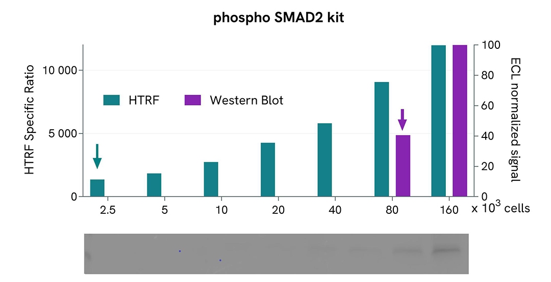
HTRF assay compared to Western Blot using Phospho-SMAD2 cellular assay on mouse C2C12 cells
Mouse C2C12 cells were cultured to 80% confluency. After hTGFβ treatment, cells were lysed and soluble supernatants were collected via centrifugation. Serial dilutions of the cell lysate were performed and 16 µL of each dilution were transferred into a 384-well low volume white microplate before finally adding Phospho-SMAD2 HTRF cellular kit reagents. A side by side comparison showed the HTRF Phospho assay is at least 32-fold more sensitive than the Western Blot.
Simplified pathway
TGF-ß signaling pathway
TGF-ß signaling is mediated by complexes of TßRI and TßRII, which activate intracellular SMAD3 and SMAD2 by phosphorylation. The binding of the TGF-ß ligand on TßRII triggers the recruitment of TßRI into the ligand-receptor complex. TßRII autophosphorylates, then transphosphorylates TßRI. Activated TßRI in turn phosphorylates SMAD2 on Ser465 and Ser467, enabling its oligomerization with SMAD4. This complex then translocates into the nucleus, and acts as a transcription factor with coactivators and corepressors to regulate the expression of multiple genes involved in cell growth, apoptosis, proliferation, migration, and differentiation, as well as in extracellular matrix remodeling and immune/inflammatory responses. Inhibitory SMAD6 and SMAD7 are involved in feedback inhibition of the pathway.

Specifications
| Application |
Cell Signaling
|
|---|---|
| Automation Compatible |
Yes
|
| Brand |
HTRF
|
| Cellular or Signaling Pathway |
Extracellular matrix remodeling / fibrosis signaling
|
| Detection Modality |
HTRF
|
| Lysis Buffer Compatibility |
Lysis Buffer 3
Lysis Buffer 4
Lysis Buffer 5
|
| Molecular Modification |
Phosphorylation
|
| Product Group |
Kit
|
| Sample Volume |
16 µL
|
| Shipping Conditions |
Shipped in Dry Ice
|
| Target |
SMAD2
|
| Target Class |
Phosphoproteins
|
| Target Species |
Human
Mouse
|
| Technology |
TR-FRET
|
| Therapeutic Area |
Cardiovascular
Metabolism/Diabetes
NASH/Fibrosis
Oncology & Inflammation
|
| Unit Size |
500 assay points
|
Video gallery
HTRF Human and Mouse Phospho-SMAD2 (Ser465/467) Detection Kit, 500 Assay Points
Resources
Are you looking for resources, click on the resource type to explore further.
Guide
Get your guide on fibrosis
A comprehensive overview of fibrosis development
Fibrosis is a main contributor to a wide range of organ failures which stems from...
Brochure
HTRF assays and reagents product list
Discover the versatility and precision of Homogeneous Time-Resolved Fluorescence (HTRF) technology. Our HTRF portfolio offers a...
Guide
HTRF solutions, guide to major applications
This guide provides you an overview of HTRF applications in several therapeutic areas.
Loading...


How can we help you?
We are here to answer your questions.

