


JP
Revvity Sites Globally
Select your location.
*e-commerce not available for this region.

HTRF Human Ki-67 Detection Kit, 500 Assay Points

HTRF Human Ki-67 Detection Kit, 500 Assay Points


Generic HTRF biomarker image
The Human Ki-67 HTRF assay kit is a fluorescent assay for quantifying the state of proliferation on cultured cells by quantitation of Ki-67 protein in cell lysates.
| Feature | Specification |
|---|---|
| Application | タンパク質定量 |
| Sample Volume | 4 µL |
The Human Ki-67 HTRF assay kit is a fluorescent assay for quantifying the state of proliferation on cultured cells by quantitation of Ki-67 protein in cell lysates.
Product variants
Unit Size: 500 assay points
Part #:
64KI67PEG
Unit Size: 10,000 assay points
Part #:
64KI67PEH
For research use only. Not for use in diagnostic procedures. All products to be used in accordance with applicable laws and regulations including without limitation, consumption, and disposal requirements under European REACH regulations (EC 1907/2006).
HTRF Human Ki-67 Detection Kit, 500 Assay Points
Generic HTRF biomarker image
Loading...
Product information
Overview
The expression of the proliferation related nuclear antigen Ki-67, also known as Ki67 or MKI67, is a cellular marker for proliferation which is only detected in dividing cells. Ki-67 is a non-histone protein characterized as a nuclear matrix-associated proliferation-related antigen present throughout the cell cycle, but not in resting, quiescent, cells (G0 phase). Ki-67 is widely used in routine pathology as a proliferation marker to measure the growth fraction of cells in human tumors. The Ki-67 protein present in the nucleus of proliferating cells is released upon cell lysis, and the Ki-67 protein extracted in the lysate is quantified using our Ki-67 HTRF assay. The Ki-67 quantitation assay is an alternative to BrdU or EdU incorporation assays, which are exogenous proliferation markers incorporated into the newly synthesized DNA of dividing cells. The scope of Ki-67 is similar to that of the proliferating cell nuclear antigen (PCNA).
How it works
Assay principle
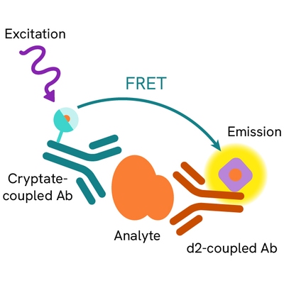
The Ki-67 quantification kit is based on a sandwich format involving two specific antibodies, respectively labelled with Cryptate (donor) and d2 (acceptor). The Ki-67 protein contained in the proliferating cell nucleus and extracted in cell lysates binds to monoclonal anti-Ki67 Eu Cryptate and monoclonal anti-Ki67 d2 conjugates. When the dyes are in close proximity, the excitation of the donor with a light source (laser or flash lamp) triggers a Fluorescence Resonance Energy Transfer (FRET) towards the acceptor, which in turn fluoresces at a specific wavelength (665 nm). The specific signal modulates positively in proportion to Ki-67 concentration.
Assay protocol
The assay protocol, using a 384-well small volume white plate or a Cisbio low volume 96-well plate (20 µL final), is described on the right. 4 µL of sample (lysate) + 12 µL of lysis buffer, or 16 µL of standard are dispensed directly into the plate for detection by HTRF reagents. The antibodies labelled with HTRF donor and acceptor can be pre-mixed and added in a single dispensing step to further streamline the assay procedure. The assay can be run in 96- to 384-well plates by simply resizing each addition volume proportionally.

Assay details
| Standard curve | 2.05 - 500 pg/mL |
| Limit of detection (LoD) | 0.41 pg/mL |
| Assay range (LoD - IC90) | 1.63 - 500 pg/mL |
| Specificity | Human Ki-67 |

Assay validation
Ki-67 protein expression on starved cultured cells upon stimulation/re-feeding with 24h 10% FCS
A serum starvation and refeeding process was used to imitate the cell cycle using human epithelial A549 cell line derived from a lung carcinoma tissue. First, Ham's F-12 medium without FCS (supplemented with 0.5% BSA, culture grade) was used to incubate A549 cells for 72 h to synchronize them (60K or 120K cells per well in 100µL medium.). The medium was then changed to complete medium in six wells (10% FCS), keeping six wells for starved control.On the first microplate, the medium was removed. Cells were then lysed in 50 µL 1X Lysis buffer and 4 µL lysate were transfered to a 384 well white microplate. 12 µL 1X lysis buffer were added, following the Ki-67 HTRF protocol.The same experiment was run in parallel on the second microplate to compare with a brdU incorporation assay, using a commercial BrdU-ELISA kit. Briefly, BrdU was added (10µL/well) after 2h incubation. Medium was removed and cells were fixed and after blocking (1% BSA 30 min). An anti-BrdU peroxidase conjugate was added (90 min incubation RT), followed by the washing and coloration steps.Ki-67 expression increased in cell lysate upon stimulation with FCS compared to the starved cell control. In parallel, BrdU incorporation increased in FCS stimulated cells. The Ki-67 expression correlates with the BrdU incorporation assay. It should be noted that the assay window was larger in the Ki-67 HTRF assay compared to BrdU assay.


Differential Ki-67 protein expression in confluent and non-confluent A549 tumor cells
A549 cells were cultured in two T175 flasks in Ham's F-12 complete medium (10% FCS), using two seeding concentrations to obtain either confluent cells or non-confluent cells. The adherent cells were washed with PBS, then dissociated (5 mL cell dissociation buffer), scraped with a rubber policeman and transferred to three 1.5 mL microtubes (10.E6 cells/tube) as technical replicates. After centrifugation (5 min, 400 RCF) the cell pellets were lysed with 500 µL of lysis buffer #4 (30 min RT vortexing). Lysates were diluted 20-fold in 1X lysis buffer and 16 µL of diluted lysate were transferred (triplicates) to a 384 small volume white microtiter plate for Ki-67 HTRF assay. The Ki-67 expression is clearly much higher in non-confluent A549 cells compared to confluent-cells.

Differential Ki-67 protein expression in confluent and non-confluent A549 tumor cells
A549 cells were cultured in two T175 flasks in Ham's F-12 complete medium (10% FCS), using two seeding concentrations to obtain either confluent cells or non-confluent cells. The adherent cells were washed with PBS, then dissociated (5 mL cell dissociation buffer), scraped with a rubber policeman and transferred to three 1.5 mL microtubes (106 cells/tube) as technical replicates. After centrifugation (5 min, 400 RCF) the cell pellets were lysed with 500 µL of lysis buffer #4 (30 min RT vortexing). Lysates were diluted 20-fold in 1X lysis buffer and 16 µL of diluted lysate were transferred (triplicates) to a 384 small volume white microtiter plate for Ki-67 HTRF assay.The Ki-67 expression is clearly much higher in non-confluent A549 cells compared to confluent-cells.
Specifications
| Application |
Protein Quantification
|
|---|---|
| Brand |
HTRF
|
| Detection Modality |
HTRF
|
| Product Group |
Kit
|
| Sample Volume |
4 µL
|
| Shipping Conditions |
Shipped in Dry Ice
|
| Target Class |
Biomarkers
|
| Target Species |
Human
|
| Technology |
TR-FRET
|
| Therapeutic Area |
Oncology & Inflammation
|
| Unit Size |
500 assay points
|
Video gallery
HTRF Human Ki-67 Detection Kit, 500 Assay Points
Resources
Are you looking for resources, click on the resource type to explore further.
Brochure
HTRF assays and reagents product list
Discover the versatility and precision of Homogeneous Time-Resolved Fluorescence (HTRF) technology. Our HTRF portfolio offers a...
Guide
HTRF solutions, guide to major applications
This guide provides you an overview of HTRF applications in several therapeutic areas.
Loading...


How can we help you?
We are here to answer your questions.

