


JP
Revvity Sites Globally
Select your location.
*e-commerce not available for this region.

HTRF HMGB1 Detection Kit, 500 Assay Points


HTRF HMGB1 Detection Kit, 500 Assay Points

Generic HTRF biomarker image
Measures the High-Mobility Group Box 1 (HMGB1) protein in cell culture medium.
| Feature | Specification |
|---|---|
| Application | タンパク質定量 |
| Sample Volume | 16 µL |
Measures the High-Mobility Group Box 1 (HMGB1) protein in cell culture medium.
Product variants
Unit Size: 500 assay points
Part #:
62HMGPEG
Unit Size: 10,000 assay points
Part #:
62HMGPEH
For research use only. Not for use in diagnostic procedures. All products to be used in accordance with applicable laws and regulations including without limitation, consumption, and disposal requirements under European REACH regulations (EC 1907/2006).
HTRF HMGB1 Detection Kit, 500 Assay Points
Generic HTRF biomarker image
Loading...
Product information
Overview
In the nucleus, High-mobility group box 1 (HMGB1) interacts with DNA and histones to determine chromatin structure. Once released, HMGB1 acquires a new role as an alarmin or a damage-associated molecular pattern (DAMP) and is capable of initiating a state of inflammation by binding TLR4 and promoting the production of cytokines.
Necrosis, pyroptosis, and apoptosis are mechanisms of regulated cell death that have been shown to lead to the release of different forms of HMGB1. In pyroptosis, HMGB1 is released together with IL-1b and IL-18.
HMGB1 is implicated in the pathology of several auto-immune diseases, including rheumatoid arthritis (RA) and systemic lupus erythematosus (SLE).
HTRF assays offer many advantages over other technologies:
- Homogeneous add-and-read format
- No wash steps
- Low background
- Straightforward miniaturization from 96- or 384-well microplates to high density assay formats such as 384-well low volume and 1536-well plates
- Stable signal, providing flexibility in time of readout or size of assays
How it works
Assay principle
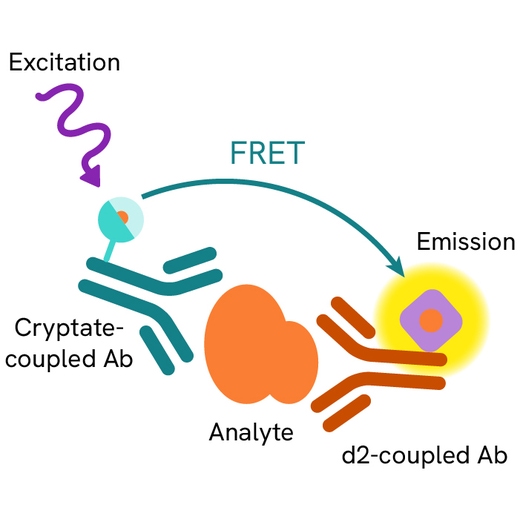
HMGB1 is measured using a sandwich immunoassay involving two specific anti-HMGB1 antibodies, respectively labeled with an HTRF donor and acceptor dyes. The intensity of the signal is directly proportional to the concentration of HMGB1 present in the sample.
Assay principle
The assay protocol, using a 384-well small volume white plate or a Revvity low volume 96-well plate (20 µL final), is described on the right. 16µl of cell supernatant, sample, or standard are dispensed directly into the detection plate for detection by HTRF® reagents. The antibodies labeled with HTRF donor and acceptor can be pre-mixed and added in a single dispensing step, to further streamline the assay procedure. The assay can be run in 96- to 384-well plates by simply resizing each addition volume proportionally.

Assay details
Product specifications
| Sample size | 16 µL |
|---|---|
| Final assay volume | 20 µL |
| Time to results | from 2h to Overnight at RT |
| Kit components | Frozen standard, frozen detection antibodies, buffers & protocol. |
| LOD & LOQ (in Diluent) | 0.2 ng/mL & 0.5 ng/mL |
| Range | 0.78 – 50 ng/mL |
| Species | Human, mouse, and rat |
| Specificity | No cross-reactivity with bovine HMGB1 |
Analytical performance
Precision
Intra assay (n=24)
| Sample | Mean [HMGB1] (ng/mL) | CV |
|---|---|---|
| 1 | 2.4 | 3.90% |
| 2 | 6.6 | 4.30% |
| 3 | 18.9 | 2.80% |
| Mean CV | 4.10% |
Each of the 3 samples was measured 24 times, and % CV was calculated for each sample.
Inter assay (n=4)
| Sample | Mean [HMGB1] (ng/mL) | Mean (delta ratio) | CV |
|---|---|---|---|
| 1 | 1.56 | 298 | 11% |
| 2 | 6.25 | 1468 | 13% |
| 3 | 25 | 7150 | 13% |
| Mean CV | 12.3 |
Each of the 3 samples was measured 24 times, and % CV was calculated for each sample.
Spike and recovery
|
Supernatant ng/mL |
spike ng/mL |
Expected ng/mL |
Measured ng/mL |
|
|
|---|---|---|---|---|---|
|
% Recovery |
|||||
|
4.1 |
10.9 |
15.0 |
13.4 |
89% |
|
|
8.2 |
10.9 |
19.1 |
16.1 |
84% |
|
|
14.4 |
10.9 |
25.3 |
21.3 |
84% |
Assay validation
HMGB1 standard curve
Recombinant HMGB1 provided in the kit (standard) was serially diluted in diluent #5, following the procedure featured in the kit’s package insert. The HTRF signal, expressed as a delta ratio, was plotted as a function of the HMGB1 concentrations tested.

Measuring HMGB1 from necroptosis induced THP-1 cells
To induce necroptosis, THP1 suspension cells were dispensed into a 96-well culture at a density of 400K cells per well in 200 µL of RPMI 1640 supplemented with 10% FCS, 2 mM L-glutamine, and 25 mM HEPES. In selected wells the caspase inhibition was induced by a 1 h treatment with Z-VAD-FMK (25 µM final). After low speed centrifugation, the medium was carefully withdrawn by gentle aspiration and new medium was added. Following caspase inhibition, TNFα (100 ng/mL final) was added either alone or in combination with the cIAP inhibitor BV-6 (5 µM final) to induce necroptosis. After overnight incubation, the supernatants were collected and tested for the presence of HMGB1, following the instructions listed in the kit’s package insert. Results are presented expressed in concentrations of HMGB1.

Measuring HMGB1 from pyroptosis induced THP-1 cells
To induce pyropoptosis, THP1 suspension cells were dispensed into a 96-well culture at a density of either 200K or 400K cells per well in 200 µL of RPMI 1640 supplemented with 10% FCS, 2 mM L-glutamine, and 25 mM HEPES. Priming of the cells was done with a final concentration of 1 µg/mL LPS. After a 3 h priming and low speed centrifugation, the medium was carefully withdrawn by gentle aspiration and new medium was added. In selected wells, Nigericin was added at a final concentration of 8 µM to induce pyroptosis. After overnight incubation, the supernatants were collected and tested for HMGB1, following the instructions listed in the kit’s package insert. Results are presented expressed in concentrations of HMGB1.

Simplified pathway
Pyroptosis & necroptosis HMGB1 signaling pathway
During pyroptosis, HMGB1 secretion is mediated by NLRP3 inflammasome. A first signal, termed 'priming', is typically provided by microbial molecules such as lipopolysaccharide (LPS). Priming induces NF-κB-dependent expression of NLRP3 and pro-IL1β. A second signal, called 'activation', is provided by danger molecules (such as extracellular ATP) or antibiotics (such as Nigericin). This leads to the activation of the inflammasome, is followed by caspase activation, and finally by the secretion of HMGB1 together with IL-18 and IL-1β. During necroptosis, which is a form of necrotic death governed by Receptor-Interacting Protein Kinase 1 and 3 (RIPK1, RIPK3), Mixed Lineage Kinase Domain-Like (MLKL), as well as caspase 8, HMGB1 is released and not IL-18 or IL-1β. TNFalpha leads to stimulation of its receptor which recruits TNFR-associated death protein (TRADD), and to TNF receptor-associated factor 2/3 (TRAF2/3) signaling to RIPK1 which in turn recruits RIPK3, thus forming the necrosome complex.

Specifications
| Application |
Protein Quantification
|
|---|---|
| Automation Compatible |
Yes
|
| Brand |
HTRF
|
| Cellular or Signaling Pathway |
Inflammasome/Pattern Recognition Receptors (PRRs)
|
| Detection Modality |
HTRF
|
| Product Group |
Kit
|
| Sample Volume |
16 µL
|
| Shipping Conditions |
Shipped in Dry Ice
|
| Target Class |
Biomarkers
|
| Technology |
TR-FRET
|
| Therapeutic Area |
Inflammation
Metabolism/Diabetes
NASH/Fibrosis
Oncology & Inflammation
|
| Unit Size |
500 assay points
|
Video gallery
HTRF HMGB1 Detection Kit, 500 Assay Points
Citations
Resources
Are you looking for resources, click on the resource type to explore further.
Brochure
HTRF assays and reagents product list
Discover the versatility and precision of Homogeneous Time-Resolved Fluorescence (HTRF) technology. Our HTRF portfolio offers a...
Guide
HTRF solutions, guide to major applications
This guide provides you an overview of HTRF applications in several therapeutic areas.
Flyer
Reagent solutions for autoimmunity research.
Advance your autoimmune disease research and benefit from Revvity broad offering of reagent technologies
Loading...


How can we help you?
We are here to answer your questions.

