
JP

Revvity Sites Globally
Select your location.
*e-commerce not available for this region.

HTRF Human Total MDM2 Detection Kit, 10,000 Assay Points
HTRF Human Total MDM2 Detection Kit, 10,000 Assay Points
generic HTRF total primary image
The HTRF human total MDM2 detection kit supports the cell-based quantitative detection of Total MDM2, in a homogeneous (no wash steps, no separation steps) format.
| Feature | Specification |
|---|---|
| Application | 細胞シグナル伝達 |
| Sample Volume | 16 µL |
The HTRF human total MDM2 detection kit supports the cell-based quantitative detection of Total MDM2, in a homogeneous (no wash steps, no separation steps) format.
Product variants
Unit Size: 500 Assay Points
Part #:
64MDMTPEG
Unit Size: 10,000 Assay Points
Part #:
64MDMTPEH
For research use only. Not for use in diagnostic procedures. All products to be used in accordance with applicable laws and regulations including without limitation, consumption and disposal requirements under European REACH regulations (EC 1907/2006).
HTRF Human Total MDM2 Detection Kit, 10,000 Assay Points
generic HTRF total primary image
Loading...
Product information
Overview
MDM2 is an oncogenic protein encoded by the MDM2 gene. It is an E3 ubiquitin ligase that negatively regulates the tumor suppressor protein p53. MDM2 binds directly to the transactivation domain of p53, inhibiting its transcriptional activity. It also promotes the proteasomal degradation of p53 through ubiquitination, maintaining low p53 levels in normal cells. The MDM2-p53 relationship forms a negative feedback loop, where p53 activates MDM2 transcription, which in turn inhibits p53.
MDM2 also possesses p53-independent functions, including roles in cell cycle progression and DNA repair. In response to genotoxic stress, MDM2 becomes phosphorylated, disrupting its interaction with p53 and allowing p53 activation. MDM2 is frequently amplified or overexpressed in many cancers, leading to suppression of p53 activity even in tumors with wild-type TP53. This enables cancer cells to evade apoptosis and cell cycle arrest. As a key oncogenic driver, MDM2 represents an attractive therapeutic target, and several inhibitors of the MDM2-p53 interaction are being developed to restore p53 function in cancer cells.
HTRF assays offer many advantages over other technologies:
- Homogeneous add-and-read format
- No wash steps
- Low background
- Straightforward miniaturization from 96- or 384-well microplates to high density assay formats such as 384-well low volume and 1536-well plates
- Stable signal, providing flexibility in readout time or assay size
How it works
Total MDM2 assay principle
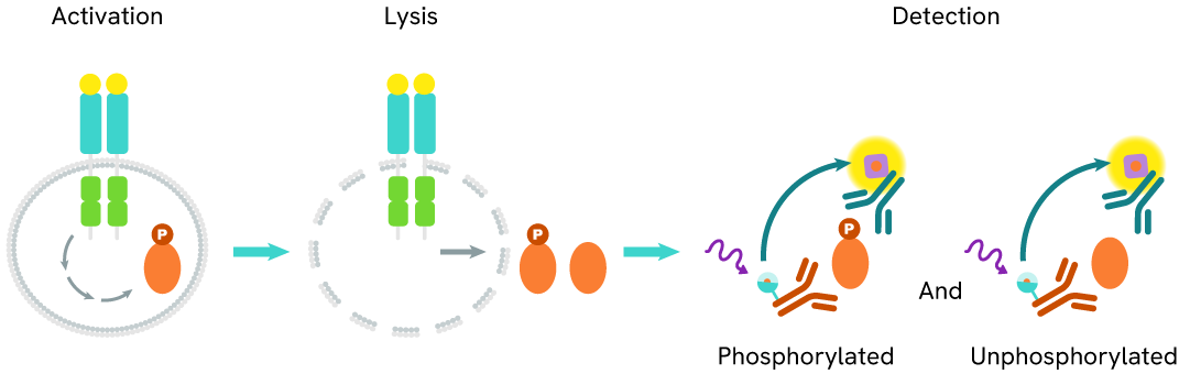
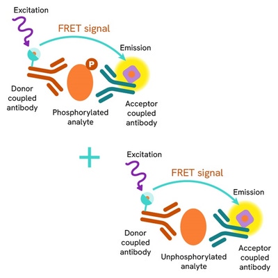
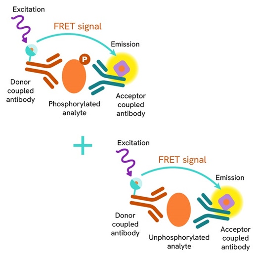
The total MDM2 assay measures MDM2 levels in cells. Unlike Western Blot, the assay is entirely plate-based and does not require gels, electrophoresis, or transfer. The assay uses 2 antibodies, one labeled with a donor fluorophore and the other with an acceptor. Both antibodies are highly specific for distinct epitopes on the MDM2 protein.
The presence of MDM2 leads to the formation of an immunocomplex involving both labeled antibodies, bringing the donor fluorophore into close proximity to the acceptor and generating a FRET signal. The signal intensity is directly proportional to the amount of total MDM2 present in the sample, providing a method for measuring the protein's phosphorylation state under a no-wash assay format.
Total MDM2 two-plate assay protocol
The two-plate protocol involves culturing cells in a 96-well plate before lysis, then transferring lysates into a 384-well low volume detection plate before the addition of total MDM2 HTRF detection reagents. This protocol allows the cells’ viability and confluence to be monitored.

Total MDM2 one-plate assay protocol
Detection of total MDM2 with HTRF reagents can be performed in a single plate used for culturing, stimulation, and lysis. No washing steps are required. This HTS designed protocol allows miniaturization while maintaining robust HTRF quality.

Assay validation
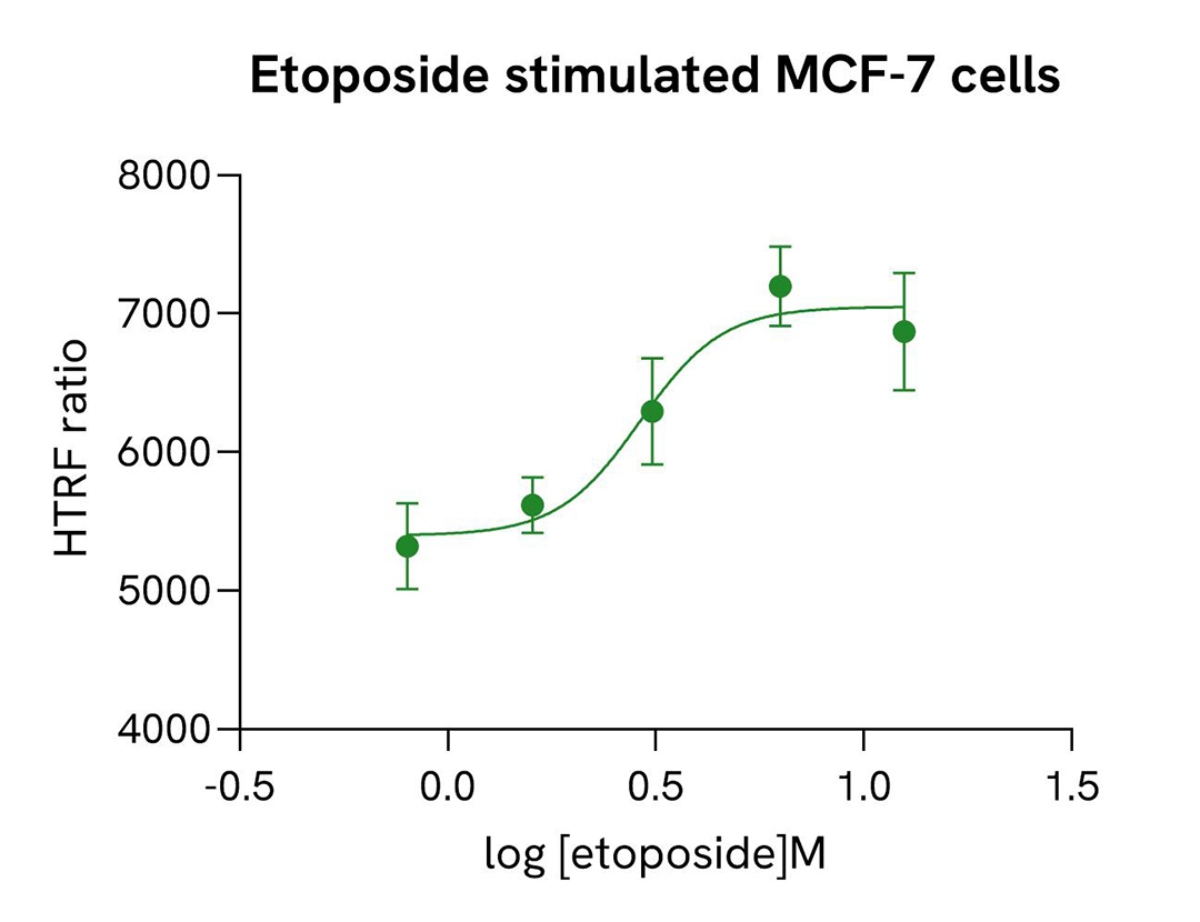
MDM2 induction by etoposide in MCF-7 cells
MCF-7 cells were seeded into 96-well plates at a density of 40,000 cells per well and cultured for 24 hours. Cells were then treated with increasing concentrations of etoposide or vehicle control for 6 hours, thereby indirectly inducing MDM2 expression through p53-dependent transcriptional activation. After treatment, cells were lysed with 50 µL of supplemented lysis buffer #4 containing 1X blocking reagent for 30 minutes at room temperature under gentle shaking. Following lysis, 16 µL of cell lysate were transferred into a 384-well low-volume white microplate, and 4 µL of HTRF Total MDM2 detection antibodies were added. The HTRF signal was recorded after overnight incubation at room temperature, using the homogeneous no-wash HTRF format for endogenous protein quantification.
As expected, etoposide treatment resulted in a dose-dependent increase in MDM2 levels. Importantly, etoposide did not induce detectable cytotoxicity under these experimental conditions, as assessed using the ATPLite cell viability assay (data not shown).
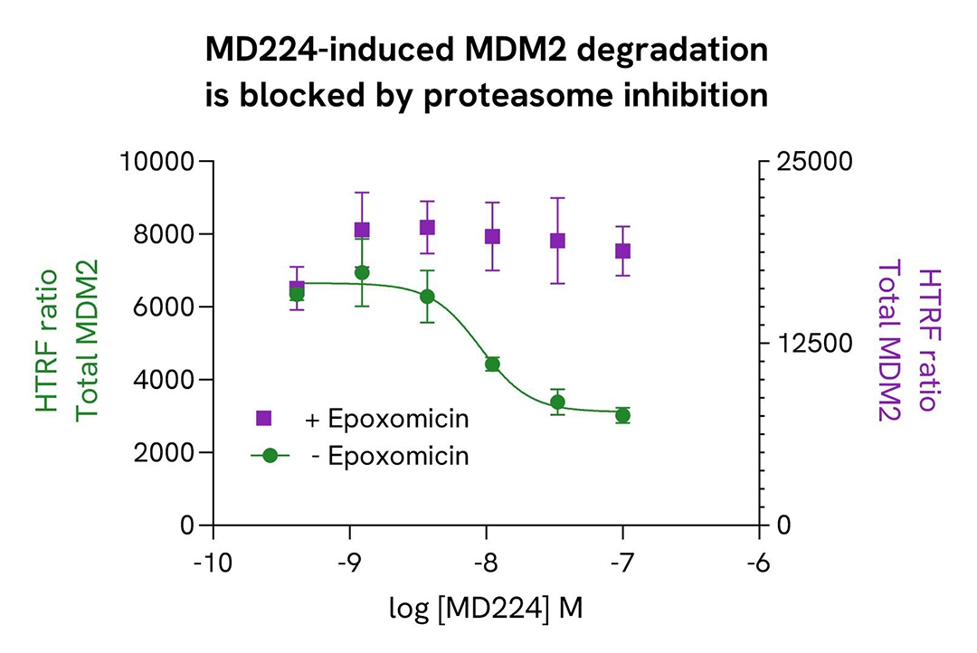
MD224-induced MDM2 degradation is blocked by proteasome inhibition
MCF-7 cells were seeded into 96-well plates at 40,000 cells per well and cultured for 24 hours. Cells were then pre-incubated for 3 hours at 37°C with MI-1061 (3.7 nM), a potent inhibitor of the MDM2-p53 interaction. MI-1061 binds to the p53-binding pocket of MDM2, disrupting the MDM2-p53 interaction and promoting p53-dependent transcriptional upregulation of MDM2.
One hour prior to PROTAC treatment, cells were incubated with 1 µM epoxomicin or vehicle control to inhibit proteasomal activity.
Cells were subsequently treated for 3 hours with increasing concentrations of MD224, a PROTAC degrader developed to induce MDM2 degradation, or with vehicle control. Following treatment, cells were lysed with 50 µL of supplemented Lysis Buffer #4 (1X) for 30 minutes at room temperature. Subsequently, 16 µL of cell lysate were transferred into a 384-well plate for Total MDM2 detection using the HTRF assay.
The HTRF signal was measured after overnight incubation at room temperature.
MD224 induced a concentration-dependent decrease in the MDM2 HTRF signal, consistent with MDM2 degradation. This effect did not exist in the presence of epoxomicin, demonstrating that MD224-mediated MDM2 degradation occurs through a proteasome-dependent mechanism.
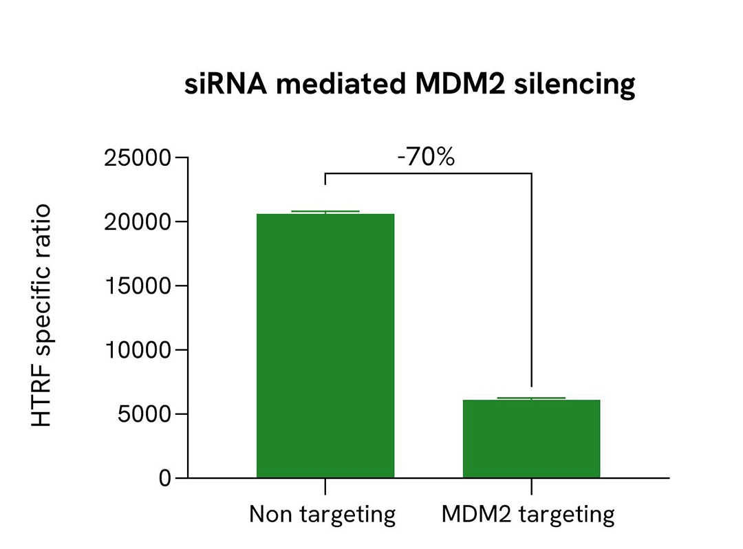
Validation of the specificity of Total MDM2 assay using siRNA
HCT116 cells were seeded into 96-well plates at a density of 10,000 cells per well and cultured for 24 hours. Cells were then transfected with either an MDM2-specific siRNA (Horizon Discovery/Revvity, #L-003279-00-0010) or a non-targeting negative control siRNA (Horizon Discovery/Revvity, #D-001810-10). Following a 48-hour incubation, cells were lysed with 50 µL of supplemented Lysis Buffer #4 (1X) containing Blocking Reagent for 30 minutes at room temperature under gentle shaking.
After lysis, 16 µL of cell lysate were transferred into a low-volume 384-well white microplate, and 4 µL of HTRF Total MDM2 detection antibodies were added. The HTRF signal was measured after overnight incubation at room temperature.
As expected, transfection with the MDM2-specific siRNA resulted in efficient knockdown of MDM2 expression, as demonstrated by an approximately 70% reduction in HTRF signal compared to cells transfected with the negative control siRNA. No significant signal modulation was observed in cells treated with the negative control siRNA.
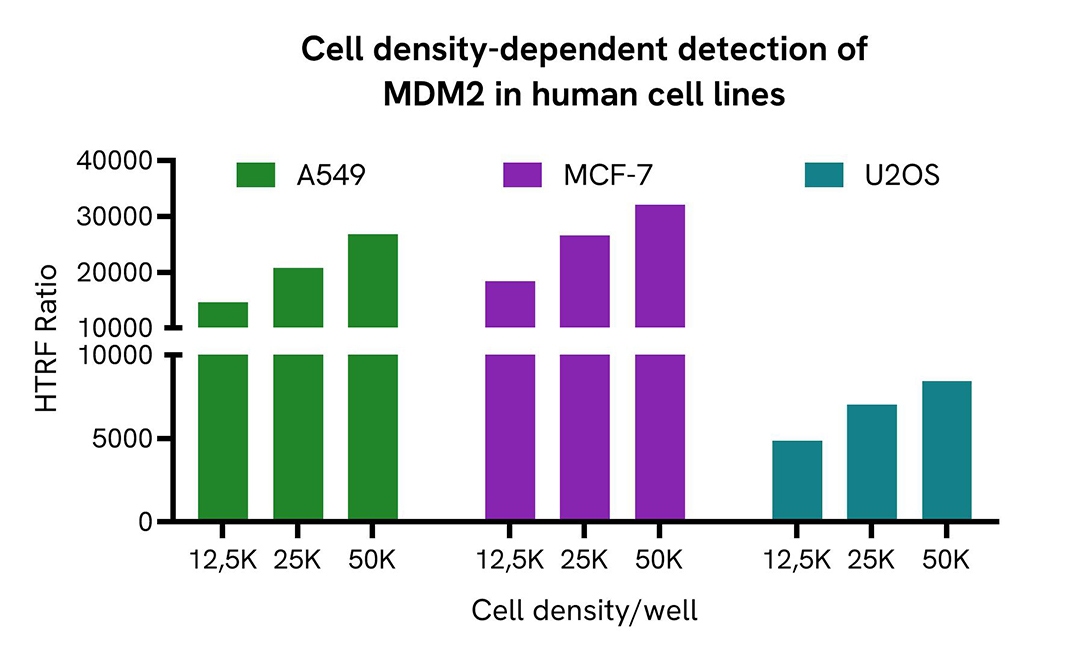
MDM2 expression level is detected in various human cell lines
Human U2OS, MCF-7, and A549 cells were seeded into 96-well plates at various cell densities and cultured for 24 hours. Cells were then treated with 10 μM of nutlin-3a or with vehicle control for 24 hours, thereby indirectly stimulating phospho-MDM2 levels through p53-dependent induction. Cells were lysed with 50 µL of supplemented Lysis Buffer #4 (1X) for 30 minutes at room temperature under gentle shaking.
Following lysis, 16 µL of cell lysate were transferred into a low-volume 384-well white microplate, and 4 µL of HTRF Total MDM2 detection antibodies were added. The HTRF signal was measured after overnight incubation at room temperature.
The Total MDM2 HTRF signal increased with cell density across all tested human cell lines, demonstrating robust detection of MDM2.
U2OS, MCF-7, and A549 cells all generated strong assay signals, confirming the suitability of these cell models for MDM2 detection. These results support the use of the HTRF Total MDM2 assay for the sensitive, reliable quantification of MDM2 expression in multiple human cell lines.
Simplified pathway
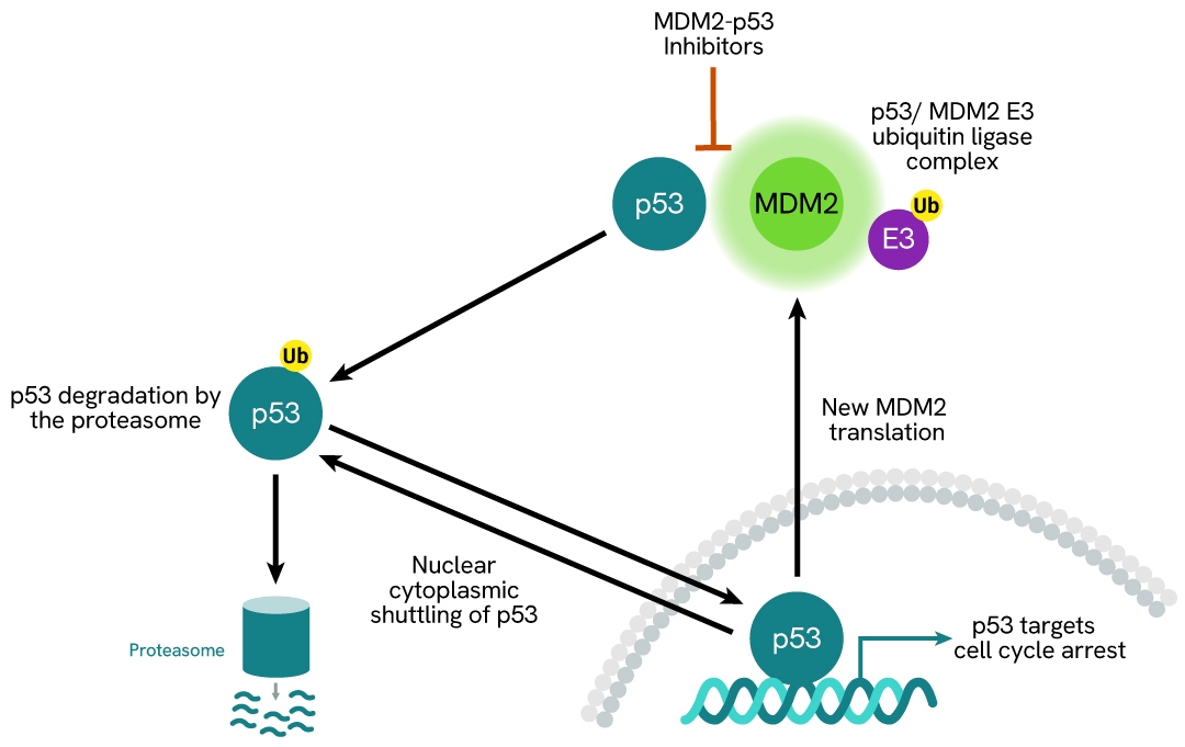
MDM2 signaling pathway
MDM2 is an oncogenic protein encoded by the MDM2 gene. It is an E3 ubiquitin ligase that negatively regulates the tumor suppressor protein p53. MDM2 binds directly to the transactivation domain of p53, inhibiting its transcriptional activity. It also promotes the proteasomal degradation of p53 via ubiquitination, maintaining low p53 levels in normal cells. The MDM2-p53 relationship forms a negative feedback loop, in which p53 activates MDM2 transcription, which in turn inhibits p53. Persistent p53 activation triggers the continuous expression of genes involved in apoptosis and cell cycle arrest in response to cellular stress, such as DNA damage. MDM2 is frequently amplified or overexpressed in a wide range of cancers, suppressing p53 even when TP53 is wild-type, thereby enabling tumor cells to evade apoptosis and cell cycle arrest. As a major oncogene, MDM2 is a prime therapeutic target, and inhibitors of the MDM2-p53 interaction are being developed to restore p53 activity in tumors.
Specifications
| Application |
Cell Signaling
|
|---|---|
| Automation Compatible |
Yes
|
| Brand |
HTRF
|
| Detection Modality |
HTRF
|
| Lysis Buffer Compatibility |
Lysis Buffer 4
|
| Molecular Modification |
Total
|
| Product Group |
Kit
|
| Sample Volume |
16 µL
|
| Shipping Conditions |
Shipped in Dry Ice
|
| Target |
MDM2
|
| Target Class |
Phosphoproteins
|
| Target Species |
Human
|
| Technology |
TR-FRET
|
| Therapeutic Area |
Inflammation
Oncology
|
| Unit Size |
10,000 Assay Points
|
Resources
Are you looking for resources, click on the resource type to explore further.
Loading...


How can we help you?
We are here to answer your questions.