


JP
Revvity Sites Globally
Select your location.
*e-commerce not available for this region.

HTRF Human Phospho-STAT2 (Tyr690) Detection Kit, 10,000 assay points

HTRF Human Phospho-STAT2 (Tyr690) Detection Kit, 10,000 assay points

generic HTRF phospho primary image
This HTRF kit allows the cell-based quantitative detection of STAT2 when phosphorylated at Tyr690.
| Feature | Specification |
|---|---|
| Application | 細胞シグナル伝達 |
| Sample Volume | 16 µL |
This HTRF kit allows the cell-based quantitative detection of STAT2 when phosphorylated at Tyr690.
Product variants
Unit Size: 500 assay points
Part #:
64STAT2Y6PEG
Unit Size: 10,000 assay points
Part #:
64STAT2Y6PEH
For research use only. Not for use in diagnostic procedures. All products to be used in accordance with applicable laws and regulations including without limitation, consumption and disposal requirements under European REACH regulations (EC 1907/2006).
HTRF Human Phospho-STAT2 (Tyr690) Detection Kit, 10,000 assay points
generic HTRF phospho primary image
Loading...
Product information
Overview
STAT2 (Signal transducer and activator of transcription 2) is a member of the cytoplasmic transcription factors family that relay signals emanating from cell-surface cytokine and growth factor receptors to the nucleus. STAT proteins control fundamental cellular processes, including cell survival, proliferation, differentiation, and immune responses. Dysregulation of STAT2 signaling has been implicated in various diseases, particularly viral infections and immune disorders.
HTRF assays offer many advantages over other technologies:
- Homogeneous add-and-read format
- No wash steps
- Low background
- Straightforward miniaturization from 96- or 384-well microplates to high density assay formats such as 384-well low volume and 1536-well plates
- Stable signal, providing flexibility in time of readout or size of assays
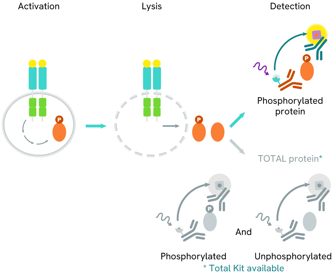
How it works
Phospho-STAT2 (Tyr690) assay principle
The Phospho-STAT2 (Tyr690) assay measures STAT2 when phosphorylated at Tyr690. Unlike Western Blot, the assay is entirely plate-based and does not require gels, electrophoresis, or transfer. The assay uses 2 antibodies, one labeled with a donor fluorophore and the other with an acceptor. The first antibody was selected for its specific binding to the phosphorylated motif on the protein, and the second for its ability to recognize the protein independently of its phosphorylation state. Protein phosphorylation leads to an immune-complex formation involving both labeled antibodies. It brings the donor fluorophore into close proximity to the acceptor, thereby generating a FRET signal. Its intensity is directly proportional to the concentration of phosphorylated protein present in the sample and provides a means of assessing the protein's phosphorylation state under a no-wash assay format.
Phospho-STAT2 (Tyr690) two-plate assay protocol
The two-plate protocol involves culturing cells in a 96-well plate before lysis, then transferring lysates into a 384-well low volume detection plate before the addition of Phospho-STAT2 (Tyr690) HTRF detection reagents. This protocol allows the cells viability and confluence to be monitored.

Phospho-STAT2 (Tyr690) one-plate assay protocol
Detection of Phosphorylated STAT2 (Tyr690) with HTRF reagents can be performed in a single plate used for culturing, stimulation, and lysis. No washing steps are required. This HTS designed protocol allows miniaturization while maintaining robust HTRF quality.

Assay validation
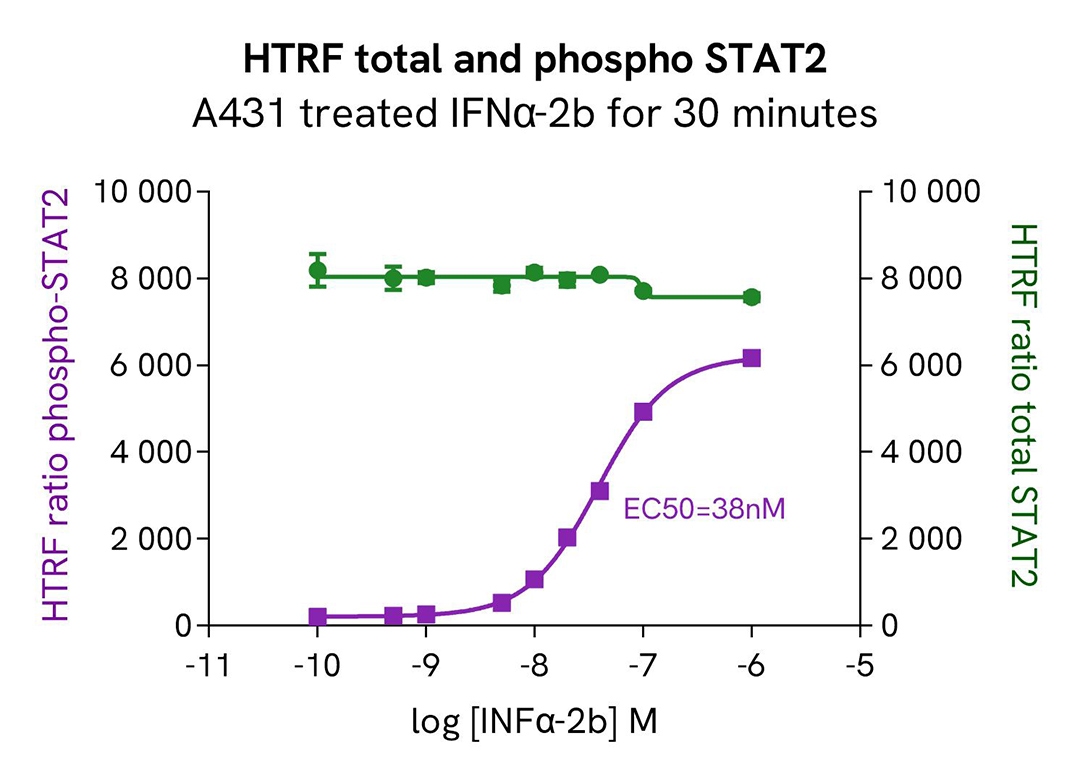
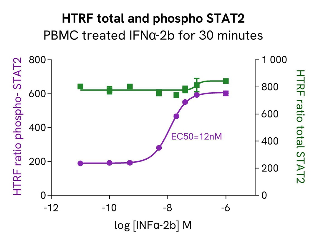
Modulation of phospho-STAT2 (Tyr690) / Total STAT2 using IFNα-2b on A431 and PBMC cells
A431 and PBMC cells were seeded in a 96-well culture plate (100,000 cells/well) in complete culture medium and incubated for 24 hours at 37°C, 5% CO2. Cells were then treated for 30 minutes with increasing concentrations of IFNα-2b.
After cell lysis, 16 µL of lysates were transferred into a 384-well low volume white microplate, and 4 µL of the HTRF Phosho-STAT2 (Tyr690) or Total STAT2 detection antibodies were added. The HTRF signal was recorded after 2 hours of incubation.
As expected, IFNα-2b triggered a dose-dependent increase in phosphorylated STAT2 at Tyr690, while the Total STAT2 protein expression level remained constant.
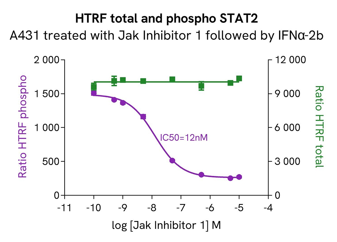
Modulation of phospho-STAT2 (Tyr690) / Total STAT2 using JAK inhibitor 1 on A431 cells
A431 cells were seeded in a 96-well culture plate (100,000 cells/well) in complete culture medium and incubated for 24 hours at 37°C, 5% CO2. Cells were then treated for 30 minutes with increasing concentrations of JAK inhibitor 1, followed by 3nM IFNα-2b treatment for 1 hour.
After cell lysis, 16 µL of lysates were transferred into a 384-well low volume white microplate, and 4 µL of the HTRF Phosho-STAT2 (Tyr690) or Total STAT2 detection antibodies were added. The HTRF signal was recorded after 2 hours of incubation.
The results indicate a clear dose-dependent inhibition of STAT2 phosphorylation Tyr690 upon treatment with JAK inhibitor 1, while the Total STAT2 protein expression level remained constant.
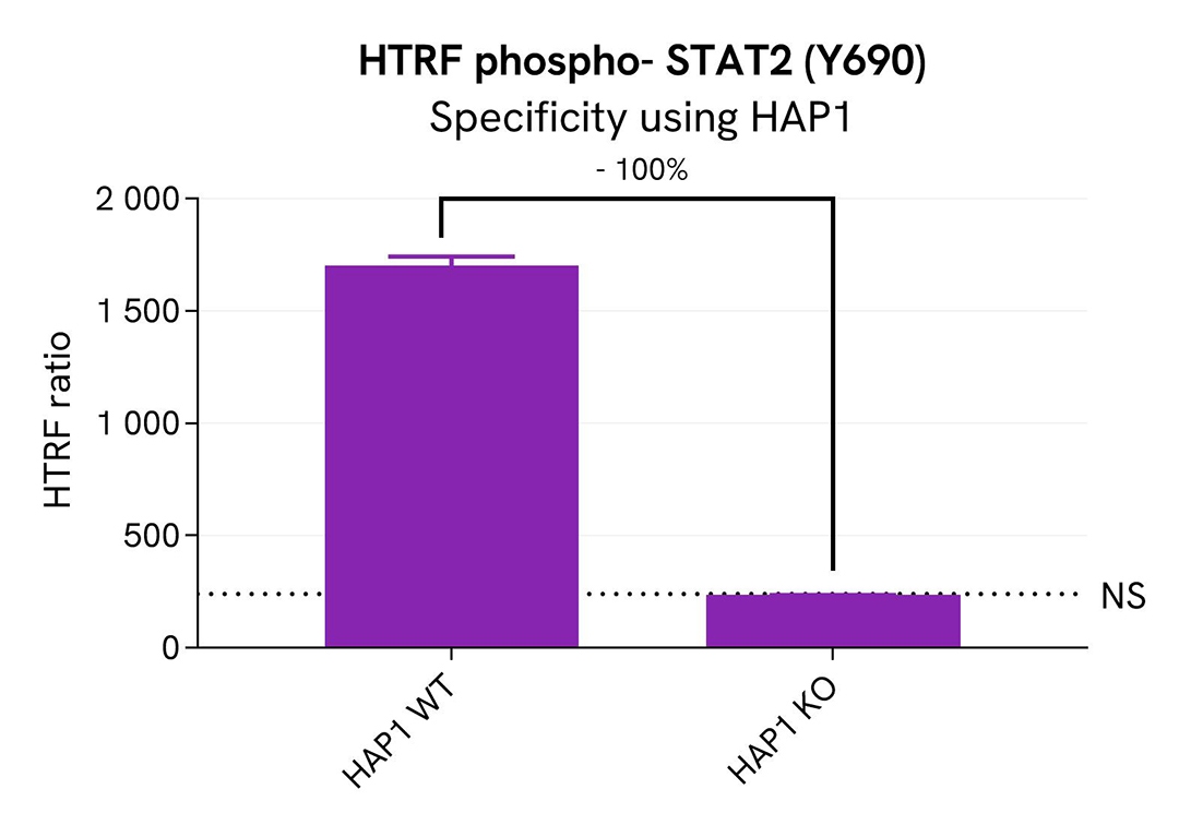
Specificity of phospho-STAT2 (Tyr690) assay using HAP1 cells knocked out for STAT2
The phospho STAT2 protein levels were assessed with the HTRF Human phospho-STAT2 (Y690) kit in HAP1 cells (WT) and a HAP1 cell line Knocked-Out for STAT2. The cell density was optimized beforehand to ensure HTRF detection within the dynamic range of the kit (data not shown).
The cells were cultured in a 96-well plate (100,000 cells/well) for 24 hours at 37°C, 5% CO2, followed by IFNα-2b treatment at 5nM for 30 minutes. The cells were then lysed with 50 µL of supplemented lysis buffer #4 (1X) for 30 minutes at RT under gentle shaking, and 16 µL of cell lysate were transferred into a low volume white microplate, followed by 4 µL of premixed phospho-STAT2 detection reagents. The HTRF signal was recorded after 2 hours of incubation at RT.
In HAP1 KO STAT2 cells, the HTRF signal decreased by 100%, indicating a significant STAT2 gene silencing, whereas the phospho- STAT2 level was well detected in the other cell line, as expected. This demonstrates that the HTRF Phospho-STAT2 (Y690) assay is specific for Phospho-STAT2 detection.
Catalog cell line references (Horizon Discovery): HAP1 Wt #C631; HAP1 KO STAT2 #HZGHC006251c008
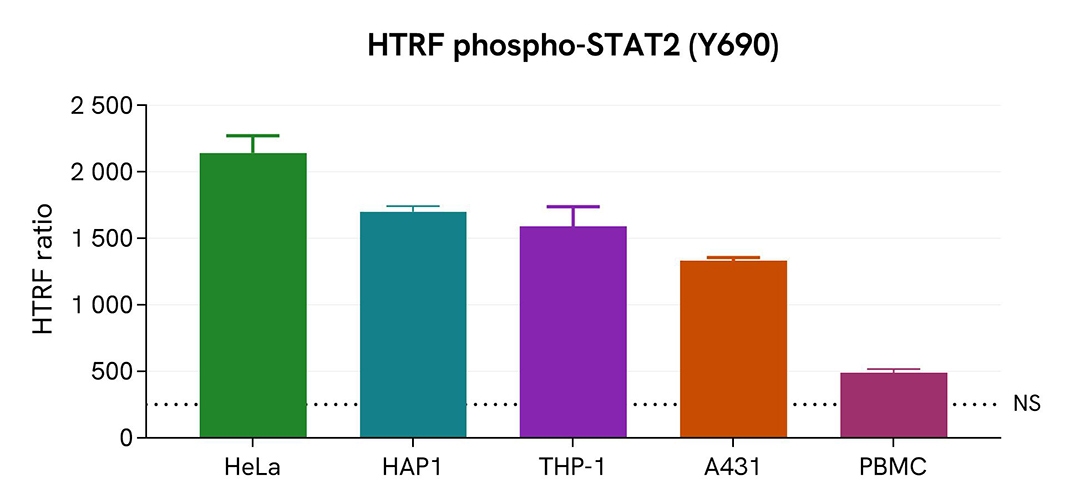
Assessment of phospho-STAT2 (Tyr690) level in various cell lines
Adherent (A431, HeLa, HAP1) and suspension (THP-1, PBMC) human cells were seeded at 100,000 cells/well in a 96-well microplate. After 24h of incubation, the cells were treated with 5nM IFNα-2b for 30 minutes. The cells were then lysed for 30 minutes with supplemented lysis buffer #4, following the protocol for adherent or suspended cells at RT under gentle shaking.
16 µL of lysate were transferred into a 384-well low volume white microplate before the addition of 4 µL of the HTRF phospho-STAT2 (Tyr690) detection reagents. The HTRF signal was recorded after 2 hours of incubation.
The HTRF phospho-STAT2 (Tyr690) assay efficiently detected phospho-STAT2 in various cellular models expressing different levels of the protein.
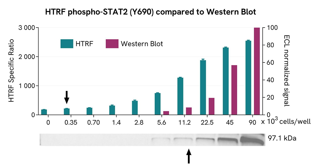
HTRF STAT2 Phospho-Y690 assay compared to Western Blot
A431 cells were grown in a T175 flask in complete culture medium at 37°C, 5% CO2, until 80% confluence. After a 24h incubation, the cells were stimulated with 5nM of IFNα-2b for 30 minutes. They were then lysed with 3 mL of supplemented lysis buffer #4 (1X) for 30 minutes at RT under gentle shaking.
Serial dilutions of the cell lysate were performed using supplemented lysis buffer, and 16 µL of each dilution were transferred into a low volume white microplate before the addition of 4 µL of HTRF Phospho STAT2 (Y690) detection reagents. Equal amounts of lysates were used for a side-by-side comparison between HTRF and Western Blot.
In these conditions, the HTRF Phospho STAT2 (Y690) assay was 32 times more sensitive than the Western Blot technique.
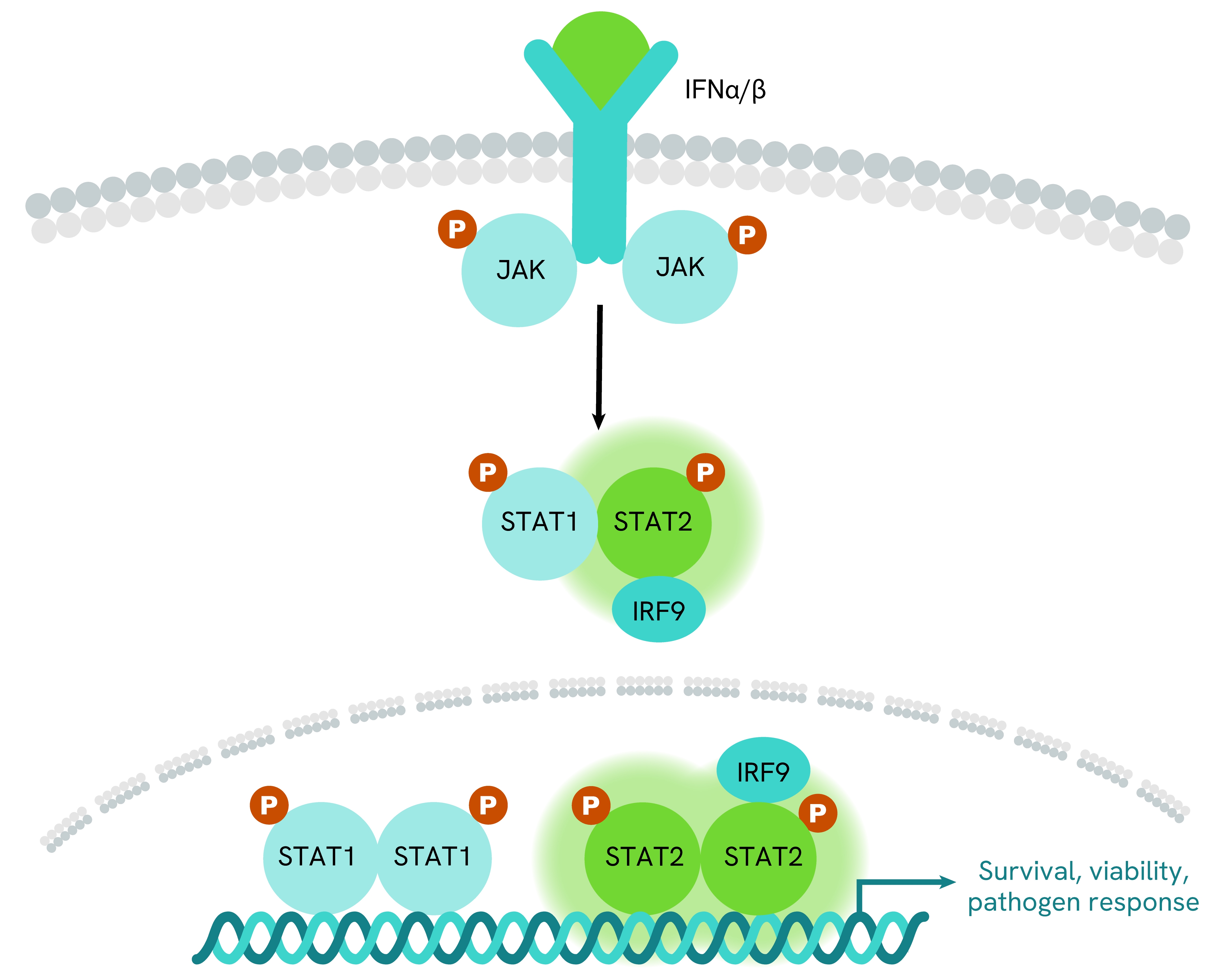
Simplified pathway
STAT2 signaling pathway
STAT2 is a key component of the type I interferon (IFN α/β) signaling pathway, and plays a central role in antiviral defense. Upon stimulation by type I interferons, the JAK1 and TYK2 kinases phosphorylate STAT2 at Tyr690. Phosphorylated STAT2 then forms a heterodimer with STAT1, and this complex subsequently associates with IRF9 (Interferon Regulatory Factor 9) to generate the ISGF3 (Interferon Stimulated Gene Factor 3) transcription factor complex.
Once formed, ISGF3 translocates into the nucleus, where it binds to ISRE (Interferon Stimulated Response Element) motifs within target gene promoters, driving the transcription of interferon stimulated genes (ISGs). These ISGs regulate a broad range of cellular antiviral mechanisms, immune-modulatory functions, and host defense pathways.
Specifications
| Application |
Cell Signaling
|
|---|---|
| Brand |
HTRF
|
| Detection Modality |
HTRF
|
| Lysis Buffer Compatibility |
Lysis Buffer 1
Lysis Buffer 2
Lysis Buffer 3
Lysis Buffer 4
|
| Molecular Modification |
Phosphorylation
|
| Product Group |
Kit
|
| Sample Volume |
16 µL
|
| Shipping Conditions |
Shipped in Dry Ice
|
| Target |
STAT2
|
| Target Class |
Phosphoproteins
|
| Target Species |
Human
|
| Technology |
TR-FRET
|
| Therapeutic Area |
Autoimmunity
Immuno-oncology
Infectious Diseases
Inflammation
Oncology
Virology
|
| Unit Size |
10,000 assay points
|
Resources
Are you looking for resources, click on the resource type to explore further.
Loading...


How can we help you?
We are here to answer your questions.

