


JP
Revvity Sites Globally
Select your location.
*e-commerce not available for this region.

HTRF Human and Mouse HTT Aggregation Kit, 10,000 Assay Points

HTRF Human and Mouse HTT Aggregation Kit, 10,000 Assay Points

TagLite GIP cell line primary image
The HTRF Human and Mouse aggregated HTT detection kit is designed for the simple and rapid quantification of aggregated HTT protein in cell/tissue lysates.
| Feature | Specification |
|---|---|
| Application | タンパク質定量 |
| Sample Volume | 10 µL |
The HTRF Human and Mouse aggregated HTT detection kit is designed for the simple and rapid quantification of aggregated HTT protein in cell/tissue lysates.
Product variants
Unit Size: 500 assay points
Part #:
64HTTAPEG
Unit Size: 10,000 assay points
Part #:
64HTTAPEH
For research use only. Not for use in diagnostic procedures. All products to be used in accordance with applicable laws and regulations including without limitation, consumption and disposal requirements under European REACH regulations (EC 1907/2006).
HTRF Human and Mouse HTT Aggregation Kit, 10,000 Assay Points
TagLite GIP cell line primary image
HTRF Human and Mouse HTT Aggregation Kit, 10,000 Assay Points
Product information
Overview
Huntingtin (HTT) is a cytoplasmic protein which is highly expressed in the brain, and whose anti-apoptotic role is critical for neuronal survival.
The wild-type (WT) protein has a functional structure, with a "normal" polyglutamine (polyQ) domain containing less than 36Q. The mutant HTT protein harbors an abnormally long polyQ tract (> 36Q) which causes the aggregation of the no longer functional protein. HTT aggregation leads to the selective neuronal cell death responsible for Huntington's Disease.
HTRF assays offer many advantages over other technologies:
- Homogeneous add-and-read format
- No wash steps
- Low background
- Straightforward miniaturization from 96- or 384-well microplates to high density assay formats such as 384-well low volume and 1536-well plates
- Stable signal, providing flexibility in time of readout or size of assays
How it works
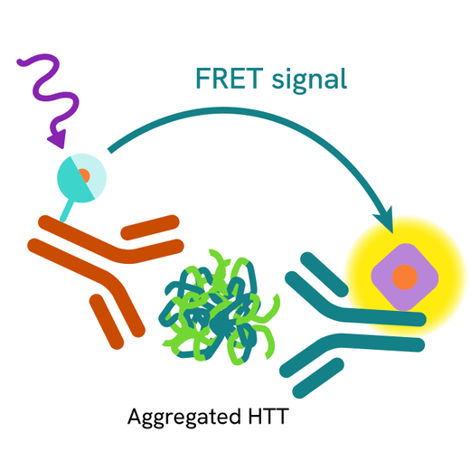
Principle of the HTRF Human & Mouse Aggregated HTT assay
The HTRF Aggregated HTT assay is based on a TR-FRET sandwich immunoassay involving two specific antibodies, one labelled with Tb3+-cryptate (donor) and the other with d2 (acceptor). One antibody is directed against the N-terminal part of the protein, and the second recognizes specifically the aggregated form. Both antibodies bind to aggregated HTT, and the donor-acceptor proximity enables a fluorescent TR-FRET signal to occur. The intensity of the signal is directly proportional to the level of HTT aggregates present in the sample (cell lysate or tissue lysate).
Protocol of the HTRF Human & Mouse Aggregated HTT assay
The HTRF Aggregated HTT assay can be run in a 96- or 384-well low volume white detection plate (20 µL final). As described here, samples (cell/tissue lysates) are dispensed directly into the assay plate for the detection of HTT aggregates by HTRF reagents. The antibodies labelled with HTRF fluorophores may be pre-mixed and added in a single dispensing step. No washing steps are needed. The protocol can be further miniaturized or upscaled by simply resizing each addition volume proportionally.

Assay details
Human & Mouse Aggregated HTT Assay details
| Sample | 10 µL |
|---|---|
| Final assay volume | 20 µL |
| Time to result | Overnight at RT |
| Kit component | Frozen detection antibodies, buffers & protocol |
| Species | Human & mouse |
Assay validation
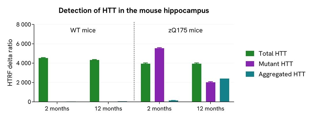
Analysis of brain tissue samples collected from WT and mutant HTT mouse models
The HTRF Aggregated HTT assay was validated on brain tissues collected from premanifest zQ175 mice and wild-type (WT) littermates, the most extensively studied preclinical knock-in mouse model used for Huntington’s Disease (Landles C. et al. Brain Commun. 2021; 3(1):fcaa231; Smith EJ. et al. Brain Commun. 2023; 5(1):fcad010; Landles et al., Brain Commun. 2024; 6(6):fcae410).
The hippocampus tissues of WT and mutant mice aged 2 or 12 months were lysed following the procedure given in the kit’s package insert. Briefly, a 10% (w/v) tissue homogenate was prepared using ice-cold 1X lysis buffer #2 supplemented with protease inhibitors. The lysates, containing aggregates and soluble HTT proteins, were analyzed using the HTRF Total HTT assay (Cat# 64HTTTPEG/H) assay, the HTRF Mutant HTT assay (Cat# 64HTTMPEG/H), and the HTRF Aggregated HTT assay. To ensure that the detected analyte was assessed at a concentration compatible with the assay’s linear range, the lysates were pre-diluted in 1X lysis buffer #2 supplemented with protease inhibitors just before detection (1/4 for Total HTT assay, 1/8 for Mutant HTT assay, and 1/2 for Aggregated HTT assay).
Results show the soluble Total HTT protein (WT and mutant forms) was measured in all lysates (green bars), and its levels were similar for all samples. No signal was obtained with the Mutant HTT assay on samples collected from WT mice, whereas the soluble mutant protein was clearly detected in hippocampus collected from premanifest zQ175 mice (purple bars). The level of soluble mutant HTT decreased in the 12-month-old mice because of mutant protein aggregation. This aggregation was well detected (blue bars), in agreement with literature.
Specifications
| Application |
Protein Quantification
|
|---|---|
| Brand |
HTRF
|
| Detection Modality |
HTRF
|
| Product Group |
Kit
|
| Sample Volume |
10 µL
|
| Shipping Conditions |
Shipped in Dry Ice
|
| Target |
Aggregated HTT
|
| Target Class |
Biomarkers
|
| Target Species |
Human
Mouse
|
| Technology |
TR-FRET
|
| Therapeutic Area |
Central Nervous System
|
| Unit Size |
10,000 assay points
|
Resources
Are you looking for resources, click on the resource type to explore further.


How can we help you?
We are here to answer your questions.

