
JP

Revvity Sites Globally
Select your location.
*e-commerce not available for this region.

HTRF Human Total IRE1 Detection Kit, 500 Assay Points
HTRF Human Total IRE1 Detection Kit, 500 Assay Points
Total IRE1 Product image
This HTRF kit allows for the cell-based quantitative detection of Total IRE1.
| Feature | Specification |
|---|---|
| Application | 細胞シグナル伝達 |
| Sample Volume | 16 µL |
This HTRF kit allows for the cell-based quantitative detection of Total IRE1.
Product variants
Unit Size: 500 assay points
Part #:
64IRE1TPEG
Unit Size: 10,000 assay points
Part #:
64IRE1TPEH
For research use only. Not for use in diagnostic procedures. All products to be used in accordance with applicable laws and regulations including without limitation, consumption, and disposal requirements under European REACH regulations (EC 1907/2006).
HTRF Human Total IRE1 Detection Kit, 500 Assay Points
Total IRE1 Product image
Loading...
Product information
Overview
IRE1 (Inositol requiring enzyme 1 alpha) is a serine/threonine kinase acting as one of three branches of the Unfolded Protein Response (UPR) signaling pathway with roles in the regulation of ER stress and UPR. IRE1 is encoded by the ERN1 gene and is ubiquitously expressed in mammals. IRE1 is a type I transmembrane protein, exhibiting an ER luminal domain that senses the protein folding status, and a catalytic kinase and RNase cytosolic domain. In recent years, IRE1 has emerged as a relevant therapeutic target in various diseases including degenerative disorders, inflammatory and metabolic pathologies, and cancer.
How it works
Total IRE1 assay principle
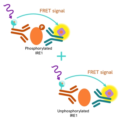
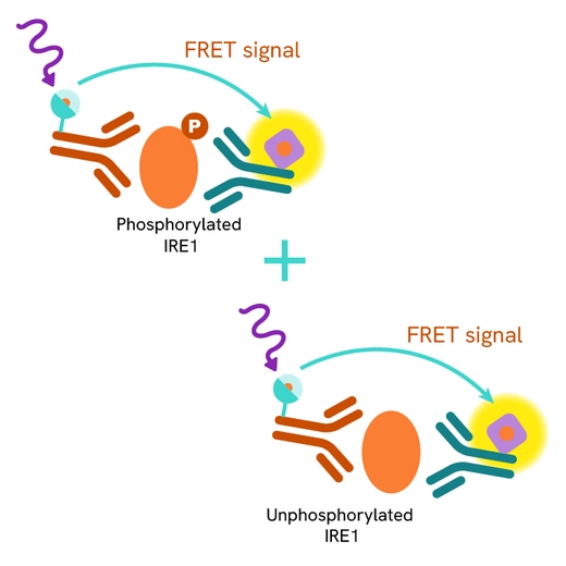
The Total IRE1 assay quantifies the expression level of IRE1 in a cell lysate. Unlike Western Blot, the assay is entirely plate-based and does not require gels, electrophoresis, or transfer. The Total IRE1 assay uses two labeled antibodies, one coupled to a donor fluorophore and the other to an acceptor. Both antibodies are highly specific for a distinct epitope on the protein. In the presence of IRE1 this enables an immune-complex formation involving both labeled antibodies, and which brings the donor fluorophore into close proximity to the acceptor, thereby generating a FRET signal.
Its intensity is directly proportional to the concentration of total protein present in the sample and provides a means of assessing the protein's phosphorylation state under a no-wash assay format.
Total IRE1 two-plate assay protocol
The two-plate protocol involves culturing cells in a 96-well plate before lysis, then transferring lysates into a 384-well low volume detection plate before the addition of HTRF Total IRE1 detection reagents. This protocol allows for the cells' viability and confluence to be monitored.

Total IRE1 one-plate assay protocol
Detection of Total IRE1 with HTRF reagents can be performed in a single plate used for culturing, stimulation, and lysis. No washing steps are required. This HTS designed protocol allows miniaturization while maintaining robust HTRF quality.

Assay validation
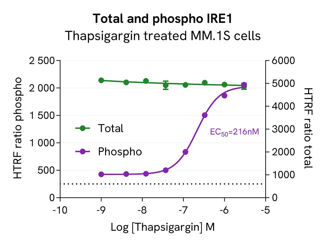
Induction of Phospho-IRE1 (Ser724) in MM.1S cells
MM.1S cells were seeded in 96-well culture plate (400,000 cells/30µL well) and treated with increasing concentrations of Thapsigargin.
After a 1 hour treatment, cells were lysed with 10 µL of supplemented lysis buffer #1 (4X) for 30 min at RT under gentle shaking. Next, 16 µL of lysate were transferred into a 384-well low volume white microplate, and 4 µL of the HTRF Phospho-IRE1 (S724) or HTRF Total IRE1 detection antibodies were added. HTRF signals were recorded after 4 hours of incubation.
Our results indicate a clear dose-dependent activation of IRE1 phosphorylation at Ser724 upon treatment with Thapsigargin, while the total IRE1 protein expression level remained constant.
Validation of Total IRE1 assay specificity
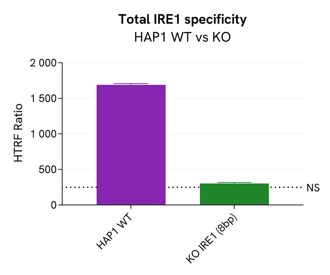
Total IRE1 protein levels were assessed with the HTRF Human Total IRE1 kit in HAP1 cells (WT) and HAP1 cell line Knocked-Out for IRE1. Cell density was optimized beforehand to ensure HTRF detection within the dynamic range of the kit (data not shown).
The different cell lines were cultured in a 96-well plate (200,000 cells/well) for 24 hours at 37°C, 5% CO2. The cells were then lysed with 25 µL of supplemented lysis buffer #1 (1X) for 30 minutes at RT under gentle shaking. Next, 16 µL of cell lysate were transferred into a low volume white microplate followed by 4 µL of premixed detection reagents. The HTRF signal was recorded after 4 hours of incubation at RT.
In HAP1 KO IRE1 cells, the HTRF signal was equivalent to the non-specific signal (dotted line), indicating a complete IRE1 gene silencing, whereas the IRE1 level was well detected in the other cell line, as expected.
Catalog cell line references (Horizon Discovery): HAP1 WT #C631; HAP1 KO IRE1 # HZGHC000742c006
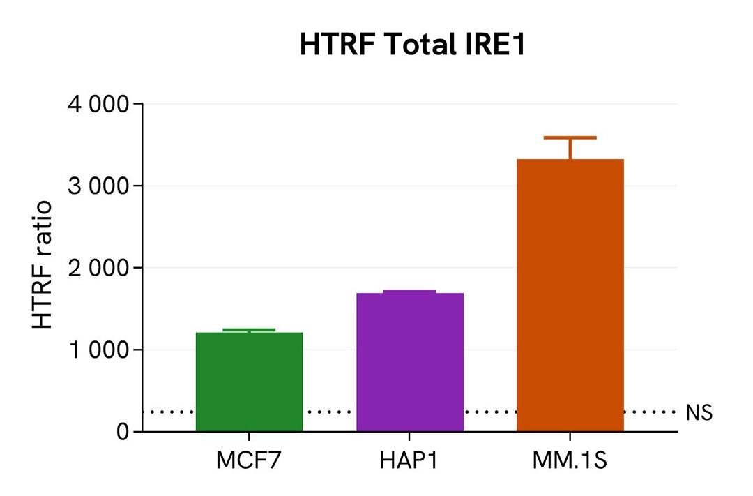
Assessment of Total IRE1 level in various cell lines
Adherent (MCF7 & HAP1) and suspension (MM.1S) human cells were seeded at 200,000 cells/well in a 96-well microplate. After 24h of incubation, the cells were lysed for 30 minutes with supplemented lysis buffer #1, following the protocol for adherent or suspended cells at RT under gentle shaking.
16 µL of lysate were transferred into a 384-well low volume white microplate before the addition of 4 µL of the HTRF Total IRE1 detection reagents. The HTRF signal was recorded after 4 hours of incubation.
The HTRF Total IRE1 assay efficiently detected Total IRE1 in various cellular models expressing different levels of the protein.
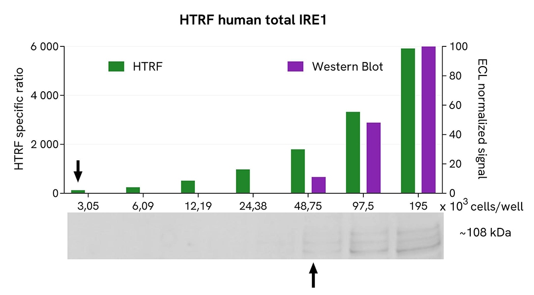
HTRF Total IRE1 assay compared to Western Blot
MM.1S cells were cultured in a T175 flask and treated with 1µM Thapsigargin for 1 hour in medium at 37°C, 5% CO2. After medium removal, the cells were lysed with 3 mL of supplemented lysis buffer #1 (1X) for 30 min at RT under gentle shaking.
Serial dilutions of the cell lysate were performed using supplemented lysis buffer #1 (1X), and 16µL of each dilution were transferred into a 384-well small volume microplate before the addition of 4µL of HTRF Total IRE1 detection antibodies. HTRF signals were recorded after 4 hours of incubation.
Equal amounts of lysates were loaded into a gel for a side-by-side comparison between HTRF and Western Blot.
In these conditions, the HTRF Total IRE1 assay is 16-fold more sensitive than the Western Blot technique.
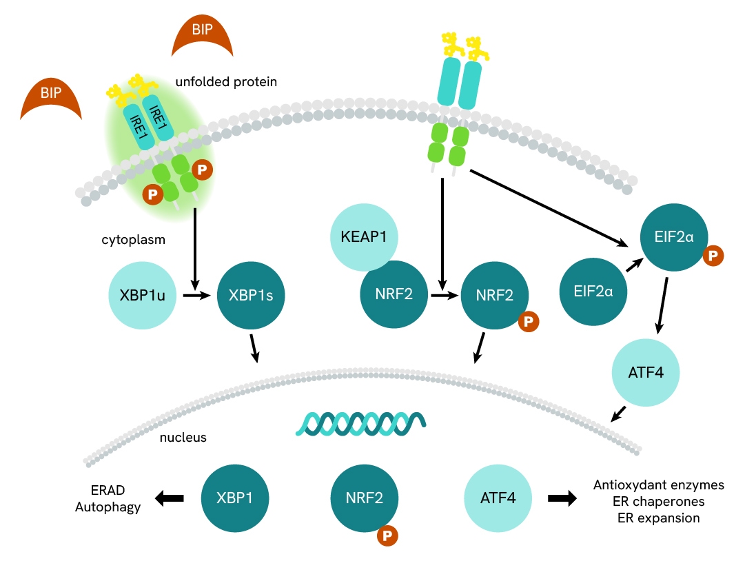
Simplified pathway
IRE1 signaling pathway
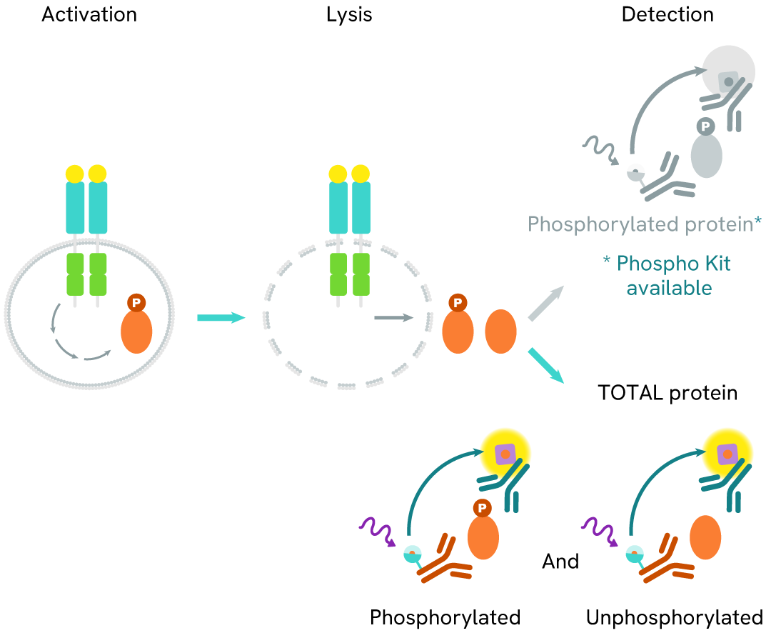
IRE1 is one of the main transducers of the Unfolded Protein Response signaling pathway with roles in regulation of ER stress and UPR (unfolded protein response). Upon ER stress and an accumulation of unfolded proteins, BiP releases its inhibitory ligand to activate IRE1. IRE1 receptor dimerizes then autophosphorylates, which activates its mRNase activity. Through this endoribonuclease activity, IRE1 degrades mRNAs (RIDD) and splices XBP-1u mRNA to XBP1s, enabling its translation and then its translocation to the nucleus. XBP-1s then acts as a transcription factor for chaperones to handle the overload of unfolded proteins.
Specifications
| Application |
Cell Signaling
|
|---|---|
| Automation Compatible |
Yes
|
| Brand |
HTRF
|
| Detection Modality |
HTRF
|
| Lysis Buffer Compatibility |
Lysis Buffer 1
|
| Molecular Modification |
Total
|
| Product Group |
Kit
|
| Sample Volume |
16 µL
|
| Shipping Conditions |
Shipped in Dry Ice
|
| Target |
IRE1
|
| Target Class |
Phosphoproteins
|
| Target Species |
Human
|
| Technology |
TR-FRET
|
| Therapeutic Area |
Central Nervous System
Inflammation
Oncology
|
| Unit Size |
500 assay points
|
Resources
Are you looking for resources, click on the resource type to explore further.

Brochure
HTRF assays and reagents product list
Discover the versatility and precision of Homogeneous Time-Resolved Fluorescence (HTRF) technology. Our HTRF portfolio offers a...
Loading...


How can we help you?
We are here to answer your questions.