


JP
Revvity Sites Globally
Select your location.
*e-commerce not available for this region.

AlphaLISA Human LC3B Detection Kit, 5,000 Assay Points
 View All
AlphaLISA Human LC3B Detection Kit, 5,000 Assay Points

AlphaLISA Sandwich Anti-analyte Conjugated Acceptor Bead
AlphaLISA™ no-wash immunoassay kit for detection of human LC3B in buffered solution or cell lysates. The antibodies in the kit detect both LC3B type I and type II. The analyte in this kit consists of recombinant LC3B fused to His-tag at the N-terminus.
| Feature | Specification |
|---|---|
| Application | タンパク質定量 |
| Dynamic Range | 7.9 - 3,000 ng/mL |
| Limit of Detection | 7.9 ng/mL |
| Limit of Quantification | 19.5 ng/mL |
| Sample Volume | 5 µL |
AlphaLISA™ no-wash immunoassay kit for detection of human LC3B in buffered solution or cell lysates. The antibodies in the kit detect both LC3B type I and type II. The analyte in this kit consists of recombinant LC3B fused to His-tag at the N-terminus.
Product variants
Unit Size: 500 assay points
Part #:
AL306C
Unit Size: 5,000 assay points
Part #:
AL306F
For research use only. Not for use in diagnostic procedures. All products to be used in accordance with applicable laws and regulations including without limitation, consumption, and disposal requirements under European REACH regulations (EC 1907/2006).
AlphaLISA Human LC3B Detection Kit, 5,000 Assay Points
AlphaLISA Sandwich Anti-analyte Conjugated Acceptor Bead
Loading...
Product information
Overview
Formats:
- Our 500 assay point kit allows you to run 500 wells in 96-well or 384-well format, using a 50 µL reaction volume (5 µL of sample).
- Our 5,000 assay point kit allows you to run 5,000 wells in 96-well or 384-well format, using a 50 µL reaction volume (5 µL of sample).
Features:
- No-wash steps, no separation steps
- ELISA alternative technology
- Sensitive detection
- Broad sample compatibility
- Small sample volume
- Results in less than 3 hours
- Half the time of an ELISA assay
LC3 represents a mammalian homologue of the yeast autophagy related gene ATG8. It was originally characterized as light chain 3 of the microtubule associated protein 1 (MAP1LC3). The protein family consists of LC3 A, B, and C and the GABARAP subfamily. Human LC3B is 125 amino acids long. After synthesis, it is cleaved by ATG4B to expose a C-terminal glycine, representing the cytosolic form LC3B I. During autophagy the C-terminus is covalently linked to autophagosomal vesicle membranes via a phospholipid anchor and this form is called LC3B II. The transformation of LC3B I to II is mediated by a ubiquitination-like process involving ATG7 (E1), ATG3 (E2) and the ATG16L complex (E3). To date, LC3B is considered as the most persistent marker of the autophagy pathway.
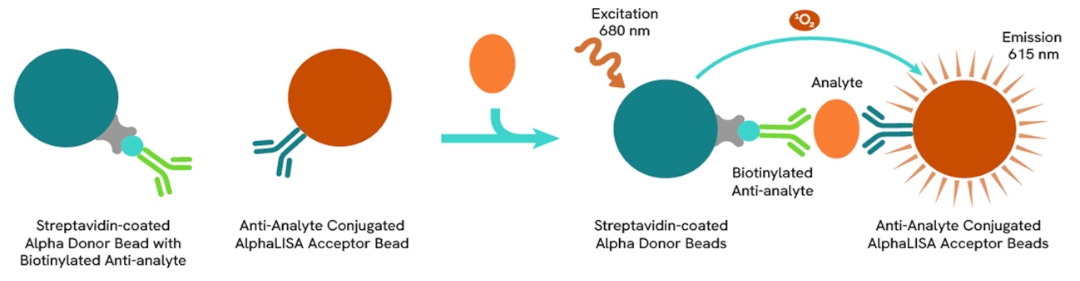
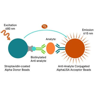
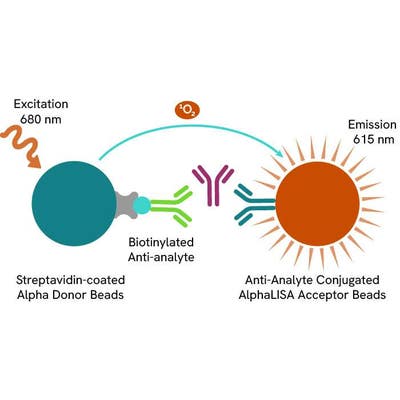
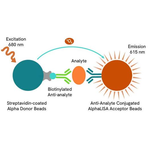
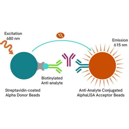
AlphaLISA technology allows the detection of molecules of interest in a no-wash, highly sensitive, quantitative assay. In an AlphaLISA assay, a biotinylated anti-analyte antibody binds to the Streptavidin-coated Donor beads while another anti-analyte antibody is conjugated to AlphaLISA Acceptor beads. In the presence of the analyte, the beads come into close proximity. The excitation of the Donor beads causes the release of singlet oxygen molecules that triggers a cascade of energy transfer in the Acceptor beads, resulting in a sharp peak of light emission at 615 nm.
How it works
Principle of the AlphaLISA assay
The AlphaLISA assay is based on an AlphaLISA sandwich immunoassay involving a biotinylated anti-analyte antibody bound to Streptavidin-coated AlphaLISA Donor beads and an anti-analyte antibody conjugated to AlphaLISA Acceptor beads. Both antibodies are directed against the analyte of interest. In the presence of the target, both antibodies bind to analyte and the beads come into proximity. The excitation of the Donor beads provokes the release of singlet oxygen molecules that triggers a cascade of energy transfer within the Acceptor beads, resulting in emission with λmax at 615 nm. The intensity of the signal is directly proportional to the concentration of analyte present in the sample.
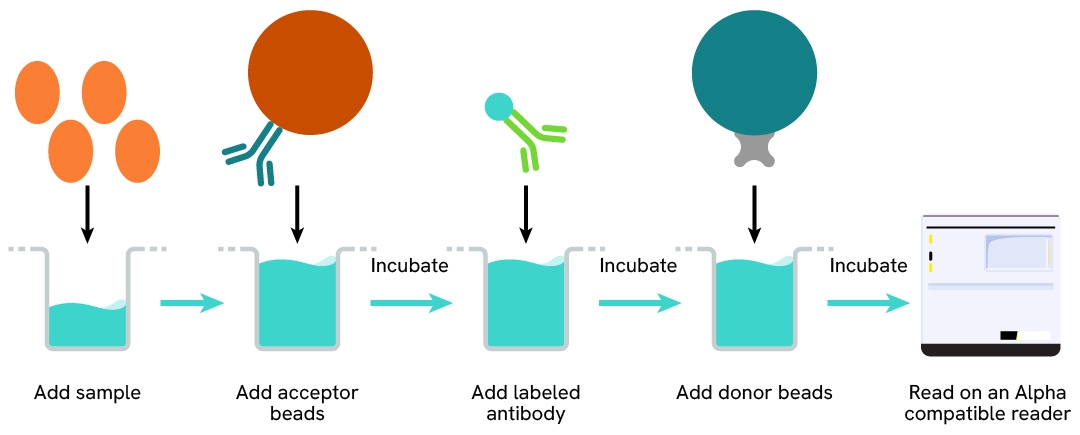
Protocol of the AlphaLISA assay
The AlphaLISA assay can be run in a 96- or 384-well detection plate (50 µL final). As described here, samples or standards are dispensed directly into the assay plate for the detection of the analyte of interest by AlphaLISA reagents. No washing steps are needed. The protocol can be further miniaturized or upscaled by simply resizing each addition volume proportionally.
Specifications
| Application |
Protein Quantification
|
|---|---|
| Automation Compatible |
Yes
|
| Brand |
AlphaLISA
|
| Detection Modality |
Alpha
|
| Dynamic Range |
7.9 - 3,000 ng/mL
|
| Limit of Detection |
7.9 ng/mL
|
| Limit of Quantification |
19.5 ng/mL
|
| Product Group |
Kit
|
| Sample Volume |
5 µL
|
| Shipping Conditions |
Shipped in Blue Ice
|
| Target |
LC3B
|
| Target Class |
Biomarkers
|
| Target Species |
Human
|
| Technology |
Alpha
|
| Unit Size |
5,000 assay points
|
Video gallery
AlphaLISA Human LC3B Detection Kit, 5,000 Assay Points
Resources
Are you looking for resources, click on the resource type to explore further.
Infographic
Autophagy lysosome axis A druggable control layer for homeostasis
Explore the autophagy–lysosome axis as a druggable therapeutic target. Download Revvity's infographic to understand autophagy...
Guide
Protein degradation awareness in a single guide
An in-depth review of molecular and cellular pathways
The maintenance of proteostasis, the biological mechanisms that control the...
Guide
Targeted Protein Degradation (TPD): a comprehensive guide
A comprehensive overview of the revolutionary TPD landscape
This guide explores Targeted Protein Degradation (TPD), the...
Loading...


How can we help you?
We are here to answer your questions.

