


JP
Revvity Sites Globally
Select your location.
*e-commerce not available for this region.

AlphaLISA SureFire Ultra Human Myd88 (Dimer) Detection Kit, 100 Assay Points
AlphaLISA SureFire Ultra Human Myd88 (Dimer) Detection Kit, 100 Assay Points
AlphaLISA SureFire Ultra aggregated
The AlphaLISA™ SureFire® Ultra™ Human MyD88 Dimer assay is a sandwich immunoassay for quantitative detection of MyD88 dimer in cellular lysates using Alpha Technology.
| Feature | Specification |
|---|---|
| Application | 細胞シグナル伝達 |
| Protocol Time | 2h at RT |
| Sample Volume | 30 µL |
The AlphaLISA™ SureFire® Ultra™ Human MyD88 Dimer assay is a sandwich immunoassay for quantitative detection of MyD88 dimer in cellular lysates using Alpha Technology.
Product variants
Unit Size: 100 assay points
Part #:
ALSU-AMYD-A-HV
Unit Size: 500 assay points
Part #:
ALSU-AMYD-A500
Unit Size: 10,000 assay points
Part #:
ALSU-AMYD-A10K
Unit Size: 50,000 assay points
Part #:
ALSU-AMYD-A50K
For research use only. Not for use in diagnostic procedures. All products to be used in accordance with applicable laws and regulations including without limitation, consumption and disposal requirements under European REACH regulations (EC 1907/2006).
AlphaLISA SureFire Ultra Human Myd88 (Dimer) Detection Kit, 100 Assay Points
AlphaLISA SureFire Ultra aggregated
Loading...
Product information
Overview
MYD88 homodimers represent the initial oligomeric state in the assembly of the myddosome signaling complex that transduces signals from TLRs and IL-1Rs. Upon receptor activation, MYD88 molecules undergo conformational changes promoting death domain interactions and dimer formation. MYD88 dimers serve as nucleation sites for higher-order oligomerization that recruit and activate IRAK family kinases. Oncogenic MYD88 L265P mutations enhance MYD88 dimerization and spontaneous myddosome formation, driving chronic NF-κB signaling in B-cell malignancies. Targeting MYD88 dimerization represents a therapeutic strategy for MYD88-mutant lymphomas.
The AlphaLISA SureFire Ultra Human MyD88 Dimer is a sandwich immunoassay for the quantitative detection of MyD88 dimer in cellular lysates, using Alpha Technology.
Formats:
- The HV (high volume) kit contains reagents to run 100 wells in 96-well format, using a 60 μL reaction volume.
- The 500-point kit contains enough reagents to run 500 wells in 384-well format, using a 20 μL reaction volume.
- The 10,000-point kit contains enough reagents to run 10,000 wells in 384-well format, using a 20 μL reaction volume.
- The 50,000-point kit contains enough reagents to run 50,000 wells in 384-well format, using a 20 μL reaction volume.
AlphaLISA SureFire Ultra kits are compatible with:
- Cell and tissue lysates
- Antibody modulators
- Biotherapeutic antibodies
AlphaLISA SureFire Ultra kits can be used for:
- Cellular kinase assays
- Receptor activation studies
- High-throughput screening for preclinical studies
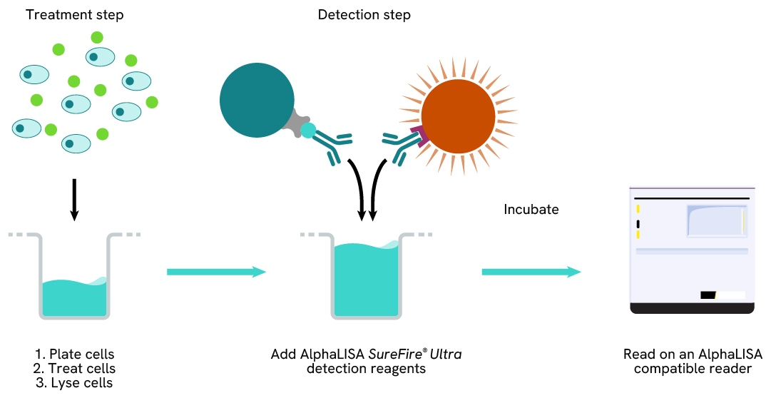
How it works
AlphaLISA SureFire Ultra protein dimer / aggregate detection assay principle
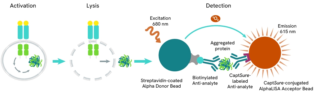
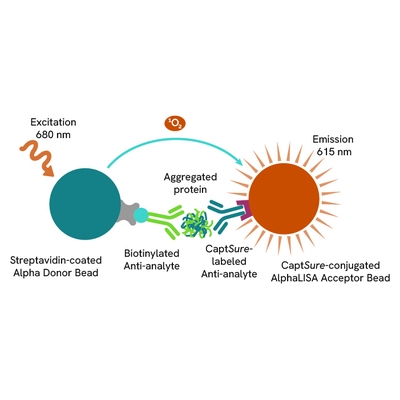
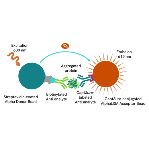
The AlphaLISA SureFire Ultra dimer/aggregate assay measures the levels of a given protein in a dimeric, multimeric or any other aggregated form in a biological sample (e.g. cell lysate).
The assay uses a single antibody clone tagged with both CaptSure™ peptide and biotin, that recognizes exposed epitopes on the multivalent target protein. AlphaLISA assays require two bead types: Acceptor and Donor Beads. Acceptor Beads are coated with a proprietary CaptSure agent to specifically immobilize the antibody labeled with a CaptSure tag. Donor Beads are coated with streptavidin to capture the same specific antibody, which is biotinylated. In the presence of the multivalent protein, the two antibodies bring the Donor and Acceptor Beads in close proximity whereby the singlet oxygen transfers energy to excite the Acceptor Bead, allowing for the generation of a luminescent Alpha signal. The amount of light emission is directly proportional to the quantity of multimeric protein present in the sample.
AlphaLISA SureFire Ultra dimer / aggregate detection two-plate assay protocol
The two-plate protocol involves culturing and treating the cells in a 96-well plate before lysis, then transferring lysates into a 384-well OptiPlate™ plate before the addition of dimer or aggregate AlphaLISA SureFire Ultra detection reagents. This protocol enables cell viability and confluence to be monitored. In addition, lysates from a single well can be used to measure multiple targets.
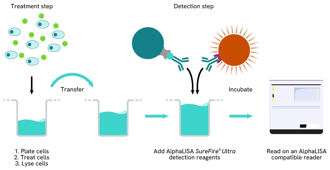
AlphaLISA SureFire Ultra protein dimer / aggregate detection one-plate assay protocol
Detection of the dimer or aggregate with AlphaLISA SureFire Ultra reagents can be performed in a single plate used for culturing, treatment, and lysis. No washing steps are required. This HTS designed protocol allows for miniaturization while maintaining robust AlphaLISA SureFire Ultra quality.
Assay validation
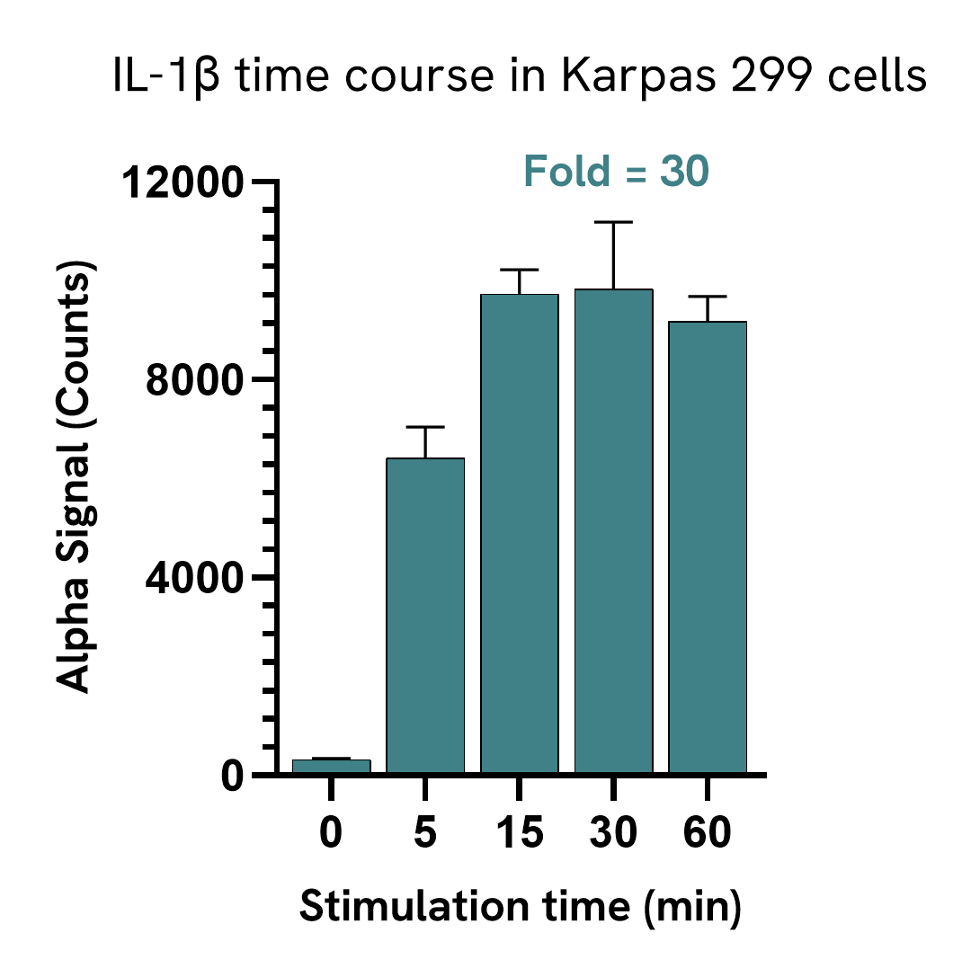
MyD88 Dimer is induced upon IL-1b stimulation
Karpas 299 cells were harvested, washed in HBSS + 0.1% BSA and seeded in a 96-well plate (400,000 cells/well). Cells were treated with 10 ng/mL IL-1b for the indicated time points.
After treatment, the cells were lysed with the addition of 50 µL of 5X Lysis Buffer for 10 minutes at RT with shaking (350 rpm). MyD88 Dimer levels were evaluated by AlphaLISA SureFire Ultra. For the detection step, 10 µL of cell lysate (approximately 16,000 cells) was transferred into a 384-well white OptiPlate, followed by 5 µL of Acceptor mix and incubated for 1 hour at RT. Finally, 5 µL of Donor mix was then added to each well and incubated for 1 hour at RT in the dark. The plate was read on an Envision using standard AlphaLISA settings.
IL-1b induced MyD88 Dimer levels within 5 minutes and the elevated signal levels persisted for up to 60 minutes.
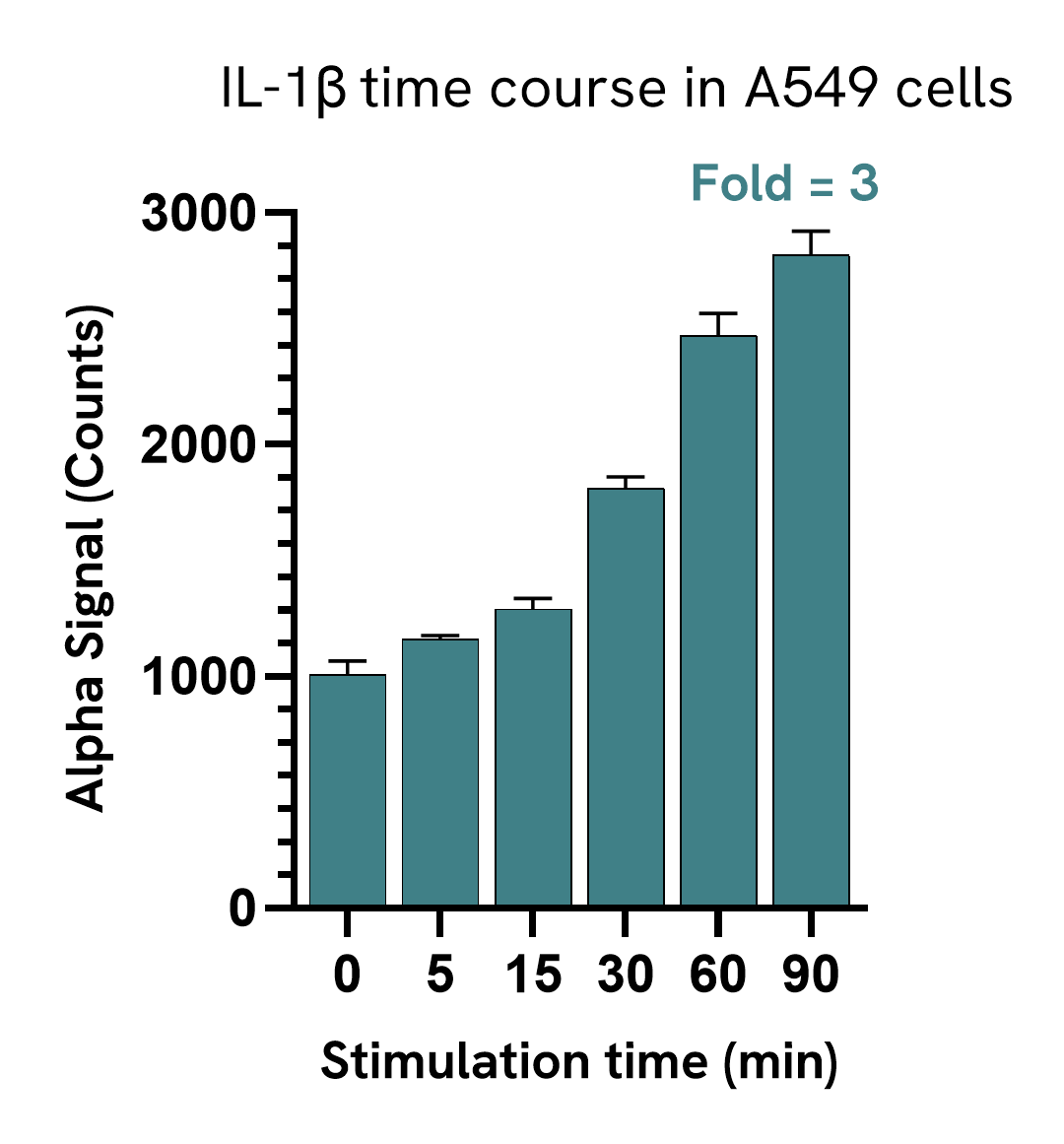
A549 cells were seeded in a 96-well plate (40,000 cells/well) in complete medium, and incubated overnight at 37°C, 5% CO2. Cells were treated with 10 ng/mL IL-1b for the indicated time points.
After treatment, the cells were lysed with 50 µL of Lysis Buffer for 10 minutes at RT with shaking (350 rpm). MyD88 Dimer levels were evaluated by AlphaLISA SureFire Ultra. For the detection step, 10 µL of cell lysate (approximately 8,000 cells) was transferred into a 384-well white OptiPlate, followed by 5 µL of Acceptor mix and incubated for 1 hour at RT. Finally, 5 µL of Donor mix was then added to each well and incubated for 1 hour at RT in the dark. The plate was read on an Envision using standard AlphaLISA settings.
IL-1b induced a modest increase in the levels of MyD88 Dimer in A549 cells.
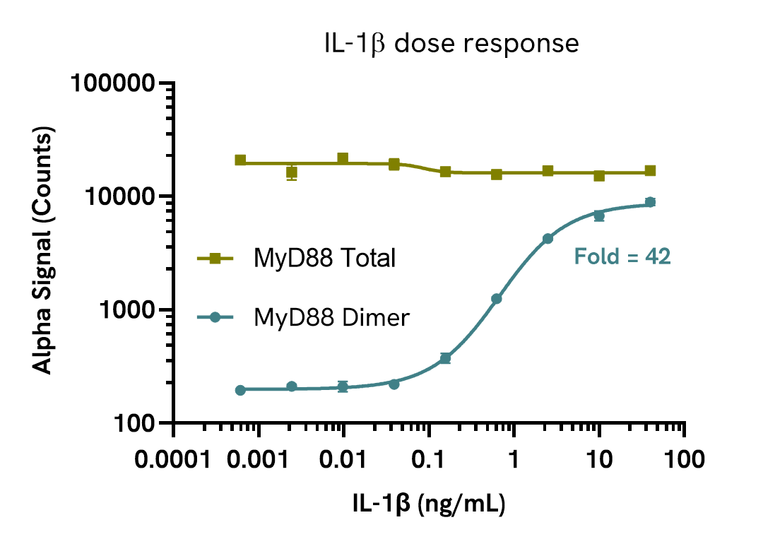
IL-1b induces MyD88 Dimer in a dose-dependent manner
Karpas 299 cells were harvested, washed in HBSS + 0.1% BSA and seeded in a 96-well plate (400,000 cells/well). Cells were treated with increasing concentrations of IL-1b for 5 minutes.
After treatment, the cells were lysed with the addition of 50 µL of 5X Lysis Buffer for 10 minutes at RT with shaking (350 rpm). MyD88 Dimer and Total MyD88 levels were evaluated using respective AlphaLISA SureFire Ultra assays. For the detection step, 10 µL of cell lysate (approximately 16,000 cells) was transferred into a 384-well white OptiPlate, followed by 5 µL of Acceptor mix and incubated for 1 hour at RT. Finally, 5 µL of Donor mix was then added to each well and incubated for 1 hour at RT in the dark. The plate was read on an Envision using standard AlphaLISA settings.
As expected, MyD88 Dimer was upregulated in a dose-dependent manner while Total MyD88 levels remained unchanged.
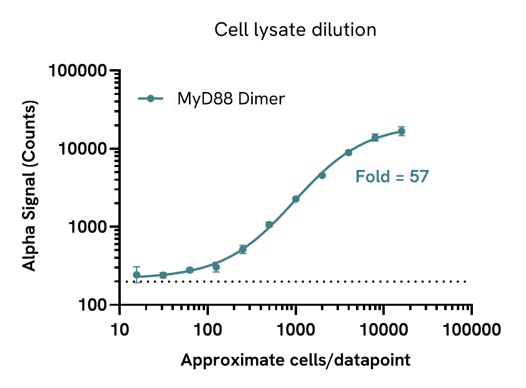
Assay sensitivity
Assay sensitivity - cell lysate dilution
Cell lysate was prepared from Karpas 299 cells prepared at 2 x 10⁶ cells/mL and stimulated with 10 ng/mL IL-1b for 10 minutes in HBSS + 0.1% BSA. Cells were lysed with the addition of 5X Lysis Buffer for 10 minutes at RT with shaking.
Lysate was serially diluted in Lysis Buffer and MyD88 Dimer levels were evaluated by AlphaLISA SureFire Ultra. For the detection step, 10 µL of lysate was transferred into a 384-well white OptiPlate, followed by 5 µL of Acceptor mix and incubated for 1 hour at room temperature. Finally, 5 µL of Donor mix was then added to each well and incubated for 1 hour at RT in the dark. The plate was read on an Envision using standard AlphaLISA settings.
Approximate number of cells per datapoint is indicated. The dotted line represents assay background. The assay can detect MyD88 Dimer down to 500 cells/datapoint.
Specifications
| Application |
Cell Signaling
|
|---|---|
| Automation Compatible |
Yes
|
| Brand |
AlphaLISA SureFire Ultra
|
| Cellular or Signaling Pathway |
Inflammasome/Pattern Recognition Receptors (PRRs)
|
| Detection Modality |
Alpha
|
| Product Group |
Kit
|
| Protocol Time |
2h at RT
|
| Sample Volume |
30 µL
|
| Shipping Conditions |
Shipped in Blue Ice
|
| Target |
Myd88
|
| Target Class |
Phosphoproteins
|
| Target Species |
Human
|
| Technology |
Alpha
|
| Therapeutic Area |
Inflammation
|
| Unit Size |
100 assay points
|
Resources
Are you looking for resources, click on the resource type to explore further.
Brochure
Alpha assays and reagents catalog
Alpha technolgy enables the rapid and straightforward mesaure of virtually any target. This includes enzymes, receptor-ligand...
Brochure
Alpha SureFire Ultra no-wash immunoassay catalog
Discover Alpha SureFire® Ultra™ assays, the no-wash cellular kinase assays leveraging Revvity's exclusive bead-based technology...
Brochure
Species compatibility for HTRF, AlphaLISA SureFire Ultra and Alpha SureFire Ultra Multiplex assays
This document includes detailed tables listing HTRF™, AlphaLISA™ SureFire® Ultra™, and Alpha SureFire® Ultra™ Multiplex assays...
Loading...


How can we help you?
We are here to answer your questions.

