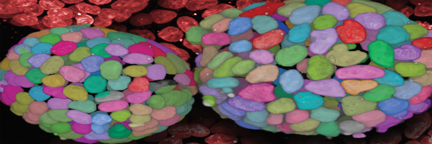
More and more researchers are turning to 3D cell models for high-content screening to better understand diseases and to test potential therapeutics.
But getting good high-content data out of these little aggregates of cells isn’t always easy – sometimes they don’t grow the same way each time you prepare them, and other times, getting good images of them and their subcellular components can be difficult because they’re just too thick. Or maybe it’s the mountains of imaging data (and the time it takes to acquire it) that’s giving you a headache?
Don’t despair – working with 3D cell models isn’t an impossible task, and the rewards can be immense. There are lots of tips and tricks to get your cell models to reveal their hidden depths.
Get the best quality images
High-quality images are the cornerstone of successful high-content analysis. For 3D cell models, confocal imaging yields the highest sensitivity, best signal-to-noise ratios, and highest X, Y, and Z resolution compared to widefield imaging, without losing throughput.
Water immersion objectives are much better than air objectives at capturing light and providing high X, Y, Z resolution, so you can capture crisp cellular details and see further into 3D structures.
If you’re working with spheroids, you can try optical clearing. It reduces light scattering and homogenizes refractive indices, so it’s easier to see the core – possibly the most interesting part –that would otherwise remain in the dark.
Consider using longer wavelength dyes – the decrease in light scattering and increase in light penetration can improve imaging depth and signal detection.
Minimize imaging time and volume of data
When you’re capturing very high-resolution images to see fine subcellular detail, it’s almost inevitable that the high volume of imaging data will slow you down. But not totally inevitable…often only part of the total well area, where the 3D model is situated, is of interest. So why waste your time acquiring data from the rest of the well?
With intelligent image acquisition technology, you can find 3D cell models at low resolution and high speed, and then image that region of interest at high-resolution, cutting down on acquisition and analysis times as well as data volume.
Read more in our guide “Harness the power of 3D cell model imaging”
 Guide
Harness the power of 3D cell model imaging


