


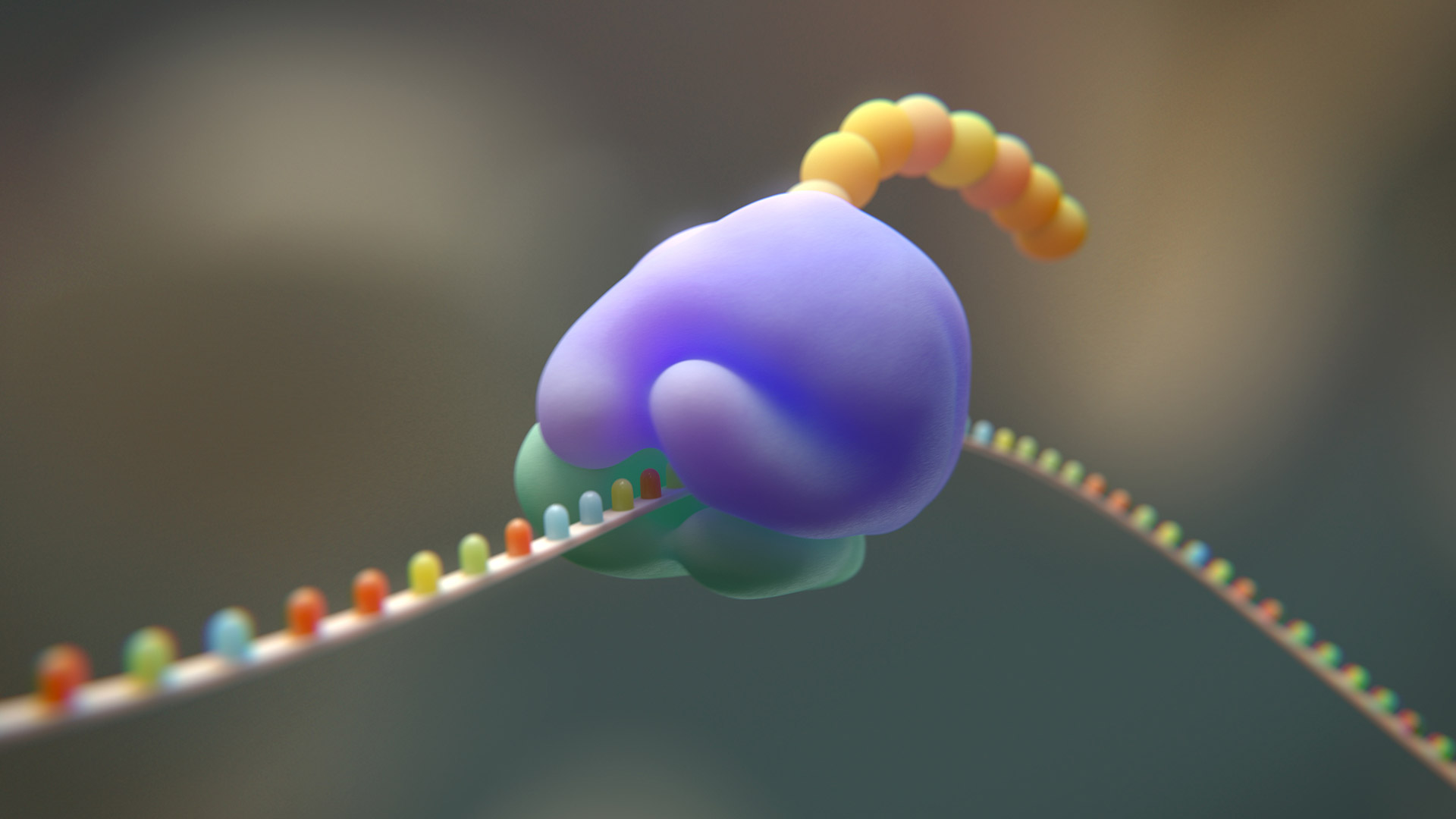

Ribosome profiling (Ribo-seq) generates short ribosome-protected fragments (RPFs), typically ~28–30 nucleotides in length in mammalian systems, which are sequenced to infer occupancy at nucleotide resolution1.
Since its original description, the method has undergone substantial methodological refinement, including modifications to nuclease digestion strategies, ribosome isolation approaches, size-selection windows, and library preparation workflows. Variants such as low-input protocols and selective ribosome capture methods2 have further expanded its applications while preserving the central principle of sequencing nuclease-protected ribosomal footprints to measure translational activity genome-wide.
As Ribo-seq has matured, a set of reproducible quality control metrics has emerged. These metrics allow researchers to assess whether the data reflect bona fide ribosome occupancy rather than RNA degradation fragments or technical artifacts introduced during nuclease digestion, ribosome isolation, or small RNA-seq library preparation.
Below we summarize the most widely accepted Ribo-seq quality control metrics, along with value ranges generally considered indicative of high-quality data in well-optimized mammalian datasets. These thresholds primarily reflect cytosolic ribosome profiling in mammalian systems using standard RNase I digestion conditions. In other organisms, including yeast, bacteria, plants or non-model species, ribosome footprint length, periodicity strength, and coding sequence enrichment patterns may differ substantially due to variations in ribosome structure, translation dynamics, nuclease sensitivity, and experimental protocol. Therefore, QC metrics should be interpreted within the biological and technical context of the system under study rather than applied as universal cutoffs.
Core quality metrics in ribosome profiling
Read length distribution
The first and most immediate metric is the read length histogram, also called a footprint length distribution plot. In mammalian systems, high-quality Ribo-seq libraries typically show a dominant peak at 28–30 nt3.
In well-digested libraries:
- ≥70–80% of mapped reads fall within a ±1 nucleotide window around the modal peak.
- The dominant peak spans only 1–2 nucleotides.
A broader distribution (spread >3–4 nt), multiple strong peaks, or absence of a clear dominant length often indicates heterogeneous RNase digestion or contamination with non-ribosomal fragments.
rRNA contamination
Ribosomes are ribonucleoprotein complexes, and therefore rRNA-derived fragments are a common technical contaminant. This is typically evaluated using a read composition bar plot showing the fraction of reads mapping to rRNA, tRNA, CDS, UTR, and intergenic regions.
After rRNA filtering4:
- High-quality libraries generally contain ≤20% rRNA-derived reads,
- Well-optimized workflows often achieve ≤10–15%.
Libraries with >30–40% rRNA reads reduce usable sequencing depth and may require improved depletion or digestion optimization.
Coding sequence (CDS) enrichment
RPFs should predominantly map to annotated coding regions. This is visualized using a feature distribution plot showing the percentage of reads in CDS, 5′UTR, 3′UTR, intronic, and intergenic regions.
Accepted standards
- ≥65–80% of uniquely mapped reads in coding sequences.
- Minimal intronic or intergenic mapping.
Values below ~60% CDS enrichment often indicate RNA degradation fragments rather than ribosome-protected fragments.
Three-nucleotide periodicity
The defining hallmark of Ribo-seq is three-nucleotide periodicity, reflecting codon-by-codon ribosome movement. This is assessed using a frame periodicity plot, which displays the proportion of reads mapping to frame 0, +1, or +2 relative to annotated coding regions.
In high-quality mammalian datasets:
- ≥60–70% of reads align to the dominant reading frame,
- Clear periodic phasing is observed across coding sequences.
Loss of periodicity or equal distribution across frames suggests contamination with non-ribosomal fragments or incorrect P-site assignment.
P-site offset calibration and metagene profiles
Accurate codon-resolution analysis requires correct inference of the ribosomal P-site position. This is determined using metagene plots centered on annotated start and stop codons.
A high-quality dataset typically shows:
- A sharp enrichment peak at annotated start codons,
- Clear periodic phasing downstream of initiation sites,
- Minimal signal extending into 3′UTRs beyond stop codons.
Diffuse initiation peaks or absence of periodic phasing indicate potential issues with digestion, alignment, or P-site calibration.
Replicate correlation
Reproducibility is evaluated using gene-level scatter plots and correlation heatmaps. In well-controlled biological replicates:
- Pearson or Spearman correlation coefficients are typically ≥0.90,
- Read length distributions and CDS enrichment metrics are consistent across replicates.
Correlations below ~0.80 suggest variability in digestion, ribosome isolation, or sample handling.
| Metric | Accepted Good Range | Warning Signs |
|---|---|---|
| Read length distribution | Dominant peak at 28–30 nt; ≥70–80% within ±1 nt | Broad distribution; multiple peaks |
| rRNA contamination | ≤20% rRNA (ideally ≤15%) | >30–40% rRNA |
| CDS enrichment | ≥65–80% reads in CDS | <60% CDS mapping |
| Three-nt periodicity | ≥60–70% reads in dominant frame | Weak/no frame enrichment |
| P-site calibration | Sharp start peak; clear phasing | Diffuse initiation signal |
| Replicate correlation | r ≥ 0.90 | r < 0.80 |
Table 1. Summary of common Ribo-seq quality metrics
Analytical pipelines
Ribosome profiling analysis pipelines typically involve a series of standardized processing steps. These include adapter trimming, removal of rRNA-derived reads, alignment to the reference genome or transcriptome, read length stratification, calibration of the ribosomal P-site offset, assessment of three-nucleotide periodicity, and calculation of translational efficiency when matched RNA-seq data are available.
Several software ecosystems support ribosome profiling analysis. Packages such as RiboTaper, RiboCode, and Plastid provide functionality for frame periodicity assessment, P-site assignment, and codon-resolution analysis. Standard analytical workflows and methodological considerations are described in detail by McGlincy and Ingolia4.
More recently, integrated environments such as Martian™ have been developed to streamline ribosome profiling analysis by generating automated quality control reports. The automation and standardization of quality control reduce user-dependent variability and simplify data processing and QC interpretation for laboratories without dedicated bioinformatics support. These reports typically include read length histograms, frame periodicity plots, start and stop codon metagene analyses, and replicate correlation matrices within standardized output formats.
Regardless of platform, the core QC principles remain consistent with those defined in early foundational studies.
Conclusions
Over more than a decade of experimental application, ribosome profiling has established a well-defined set of quality benchmarks. Tight footprint length distribution, strong three-nucleotide periodicity, high CDS enrichment, low rRNA contamination, accurate P-site calibration, and strong replicate reproducibility are now widely accepted indicators of high-quality Ribo-seq data.
For researchers implementing ribosome profiling in drug discovery or perturbation studies, these metrics provide a practical framework for judging whether a Ribo-seq library is likely to reflect bona fide ribosome occupancy, while still requiring interpretation in the context of sample type, protocol design, and analysis parameters.
References:
- Ingolia, N.T., et al. (2009). Genome-wide analysis in vivo of translation with nucleotide resolution using ribosome profiling. Science. 324(5924):218-23. doi: 10.1126/science.1168978.
- Clamer, M., et al. (2018). Active Ribosome Profiling with RiboLace. Cell Rep. 25(4):1097-1108.e5. doi: 10.1016/j.celrep.2018.09.084.
- Ingolia, N.T., et al. (2011). Ribosome profiling of mouse embryonic stem cells reveals the complexity and dynamics of mammalian proteomes. Cell. 147(4):789-802. doi: 10.1016/j.cell.2011.10.002.
- McGlincy, N.J., Ingolia, N.T. (2017). Transcriptome-wide measurement of translation by ribosome profiling. Methods. 126:112-129. doi: 10.1016/j.ymeth.2017.05.028.
For research use only. Not for use in diagnostic procedures.


