


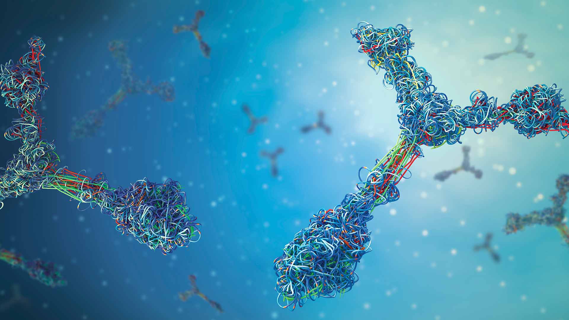

Extracellular vesicles (EVs) are membrane-bound particles secreted by virtually all cell types. They carry a diverse array of biomolecules including lipids, proteins, RNAs, and occasionally DNA. Their content reflects the physiological state of their cell of origin. EVs are increasingly explored for biomarker discovery , with their RNA cargo commonly analyzed through small RNA sequencing to capture disease- or tissue-specific signals.
Human plasma is known to be a molecular “mirror” of the internal organs, providing a snapshot of cellular activity across the entire body. Because of this, it is a compelling source for EVs , particularly when direct tissue sampling is invasive or unfeasible. The challenge is that in human plasma ~ 99.8% of EVs derive from hematopoietic (blood) cells, with only ~ 0.2% attributed to solid organs or other non-blood tissues1,2. This cellular bias introduces significant background noise into EV-derived miRNA datasets, masking signals from tissues of interest, and limiting reproducibility across studies.
Targeted immunocapture with NEXPLOR
A recent study addressed this problem with Mursla Bio’s NEXPLOR® (Novel EXtracellular vesicle PopuLation and Omics Revealer), a magnetic bead-based system that captures EVs from a specific tissue by using a validated cocktail of surface antibodies. NEXPLOR® improves tissue specificity to EVs datasets and represents a critical step toward resolving the context problem that has long challenged the EV field3
For liver EVs, a panel of four hepatocyte-specific markers (ASGR1, ASGR2, TFR2, and SLCO1B1) was identified through bioinformatics and experimental validation, enabling enrichment of hepatocyte-derived EVs (h-EVs) directly from plasma. In the second half of the study, h-EVs were isolated from plasma donors with hepatocellular carcinoma and cirrhosis, and healthy controls. Small RNA libraries were then prepared using the NEXTFLEX Small RNA-Seq Kit v4, and sequenced on the Illumina NextSeq® 2000 platform.
Bulk EVs yielded a much broader miRNA repertoire (250 miRNAs detected) but included many non-specific species likely from hematopoietic cells. In contrast, h-EVs contained only 39 miRNAs, but these were more informative and tightly associated with liver biology and hepatocellular carcinoma.
Specifically, miR-124-3p and miR-23b-3p, both known tumor suppressors in hepatocellular carcinoma, were only detected in h-EVs, not in bulk EVs. Differential expression analysis revealed five miRNAs significantly altered between hepatocellular carcinoma and cirrhosis samples exclusively in h-EVs, including miR-22-3p and miR-23a-3p. Finally, it was found that h-EV–associated miRNAs showed specific activation of liver cancer–relevant pathways like HIF-1 signalling, glycolysis/gluconeogenesis, and hepatocyte growth factor receptor signalling, which were not enriched in bulk EVs.
The implications are profound: while bulk EVs offer quantity, h-EVs offer quality and biological resolution, especially for miRNA-based biomarkers. This is analogous to how single-cell sequencing enhances resolution within bulk transcriptomic data, by disentangling complex mixtures to reveal signals from defined cellular or tissue source.
Why this matters for EV small RNA research
This work sets a new standard toward reliable, multi-omic liquid biopsies. Small RNA-seq data from enriched EV subsets reflect true tissue biology, reducing the noise of systemic circulation and facilitating meaningful interpretation of miRNA profiles.
Learn more about Revvity’s Small RNA Solution
References
- Alberro, A. et al. (2021). Extracellular Vesicles in Blood: Sources, Effects, and Applications. Int. J. Mol. Sci. 22, 8163. doi: 10.3390/ijms22158163.
- Auber, M., Svenningsen, P. (2022). An estimate of extracellular vesicle secretion rates of human blood cells. J Extracell Biol. 1(6):e46. doi: 10.1002/jex2.46.
- Figueiras, R., et al. (2025). Organ-specific isolation of hepatocyte extracellular vesicles from human plasma enables tissue-resolved proteomic and miRNA profiling. bioRxiv 2025.06.13.658908; doi: 10.1101/2025.06.13.658908.


