


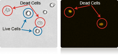
Examples of cell viability measurement using Propidium Iodide
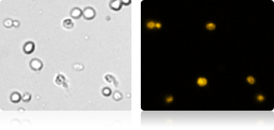
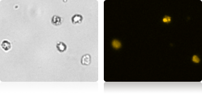
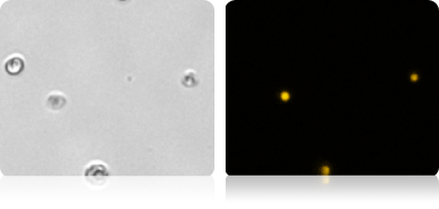
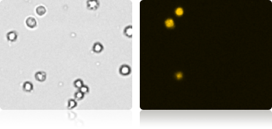
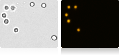
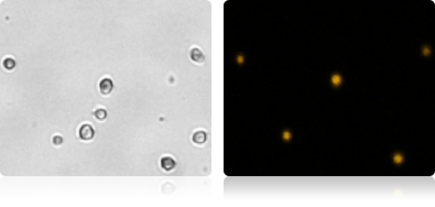
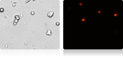
Below are images of cultured cells that have been stained with propidium iodide (PI) and analyzed using a Cellometer Vision* instrument. For each example a brightfield and a fluorescent image is provided. In brightfield, live cells appear round with well-defined membranes and bright centers (example cells circled in blue). The cell morphology of dead cells is often distinctly different from live cells. Non-viable cells often have poorly defined faint cell membranes and centers (example cells circled in red). Only the non-viable cells are stained with PI and are seen in the fluorescent images.
*The Cellometer Vision has been superseded by the Cellometer Spectrum instrument.
3T3 cells
HEK293 cells
K562 cells
PC3 cells
CHO cells
Hi5 cells
LnCap cells
Jurkat cells
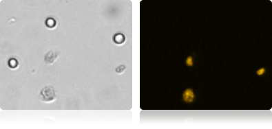
Viability measurement in primary cells
Detection of viable cells in primary samples is an important step in evaluating the sample quality and percent of viable cells.
Tumor digest cell images
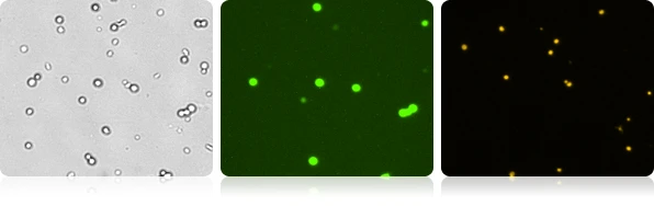
Viability measurement in GFP expressing cells
By staining the cells with propidium iodide, in a single assay, we can monitor the quality of the cell culture during GFP expression. In this example, healthy mouse embryonic stem cells that are expressing GFP are shown to be negative for PI staining, while other cells in the culture are PI positive.
GFP expressing mouse embryonic stem cells


For research use only. Not for use in diagnostic procedures.