メニュー
Revvity logo
Contact us
JP
Revvity Sites Globally

Select your location.

*e-commerce not available for this region.

australia.webp Australia
austria.webp Austria
belgium.webp Belgium
brazil.webp Brazil *
canada.webp Canada
china.webp China *
denmark.webp Denmark
finland.webp Finland
france.webp France
germany.webp Germany
hong-kong.webp Hong Kong (China) *
india.webp India *
ireland.webp Ireland
italy.webp Italy
japan.webp Japan *
luxembourg.webp Luxembourg
mexico.webp Mexico *
netherlands.webp Netherlands
norway.webp Norway
philippines.webp Philippines *
republic of korea.webp Republic of Korea *
singapore.webp Singapore *
spain.webp Spain
sweden.webp Sweden
switzerland.webp Switzerland
thailand.webp Thailand *
uk.webp United Kingdom
usa.webp United States
Breadcrumb
...
  • ホーム
  • Knowledge Base
  • CellCountingSub Section
  • CPE of ECMV on Baby Hamster Kidney Cells
Cell Counting and Image Cytometry

CPE of ECMV on Baby Hamster Kidney Cells

Section
Modern Virology Assays
Celigo Applications
Cell Counting Method Selection
Cell Counting and Image Cytometry FAQs
Cell-based Assays for Bioprocessing
Cell-based Assays for Gene Therapy Development
Cellometer Applications
Modern Virology Assays
Sub Section
Cytopathic Effects
Antibody Binding Inhibition
Antibody Neutralization
Cytopathic Effects
Viral Titre
Topic
CPE of ECMV on Baby Hamster Kidney Cells
CPE of SV-5 Virus on CHO-K1 Cells
CPE of Zika Virus on HMC3 and JEG-3 Cells
CPE of Measles Virus on MRC-5 Cells
CPE of PI3 Virus on Vero Cells

Cytopathic effects (CPE) of encephalomyocalditis virus (ECMV) on baby hamster kidney cells (BSR)

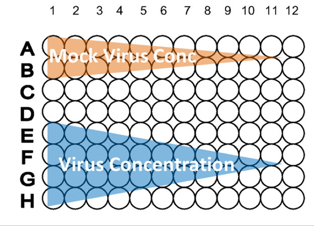
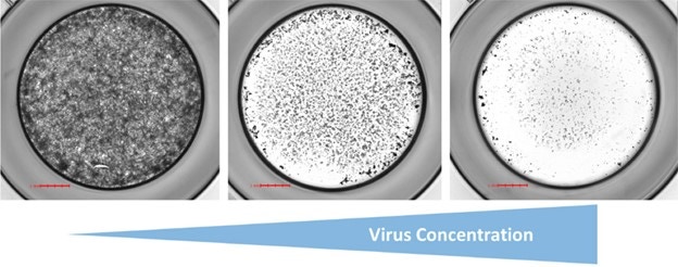
The BSR host cells are seeded in a 96-well plate and allowed to reach 100% confluence in the wells. The host cells are then infected with ECMV and a mock virus at 1:10 titrations following the plate map shown below. After the incubation, the monolayer of cells is fixed and stained with crystal violet. As the virus concentration increase, the host cells are killed and reducing the monolayer in the wells. The Celigo™ image cytometer automatically quantifies the percentage of host cells remaining in each well which, in this example, contain serial dilutions of ECMV.

CPE of ECMV on baby hamster kidney cells-1

Plate map showing host cells infected with ECMV and a mock virus at 1:10 titrations
CPE of ECMV on baby hamster kidney cells-2

Virus concentration-dependent CPE response showing a reduction in host cell confluence as virus concentration increases. From left, monolayer remaining is 99%, 40% and 10%.
CPE of ECMV on baby hamster kidney cells-3

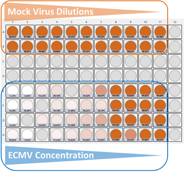
Celigo generated heat-map for immediate viewing of trends and reproducibility
CPE of ECMV on baby hamster kidney cells-4

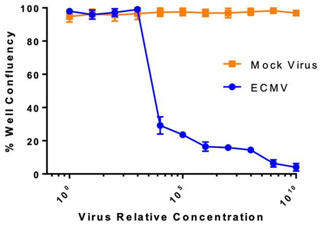
The confluence percentage data can be plotted to produce viral titer curves and determine TCID50s

For research use only. Not for use in diagnostic procedures.

ON THIS PAGE
Revvity Logo

©2025 Revvity - All rights reserved

Revvity is a trademark of Revvity, Inc. All other trademarks are the property of their respective owners.